Advertisement

Main menu

  • Home
  • Content
    • Current Issue
    • Accepted Manuscripts
    • Article Preview
    • Past Issue Archive
    • Video Articles
    • AJNR Case Collection
    • Case of the Week Archive
    • Case of the Month Archive
    • Classic Case Archive
  • Special Collections
    • AJNR Awards
    • Low-Field MRI
    • Alzheimer Disease
    • ASNR Foundation Special Collection
    • Photon-Counting CT
    • View All
  • Multimedia
    • AJNR Podcasts
    • AJNR SCANtastic
    • Trainee Corner
    • MRI Safety Corner
    • Imaging Protocols
  • For Authors
    • Submit a Manuscript
    • Submit a Video Article
    • Submit an eLetter to the Editor/Response
    • Manuscript Submission Guidelines
    • Statistical Tips
    • Fast Publishing of Accepted Manuscripts
    • Graphical Abstract Preparation
    • Imaging Protocol Submission
    • Author Policies
  • About Us
    • About AJNR
    • Editorial Board
    • Editorial Board Alumni
  • More
    • Become a Reviewer/Academy of Reviewers
    • Subscribers
    • Permissions
    • Alerts
    • Feedback
    • Advertisers
    • ASNR Home

User menu

  • Alerts
  • Log in

Search

  • Advanced search
American Journal of Neuroradiology

American Journal of Neuroradiology

ASHNR American Society of Functional Neuroradiology ASHNR American Society of Pediatric Neuroradiology ASSR
  • Alerts
  • Log in

Advanced Search

  • Home
  • Content
    • Current Issue
    • Accepted Manuscripts
    • Article Preview
    • Past Issue Archive
    • Video Articles
    • AJNR Case Collection
    • Case of the Week Archive
    • Case of the Month Archive
    • Classic Case Archive
  • Special Collections
    • AJNR Awards
    • Low-Field MRI
    • Alzheimer Disease
    • ASNR Foundation Special Collection
    • Photon-Counting CT
    • View All
  • Multimedia
    • AJNR Podcasts
    • AJNR SCANtastic
    • Trainee Corner
    • MRI Safety Corner
    • Imaging Protocols
  • For Authors
    • Submit a Manuscript
    • Submit a Video Article
    • Submit an eLetter to the Editor/Response
    • Manuscript Submission Guidelines
    • Statistical Tips
    • Fast Publishing of Accepted Manuscripts
    • Graphical Abstract Preparation
    • Imaging Protocol Submission
    • Author Policies
  • About Us
    • About AJNR
    • Editorial Board
    • Editorial Board Alumni
  • More
    • Become a Reviewer/Academy of Reviewers
    • Subscribers
    • Permissions
    • Alerts
    • Feedback
    • Advertisers
    • ASNR Home
  • Follow AJNR on Twitter
  • Visit AJNR on Facebook
  • Follow AJNR on Instagram
  • Join AJNR on LinkedIn
  • RSS Feeds

AJNR Awards, New Junior Editors, and more. Read the latest AJNR updates

Research ArticleHead and Neck Imaging

Nonbifurcating Cervical Carotid Artery Diagnosed by MR Angiography

A. Uchino, N. Saito, T. Watadani, W. Mizukoshi and R. Nakajima
American Journal of Neuroradiology June 2011, 32 (6) 1119-1122; DOI: https://doi.org/10.3174/ajnr.A2462
A. Uchino
  • Find this author on Google Scholar
  • Find this author on PubMed
  • Search for this author on this site
N. Saito
  • Find this author on Google Scholar
  • Find this author on PubMed
  • Search for this author on this site
T. Watadani
  • Find this author on Google Scholar
  • Find this author on PubMed
  • Search for this author on this site
W. Mizukoshi
  • Find this author on Google Scholar
  • Find this author on PubMed
  • Search for this author on this site
R. Nakajima
  • Find this author on Google Scholar
  • Find this author on PubMed
  • Search for this author on this site
  • Article
  • Figures & Data
  • Info & Metrics
  • Responses
  • References
  • PDF
Loading

Article Figures & Data

Figures

  • Tables
  • Fig 1.
    • Download figure
    • Open in new tab
    • Download powerpoint
    Fig 1.

    Case 2. Anteroposterior (A) and lateral (B) partial MIP images of the left carotid artery show a separately arising F-L trunk, a distal ECA, and the OA. There is no physiologic dilation of the ICA at its origin. Arrowheads and arrows in A indicate the most proximally arising F-L trunk and in B, the most distally arising OA.

  • Fig 2.
    • Download figure
    • Open in new tab
    • Download powerpoint
    Fig 2.

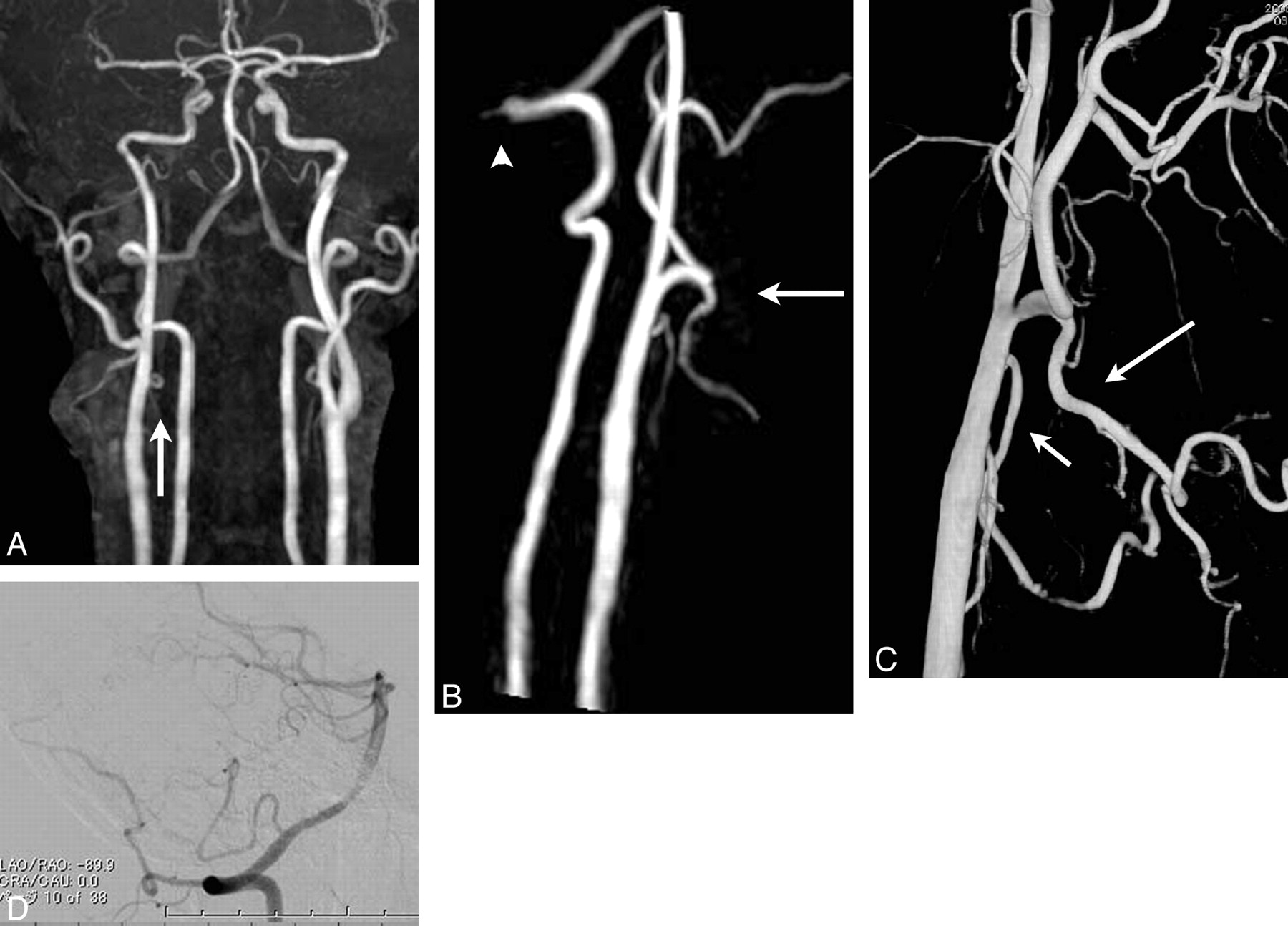
    Case 6. Anteroposterior MIP image (A) and lateral partial MIP image of the right carotid and vertebrobasilar arteries (B) show a straight ICA without physiologic dilation and a separately arising LA. The arrow in A indicates the LA. The FA originates from the distal ECA (arrow in B), resembling high bifurcation of the carotid artery. However, the OA arises from the right VA (arrowhead in B). C, 3D DSA of the right CCA clearly demonstrates the anomalously branching pattern. D, The short arrow indicates the LA, and the long arrow indicates the FA. DSA of a lateral projection of the right VA shows the OA arising from the level of the craniovertebral junction (arrowhead).

  • Fig 3.
    • Download figure
    • Open in new tab
    • Download powerpoint
    Fig 3.

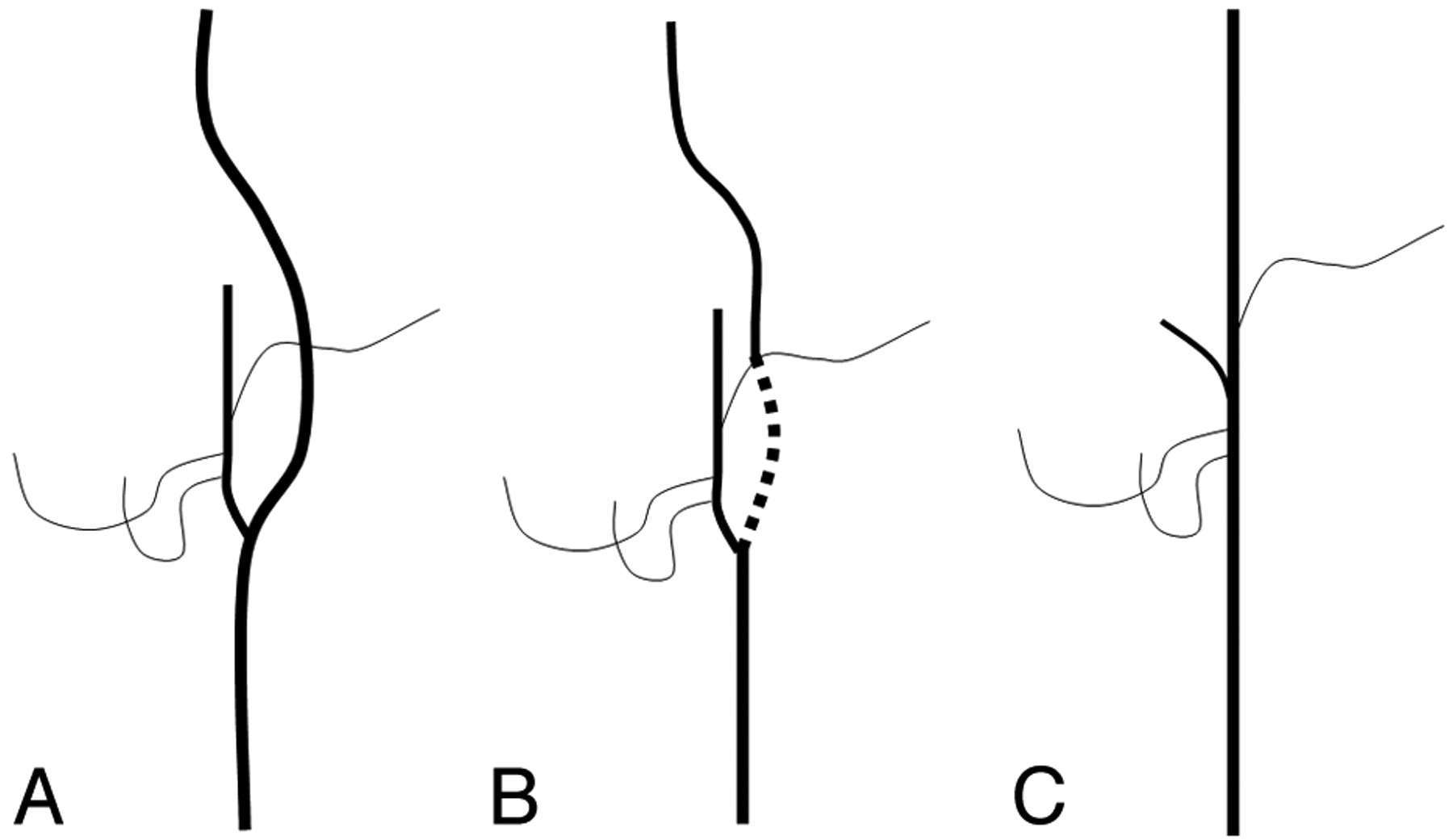
    Schematic illustration of our hypothesis of a nonbifurcating cervical carotid artery in a lateral projection. A, Normal development. B, Agenesis of the most proximal ICA. C, Final configuration of this anomaly. The proximal OA or APA probably plays an important role in forming the nonbifurcating cervical carotid artery.

Tables

  • Figures
    • View popup
    Table 1:

    Nonbifurcating cervical carotid artery in this study

    Case No./Age (yr)/Sex/Left or RightBranching Order (Proximal to Distal)Diagnostic Modality
    1/76/F/leftF-L trunk, main trunk of ECA, OAMRA
    2/60/M/leftF-L trunk, main trunk of ECA, OAMRA
    3/78/M/rightF-L trunk, main trunk of ECA, OAMRA
    4/72/M/rightF-L trunk, main trunk of ECA, OAMRA
    5/78/M/rightLA, common trunk of FA, ECA, OAMRA
    6/48/M/rightLA, common trunk of FA, ECAaMRA/angiography
    • a OA arising from the VA.

    • View popup
    Table 2:

    Reported cases of a nonbifurcating cervical carotid artery in the English language literature

    Reference No./Age (yr)/Sex/Left or RightBranching Order (Proximal to Distal)Diagnostic Modality
    1/52/M/leftLA, FA, main trunk of ECA, OAAutopsy
    2/43/M/leftF-L trunk, main trunk of ECAaAngiography/surgery
    3/55/F/leftLA, FA, main trunk of ECA, OAAngiography
    4/76/M/leftF-L trunk, main trunk of ECAaAngiography
    5/67/M/leftLA, FA, main trunk of ECA, OAAutopsy
    6/66/M/rightLA, FA, main trunk of ECAaAngiography/MRA/surgery
    7/75/F/leftF-L trunk, OA, main trunk of ECAAngiography
    7/67/F/leftF-L trunk, main trunk of ECA, OAAngiography
    7/65/M/leftF-L trunk, main trunk of ECA, OAAngiography
    8/64/F/rightLA, FA, main trunk of ECAaAngiography
    9/71/M/rightLA, FA, main trunk of ECA, OAAngiography
    • a OA arising from the carotid artery could not be identified.

PreviousNext
Back to top

In this issue

American Journal of Neuroradiology: 32 (6)
American Journal of Neuroradiology
Vol. 32, Issue 6
1 Jun 2011
  • Table of Contents
  • Index by author
Advertisement
Print
Download PDF
Email Article

Thank you for your interest in spreading the word on American Journal of Neuroradiology.

NOTE: We only request your email address so that the person you are recommending the page to knows that you wanted them to see it, and that it is not junk mail. We do not capture any email address.

Enter multiple addresses on separate lines or separate them with commas.
Nonbifurcating Cervical Carotid Artery Diagnosed by MR Angiography
(Your Name) has sent you a message from American Journal of Neuroradiology
(Your Name) thought you would like to see the American Journal of Neuroradiology web site.
CAPTCHA
This question is for testing whether or not you are a human visitor and to prevent automated spam submissions.
Cite this article
A. Uchino, N. Saito, T. Watadani, W. Mizukoshi, R. Nakajima
Nonbifurcating Cervical Carotid Artery Diagnosed by MR Angiography
American Journal of Neuroradiology Jun 2011, 32 (6) 1119-1122; DOI: 10.3174/ajnr.A2462

Citation Manager Formats

  • BibTeX
  • Bookends
  • EasyBib
  • EndNote (tagged)
  • EndNote 8 (xml)
  • Medlars
  • Mendeley
  • Papers
  • RefWorks Tagged
  • Ref Manager
  • RIS
  • Zotero
0 Responses
Respond to this article
Share
Bookmark this article
Nonbifurcating Cervical Carotid Artery Diagnosed by MR Angiography
A. Uchino, N. Saito, T. Watadani, W. Mizukoshi, R. Nakajima
American Journal of Neuroradiology Jun 2011, 32 (6) 1119-1122; DOI: 10.3174/ajnr.A2462
del.icio.us logo Twitter logo Facebook logo Mendeley logo
  • Tweet Widget
  • Facebook Like
  • Google Plus One
Purchase

Jump to section

  • Article
    • Abstract
    • Abbreviations
    • Materials and Methods
    • Results
    • Discussion
    • Conclusions
    • Acknowledgments
    • References
  • Figures & Data
  • Info & Metrics
  • Responses
  • References
  • PDF

Related Articles

  • No related articles found.
  • PubMed
  • Google Scholar

Cited By...

  • No citing articles found.
  • Crossref (22)
  • Google Scholar

This article has been cited by the following articles in journals that are participating in Crossref Cited-by Linking.

  • Variation of the origin of the left common carotid artery diagnosed by CT angiography
    Akira Uchino, Naoko Saito, Yoshitaka Okada, Eito Kozawa, Naoko Nishi, Waka Mizukoshi, Reiko Nakajima, Masahiro Takahashi, Yusuke Watanabe
    Surgical and Radiologic Anatomy 2013 35 4
  • Congenital external carotid–internal carotid artery anastomosis associated with contralateral non-bifurcating cervical carotid artery
    Kanji Nakai, Tatsumi Kaji, Akira Uchino, Toshio Kawauchi, Chiharu Tamura, Naoki Otani, Hiroshi Nawashiro
    Neuroradiology 2012 54 5
  • Occipital artery arising from the cervical internal carotid artery at the level of the C2 vertebral body: three cases detected utilizing magnetic resonance angiography
    Akira Uchino, Naoko Saito
    Surgical and Radiologic Anatomy 2020 42 7
  • Aberrant internal carotid artery associated with occipital artery arising from the internal carotid artery
    Akira Uchino, Naoko Saito, Nanami Okano, Yoshiaki Kakehi
    Surgical and Radiologic Anatomy 2015 37 9
  • Congenital External Carotid‐Internal Carotid Artery Anastomosis Diagnosed by MR Angiography
    Akira Uchino, Naoko Saito, Takeyuki Watadani
    Journal of Neuroimaging 2013 23 1
  • Carotid Endarterectomy for Atherosclerotic Stenosis Associated with Non-bifurcating Cervical Carotid Artery: A Case Report
    Shinsuke Yoshida, Takahiro Ota
    NMC Case Report Journal 2016 3 3
  • Asymptomatic Non-Bifurcating Carotid Artery
    Pedro Lourenco, Manraj Heran
    The Neuroradiology Journal 2014 27 4
  • Dissection of a non-bifurcating cervical carotid artery
    Omer Fatih Nas, Zeynel Karakullukcuoglu, Bahattin Hakyemez, Cuneyt Erdogan
    The Neuroradiology Journal 2016 29 3
  • Right vertebral artery entering the third transverse foramen diagnosed by magnetic resonance angiography: a report of two cases
    Akira Uchino, Atsuko Mochizuki, Shoichiro Ishihara
    Surgical and Radiologic Anatomy 2021 43 3
  • An Anatomical Variation in the Cervical Carotid Artery of a Young Stroke Patient
    Tatsunori Natori, Shinsuke Narumi, Takafumi Suzuki, Mitsunobu Sato, Keisuke Tsuda, Asami Kamada, Makiko Yoshida, Kiyotaka Oi, Yoshio Suzuki, Yasuo Terayama
    Internal Medicine 2019 58 1

More in this TOC Section

  • Hydrops Herniation into the Semicircular Canals
  • DIVr of NPC Predicted Outcome
  • MRI of the Parasellar Ligaments
Show more HEAD AND NECK IMAGING

Similar Articles

Advertisement

Indexed Content

  • Current Issue
  • Accepted Manuscripts
  • Article Preview
  • Past Issues
  • Editorials
  • Editor's Choice
  • Fellows' Journal Club
  • Letters to the Editor
  • Video Articles

Cases

  • Case Collection
  • Archive - Case of the Week
  • Archive - Case of the Month
  • Archive - Classic Case

More from AJNR

  • Trainee Corner
  • Imaging Protocols
  • MRI Safety Corner
  • Book Reviews

Multimedia

  • AJNR Podcasts
  • AJNR Scantastics

Resources

  • Turnaround Time
  • Submit a Manuscript
  • Submit a Video Article
  • Submit an eLetter to the Editor/Response
  • Manuscript Submission Guidelines
  • Statistical Tips
  • Fast Publishing of Accepted Manuscripts
  • Graphical Abstract Preparation
  • Imaging Protocol Submission
  • Evidence-Based Medicine Level Guide
  • Publishing Checklists
  • Author Policies
  • Become a Reviewer/Academy of Reviewers
  • News and Updates

About Us

  • About AJNR
  • Editorial Board
  • Editorial Board Alumni
  • Alerts
  • Permissions
  • Not an AJNR Subscriber? Join Now
  • Advertise with Us
  • Librarian Resources
  • Feedback
  • Terms and Conditions
  • AJNR Editorial Board Alumni

American Society of Neuroradiology

  • Not an ASNR Member? Join Now

© 2025 by the American Society of Neuroradiology All rights, including for text and data mining, AI training, and similar technologies, are reserved.
Print ISSN: 0195-6108 Online ISSN: 1936-959X

Powered by HighWire