
Article Figures & Data
Figures
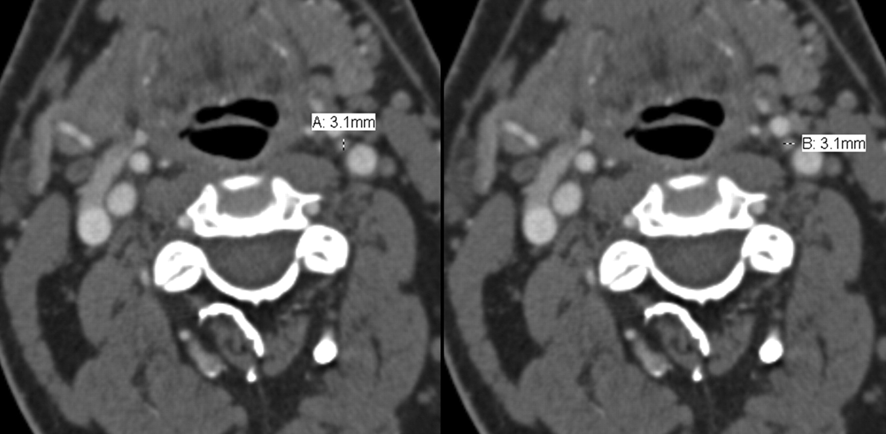
- Fig 1.
Representative axial CTA images demonstrate the method of orthogonal measurements, which were performed bilaterally.
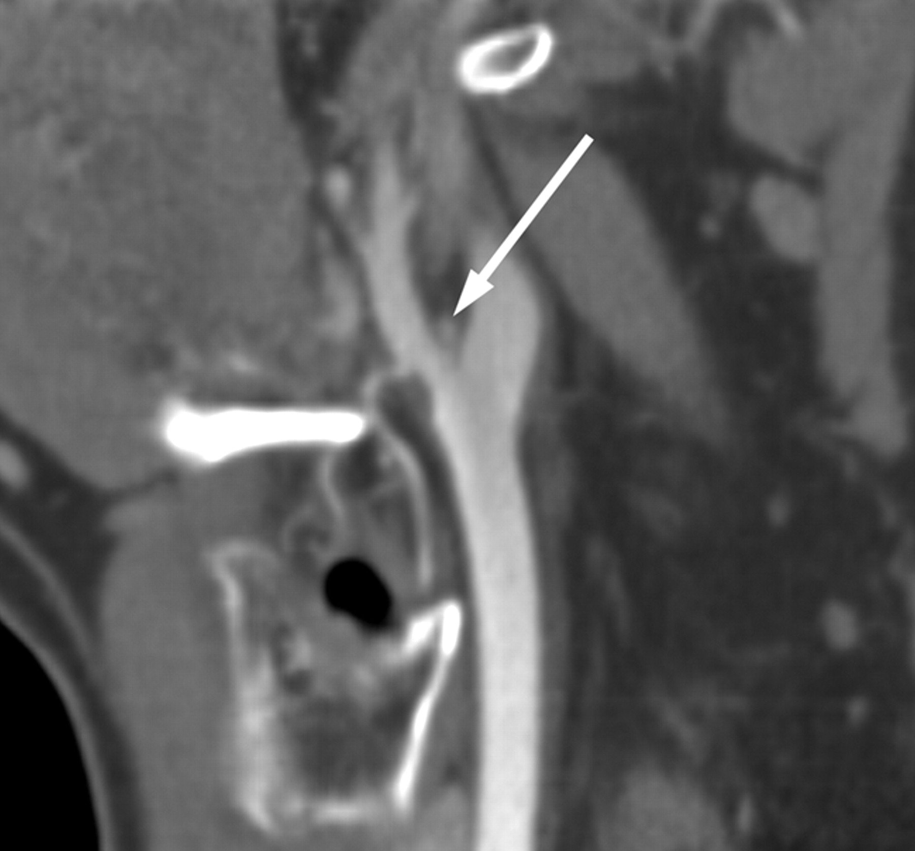
- Fig 2.
Oblique sagittal reformations were performed with a B20 kernel at a 2-mm thickness and at 2-mm increments. The normal carotid body is seen at the carotid bifurcation.
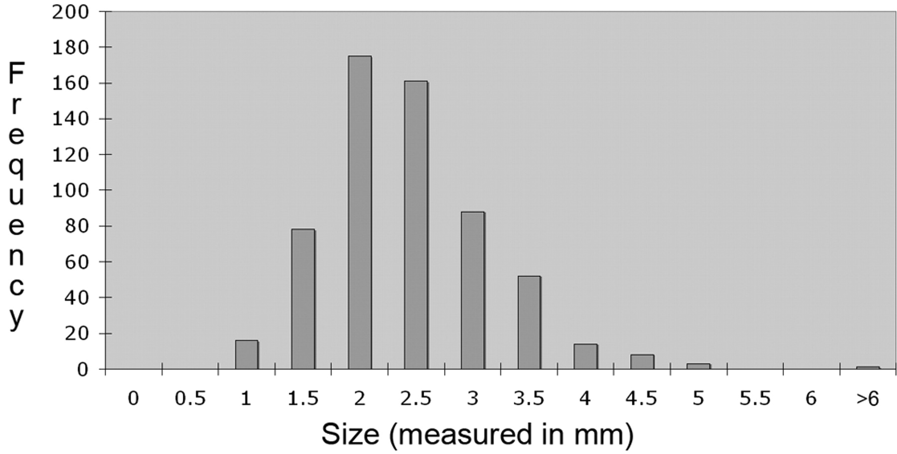
- Fig 3.
Bar graph shows the distribution in size of the carotid body based on measurements of 358 carotid bodies. The right carotid body measures an average of 2.4 (transverse) × 2.0 (AP) ± 0.8 (transverse) and 0.6 (AP) mm. The left carotid body measures, on average, 2.2 (transverse) × 2.1 (AP) ± 0.7 (transverse) and 0.5 (AP) mm. The range of normal defined as 2 SDs is 1.1–3.9 mm for the carotid body.
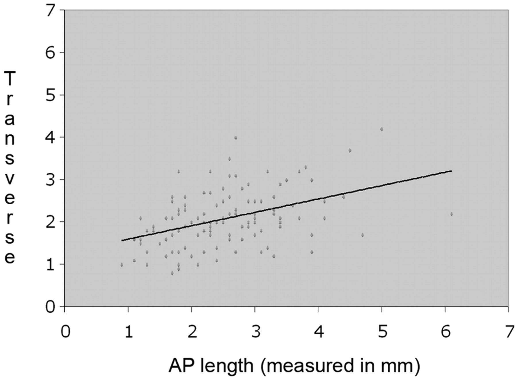
- Fig 4.
Line graph shows that the Pearson coefficient of the transverse dimension of the right carotid body to the left is 0.52 with a P value of <.0001. The data indicate a correlation between the AP and transverse dimensions of the right and left carotid bodies.
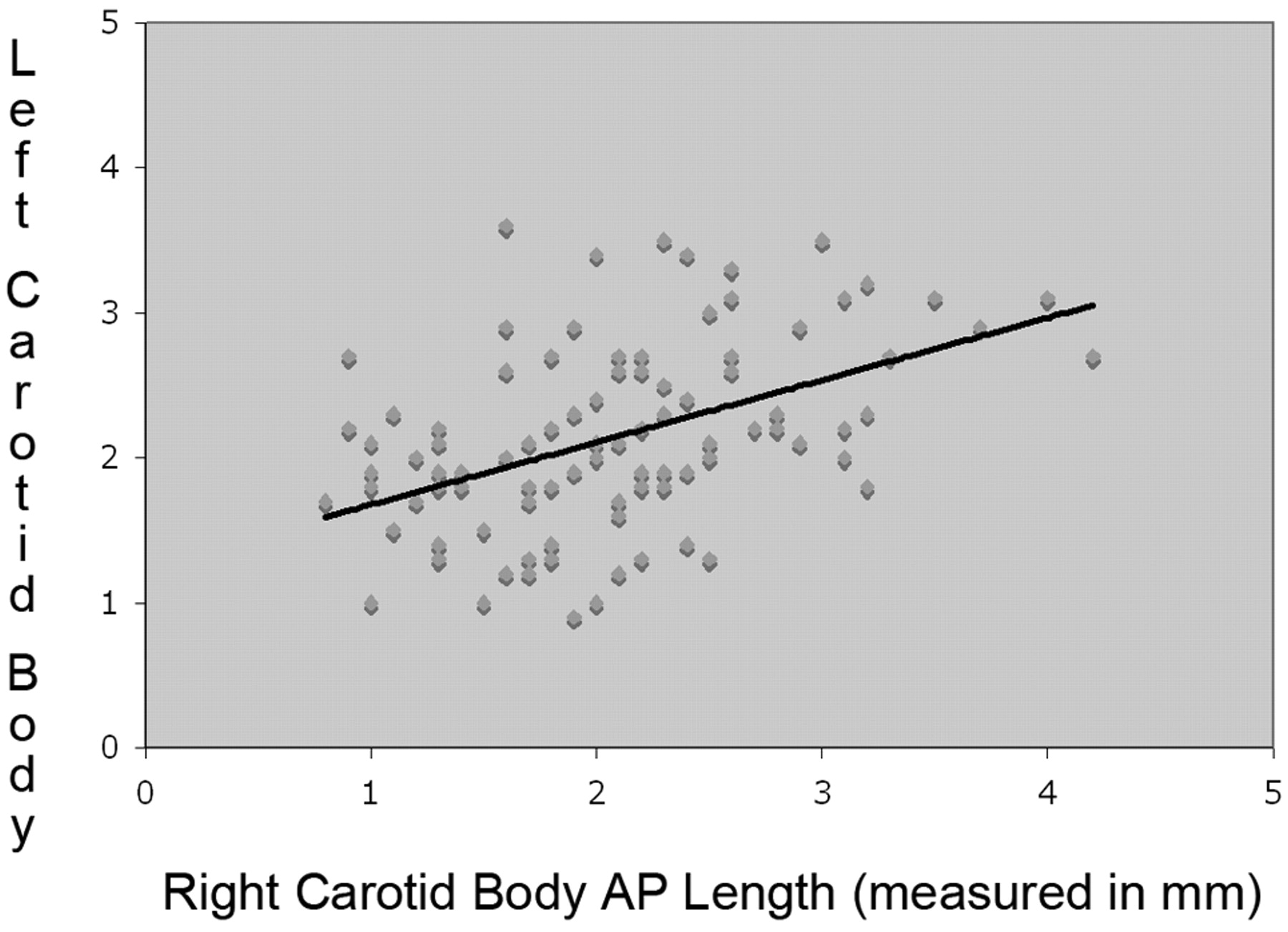
- Fig 5.
Line graph shows that the Pearson coefficient of the AP dimension of the right carotid body to the left is 0.43 with a P value of <.0001. This illustrates a correlation between the size of the right and left carotid bodies.
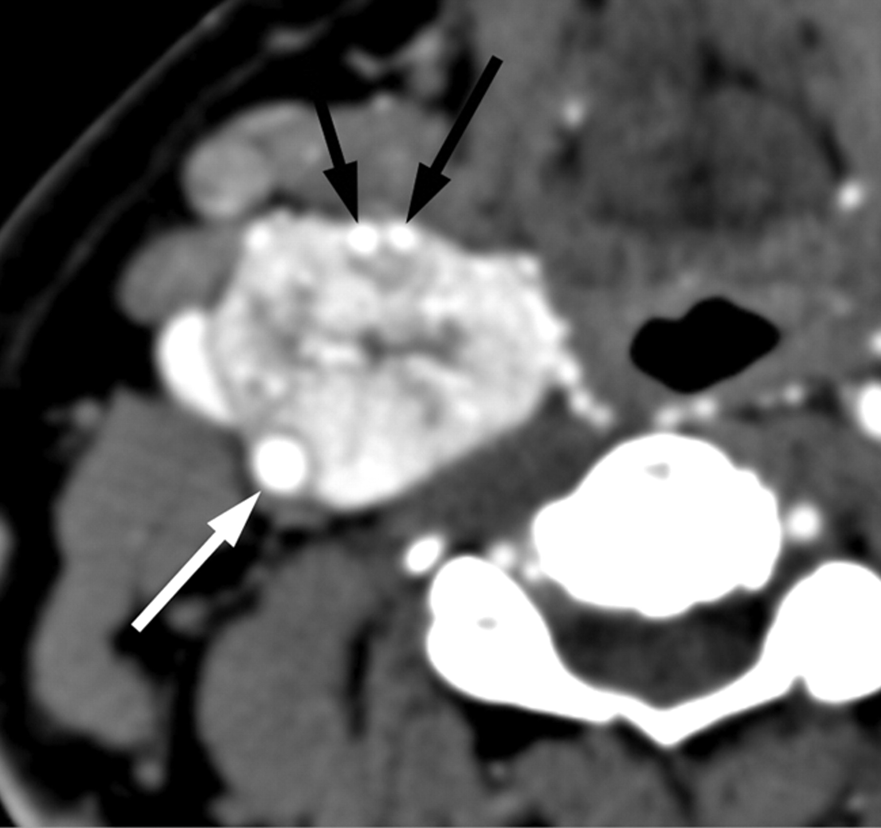
- Fig 6.
Axial CTA image depicts an avidly enhancing mass at the right carotid bifurcation splaying the internal (white arrow) and external carotid arteries (black arrows). There are numerous central and peripheral enlarged feeding arterial branches within this carotid body tumor.
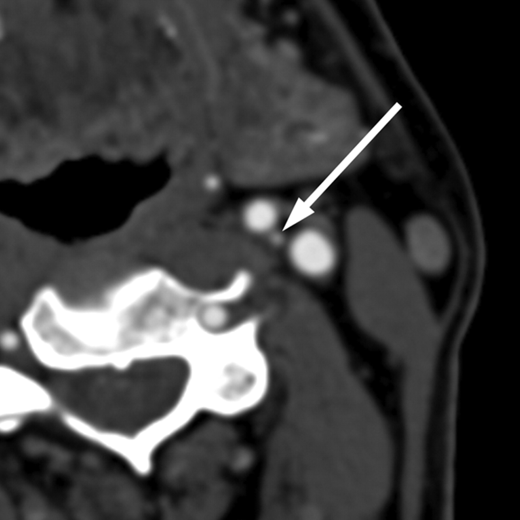
- Fig 7.
Axial CTA exhibits a small enhancing structure at the left carotid bifurcation, corresponding to a normal carotid body.


