Advertisement

Main menu

  • Home
  • Content
    • Current Issue
    • Accepted Manuscripts
    • Article Preview
    • Past Issue Archive
    • Video Articles
    • AJNR Case Collection
    • Case of the Week Archive
    • Case of the Month Archive
    • Classic Case Archive
  • Special Collections
    • AJNR Awards
    • Low-Field MRI
    • Alzheimer Disease
    • ASNR Foundation Special Collection
    • Photon-Counting CT
    • View All
  • Multimedia
    • AJNR Podcasts
    • AJNR SCANtastic
    • Trainee Corner
    • MRI Safety Corner
    • Imaging Protocols
  • For Authors
    • Submit a Manuscript
    • Submit a Video Article
    • Submit an eLetter to the Editor/Response
    • Manuscript Submission Guidelines
    • Statistical Tips
    • Fast Publishing of Accepted Manuscripts
    • Graphical Abstract Preparation
    • Imaging Protocol Submission
    • Author Policies
  • About Us
    • About AJNR
    • Editorial Board
    • Editorial Board Alumni
  • More
    • Become a Reviewer/Academy of Reviewers
    • Subscribers
    • Permissions
    • Alerts
    • Feedback
    • Advertisers
    • ASNR Home

User menu

  • Alerts
  • Log in

Search

  • Advanced search
American Journal of Neuroradiology

American Journal of Neuroradiology

ASHNR American Society of Functional Neuroradiology ASHNR American Society of Pediatric Neuroradiology ASSR
  • Alerts
  • Log in

Advanced Search

  • Home
  • Content
    • Current Issue
    • Accepted Manuscripts
    • Article Preview
    • Past Issue Archive
    • Video Articles
    • AJNR Case Collection
    • Case of the Week Archive
    • Case of the Month Archive
    • Classic Case Archive
  • Special Collections
    • AJNR Awards
    • Low-Field MRI
    • Alzheimer Disease
    • ASNR Foundation Special Collection
    • Photon-Counting CT
    • View All
  • Multimedia
    • AJNR Podcasts
    • AJNR SCANtastic
    • Trainee Corner
    • MRI Safety Corner
    • Imaging Protocols
  • For Authors
    • Submit a Manuscript
    • Submit a Video Article
    • Submit an eLetter to the Editor/Response
    • Manuscript Submission Guidelines
    • Statistical Tips
    • Fast Publishing of Accepted Manuscripts
    • Graphical Abstract Preparation
    • Imaging Protocol Submission
    • Author Policies
  • About Us
    • About AJNR
    • Editorial Board
    • Editorial Board Alumni
  • More
    • Become a Reviewer/Academy of Reviewers
    • Subscribers
    • Permissions
    • Alerts
    • Feedback
    • Advertisers
    • ASNR Home
  • Follow AJNR on Twitter
  • Visit AJNR on Facebook
  • Follow AJNR on Instagram
  • Join AJNR on LinkedIn
  • RSS Feeds

AJNR Awards, New Junior Editors, and more. Read the latest AJNR updates

Research ArticleFunctional

Comparison of Hypothesis- and a Novel Hybrid Data/Hypothesis-Driven Method of Functional MR Imaging Analysis in Patients with Brain Gliomas

M. Caulo, R. Esposito, D. Mantini, C. Briganti, C. Sestieri, P.A. Mattei, C. Colosimo, G.L. Romani and A. Tartaro
American Journal of Neuroradiology June 2011, 32 (6) 1056-1064; DOI: https://doi.org/10.3174/ajnr.A2428
M. Caulo
  • Find this author on Google Scholar
  • Find this author on PubMed
  • Search for this author on this site
R. Esposito
  • Find this author on Google Scholar
  • Find this author on PubMed
  • Search for this author on this site
D. Mantini
  • Find this author on Google Scholar
  • Find this author on PubMed
  • Search for this author on this site
C. Briganti
  • Find this author on Google Scholar
  • Find this author on PubMed
  • Search for this author on this site
C. Sestieri
  • Find this author on Google Scholar
  • Find this author on PubMed
  • Search for this author on this site
P.A. Mattei
  • Find this author on Google Scholar
  • Find this author on PubMed
  • Search for this author on this site
C. Colosimo
  • Find this author on Google Scholar
  • Find this author on PubMed
  • Search for this author on this site
G.L. Romani
  • Find this author on Google Scholar
  • Find this author on PubMed
  • Search for this author on this site
A. Tartaro
  • Find this author on Google Scholar
  • Find this author on PubMed
  • Search for this author on this site
  • Article
  • Figures & Data
  • Info & Metrics
  • Responses
  • References
  • PDF
Loading

Article Figures & Data

Figures

  • Tables
  • Fig 1.
    • Download figure
    • Open in new tab
    • Download powerpoint
    Fig 1.

    Outline of the ICA-based GLM analysis. The procedure can be divided into 3 steps: 1) The fMRI time courses are decomposed by means of spatial ICA, and the IC showing the largest correspondence with the predictor is selected; 2) a spatial mask is created by thresholding the IC map, which is applied to the fMRI data; and 3) GLM analysis is performed on the masked fMRI time course as brain response data, by using the IC model.

  • Fig 2.
    • Download figure
    • Open in new tab
    • Download powerpoint
    Fig 2.

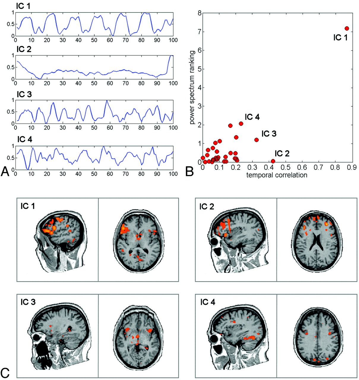
    Results of the ICA decomposition on a patient with a brain tumor. A, Time courses of the first 4 ranked ICs. B, Comparison between the results of power spectrum ranking and temporal correlation on all the separated ICs. C, Spatial maps of the first 4 ranked ICs, thresholded at z > 2.

  • Fig 3.
    • Download figure
    • Open in new tab
    • Download powerpoint
    Fig 3.

    GLM (left column) and ICA-GLM (right column) activations (P < .001, uncorrected) during VGt in representative controls and patients. The left hemispheres are displayed with different Talairach x-values, to visualize the IFG (Broca area) and TPJ (Wernicke area). The bar charts in the lower left-hand corners show the correlation coefficients of the first 4 ICs and the task predictor; the chosen IC is displayed in red. In controls, both the GLM and ICA-GLM show activation in the IFG and TPJ. In patient 1 (left parietal glioma), the ICA-GLM activates both the IFG and TPJ, whereas the GLM activates only the IFG. In patient 2 (left temporal glioma), the ICA-GLM activates both the IFG and TPJ, whereas no language regions are detected with the GLM. In patient 3 (left temporal glioma), the ICA-GLM activates only the IFG, whereas no language regions are detected with the GLM.

  • Fig 4.
    • Download figure
    • Open in new tab
    • Download powerpoint
    Fig 4.

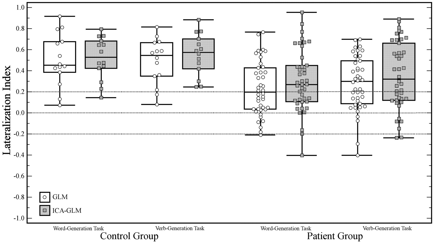
    Comparison of LI values obtained with the classic GLM and ICA-GLM of controls and patients for the following tasks: With WGt, 2 controls presented a bilateral LI (−0.2 < LI < 0.2) with the GLM. The same 2 subjects showed a left dominance with ICA-GLM, while the third showed a bilateral organization with ICA-GLM. With VGt, the same 2 controls with bilateral LI with GLM. Both presented a left dominance with ICA-GLM. With WGt, 20 patients had an LI indicating a bilateral language organization with the GLM: Seven presented left dominance with ICA-GLM and 13 remained bilateral. Three patients showed a left dominance with the GLM but a bilateral dominance with ICA-GLM. Eighteen of the remaining patients had a left dominance with both the GLM and ICA-GLM, and 1, a concordant right dominance. With VGt, 16 patients presented a bilateral LI indicated by the GLM, of which 6 had left and 1 right dominance with ICA-GLM. Four patients showed a left dominance with the GLM but a bilateral dominance with ICA-GLM. One patient presented a right dominance with the GLM but a bilateral dominance with ICA-GLM. Of the remaining patients, 20 had a left dominance with both the GLM and ICA-GLM and 1 patient had a concordant right dominance.

  • Fig 5.
    • Download figure
    • Open in new tab
    • Download powerpoint
    Fig 5.

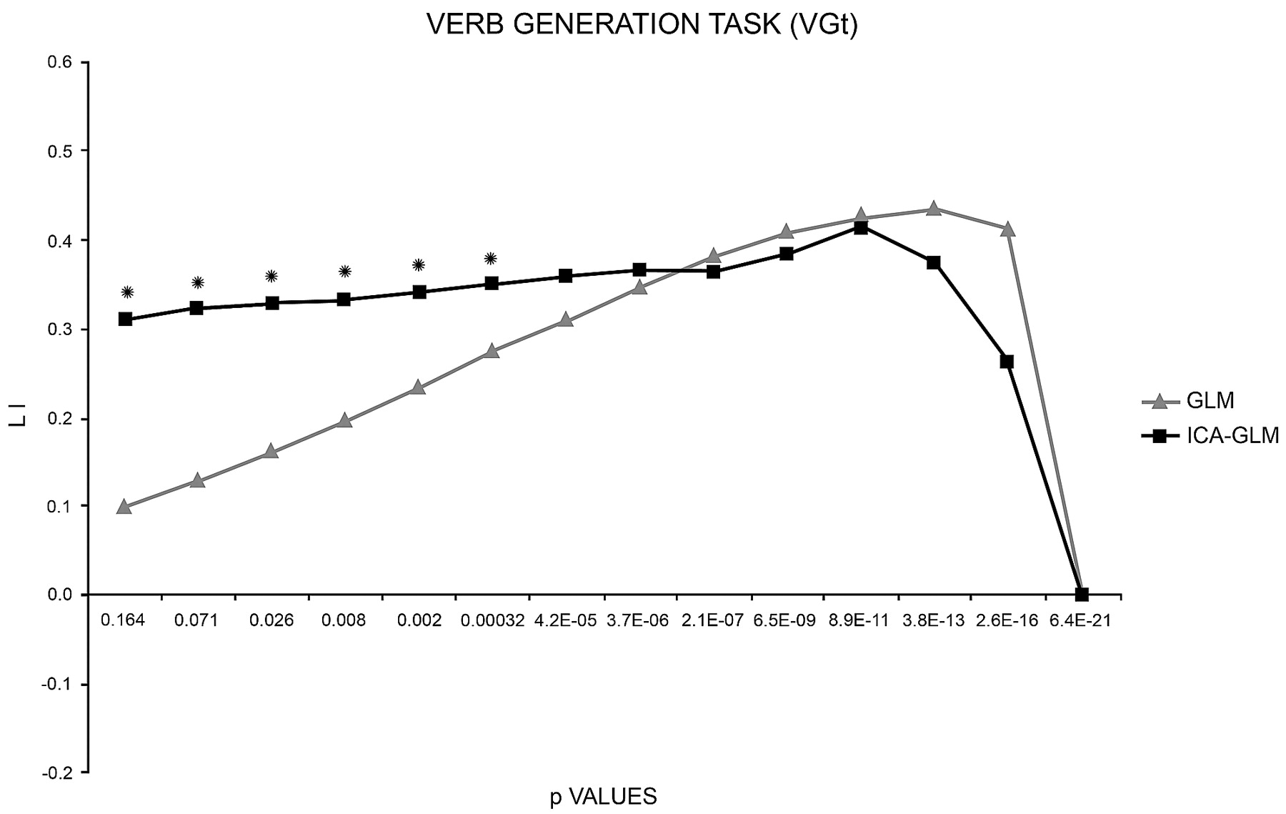
    LI curves of patients performing the VGt analyzed with the 2 methods (GLM and ICA-GLM). With the GLM, the LI curve starts near low values and increases as the threshold rises toward stricter values. With ICA-GLM, the LI curve starts from higher values and remains constant toward stricter values. Both curves eventually drop to zero when no activated voxels resist. Asterisks indicate the threshold values at which the 2 methods are statistically different.

Tables

  • Figures
  • Comparison of the GLM and ICA-GLMa

    Left IFGLeft TPJRight IFGRight TPJ
    PatientsControlsPatientsControlsPatientsControlsPatientsControls
    GLM40 (95)14 (100)27 (64)13 (93)36 (86)14 (100)13 (31)27 (64)
    ICA-GLM42 (100)14 (100)38 (90)13 (93)36 (86)13 (93)13 (31)10 (71)
    • a Number (percentage) of subjects with BOLD activity detected in the 4 classic language regions.

PreviousNext
Back to top

In this issue

American Journal of Neuroradiology: 32 (6)
American Journal of Neuroradiology
Vol. 32, Issue 6
1 Jun 2011
  • Table of Contents
  • Index by author
Advertisement
Print
Download PDF
Email Article

Thank you for your interest in spreading the word on American Journal of Neuroradiology.

NOTE: We only request your email address so that the person you are recommending the page to knows that you wanted them to see it, and that it is not junk mail. We do not capture any email address.

Enter multiple addresses on separate lines or separate them with commas.
Comparison of Hypothesis- and a Novel Hybrid Data/Hypothesis-Driven Method of Functional MR Imaging Analysis in Patients with Brain Gliomas
(Your Name) has sent you a message from American Journal of Neuroradiology
(Your Name) thought you would like to see the American Journal of Neuroradiology web site.
CAPTCHA
This question is for testing whether or not you are a human visitor and to prevent automated spam submissions.
Cite this article
M. Caulo, R. Esposito, D. Mantini, C. Briganti, C. Sestieri, P.A. Mattei, C. Colosimo, G.L. Romani, A. Tartaro
Comparison of Hypothesis- and a Novel Hybrid Data/Hypothesis-Driven Method of Functional MR Imaging Analysis in Patients with Brain Gliomas
American Journal of Neuroradiology Jun 2011, 32 (6) 1056-1064; DOI: 10.3174/ajnr.A2428

Citation Manager Formats

  • BibTeX
  • Bookends
  • EasyBib
  • EndNote (tagged)
  • EndNote 8 (xml)
  • Medlars
  • Mendeley
  • Papers
  • RefWorks Tagged
  • Ref Manager
  • RIS
  • Zotero
0 Responses
Respond to this article
Share
Bookmark this article
Comparison of Hypothesis- and a Novel Hybrid Data/Hypothesis-Driven Method of Functional MR Imaging Analysis in Patients with Brain Gliomas
M. Caulo, R. Esposito, D. Mantini, C. Briganti, C. Sestieri, P.A. Mattei, C. Colosimo, G.L. Romani, A. Tartaro
American Journal of Neuroradiology Jun 2011, 32 (6) 1056-1064; DOI: 10.3174/ajnr.A2428
del.icio.us logo Twitter logo Facebook logo Mendeley logo
  • Tweet Widget
  • Facebook Like
  • Google Plus One
Purchase

Jump to section

  • Article
    • Abstract
    • Abbreviations
    • Materials and Methods
    • Results
    • Discussion
    • Conclusions
    • References
  • Figures & Data
  • Info & Metrics
  • Responses
  • References
  • PDF

Related Articles

  • No related articles found.
  • PubMed
  • Google Scholar

Cited By...

  • Reliability of Functional and Diffusion MR Imaging Near Cerebral Cavernous Malformations
  • Reduction of Motion Artifacts and Noise Using Independent Component Analysis in Task-Based Functional MRI for Preoperative Planning in Patients with Brain Tumor
  • Reorganization of Functional Connectivity of the Language Network in Patients with Brain Gliomas
  • Localizing Seizure-Onset Zones in Presurgical Evaluation of Drug-Resistant Epilepsy by Electroencephalography/fMRI: Effectiveness of Alternative Thresholding Strategies
  • Crossref (14)
  • Google Scholar

This article has been cited by the following articles in journals that are participating in Crossref Cited-by Linking.

  • Reorganization of Functional Connectivity of the Language Network in Patients with Brain Gliomas
    C. Briganti, C. Sestieri, P.A. Mattei, R. Esposito, R.J. Galzio, A. Tartaro, G.L. Romani, M. Caulo
    American Journal of Neuroradiology 2012 33 10
  • Modifications of Default-Mode Network Connectivity in Patients with Cerebral Glioma
    Roberto Esposito, Peter A. Mattei, Chiara Briganti, Gian Luca Romani, Armando Tartaro, Massimo Caulo, Yu-Feng Zang
    PLoS ONE 2012 7 7
  • Multimodal assessment of hemispheric lateralization for language and its relevance for behavior
    C. Piervincenzi, A. Petrilli, A. Marini, M. Caulo, G. Committeri, C. Sestieri
    NeuroImage 2016 142
  • Narrative comprehension beyond language: Common brain networks activated by a movie and its script
    Pia Tikka, Janne Kauttonen, Yevhen Hlushchuk, Emmanuel Andreas Stamatakis
    PLOS ONE 2018 13 7
  • Localizing Seizure-Onset Zones in Presurgical Evaluation of Drug-Resistant Epilepsy by Electroencephalography/fMRI: Effectiveness of Alternative Thresholding Strategies
    M. Hauf, K. Jann, K. Schindler, O. Scheidegger, K. Meyer, C. Rummel, L. Mariani, T. Koenig, R. Wiest
    American Journal of Neuroradiology 2012 33 9
  • Functional and effective connectivity analysis of drug-resistant epilepsy: a resting-state fMRI analysis
    Eric Jacob Bacon, Chaoyang Jin, Dianning He, Shuaishuai Hu, Lanbo Wang, Han Li, Shouliang Qi
    Frontiers in Neuroscience 2023 17
  • Reduction of Motion Artifacts and Noise Using Independent Component Analysis in Task-Based Functional MRI for Preoperative Planning in Patients with Brain Tumor
    E.H. Middlebrooks, C.J. Frost, I.S. Tuna, I.M. Schmalfuss, M. Rahman, A. Old Crow
    American Journal of Neuroradiology 2017 38 2
  • Evaluation of the factors influencing brain language laterality in presurgical planning
    Seyed Amir Hossein Batouli, Nafiseh Hasani, Sara Gheisari, Ebrahim Behzad, Mohammad Ali Oghabian
    Physica Medica 2016 32 10
  • The influence of mental fatigue on the face and word encoding activations
    Seyed Amir Hossein Batouli, Razieh Alemi, Haleh Khoshkhouy Delshad, Mohammad Ali Oghabian
    Clinical Neurology and Neurosurgery 2020 189
  • Functional Connectivity MRI and Post-Operative Language Performance in Temporal Lobe Epilepsy: Initial Experience
    Emanuele Pravatà, Carlo Sestieri, Gabriella Colicchio, Cesare Colosimo, Gian Luca Romani, Massimo Caulo
    The Neuroradiology Journal 2014 27 2

More in this TOC Section

  • Kurtosis and Epileptogenic Tubers: A Pilot Study
  • Glutaric Aciduria Type 1: DK vs. Conventional MRI
  • WM Neuroaxonal Loss in Type 1 Diabetes
Show more Functional

Similar Articles

Advertisement

Indexed Content

  • Current Issue
  • Accepted Manuscripts
  • Article Preview
  • Past Issues
  • Editorials
  • Editor's Choice
  • Fellows' Journal Club
  • Letters to the Editor
  • Video Articles

Cases

  • Case Collection
  • Archive - Case of the Week
  • Archive - Case of the Month
  • Archive - Classic Case

More from AJNR

  • Trainee Corner
  • Imaging Protocols
  • MRI Safety Corner
  • Book Reviews

Multimedia

  • AJNR Podcasts
  • AJNR Scantastics

Resources

  • Turnaround Time
  • Submit a Manuscript
  • Submit a Video Article
  • Submit an eLetter to the Editor/Response
  • Manuscript Submission Guidelines
  • Statistical Tips
  • Fast Publishing of Accepted Manuscripts
  • Graphical Abstract Preparation
  • Imaging Protocol Submission
  • Evidence-Based Medicine Level Guide
  • Publishing Checklists
  • Author Policies
  • Become a Reviewer/Academy of Reviewers
  • News and Updates

About Us

  • About AJNR
  • Editorial Board
  • Editorial Board Alumni
  • Alerts
  • Permissions
  • Not an AJNR Subscriber? Join Now
  • Advertise with Us
  • Librarian Resources
  • Feedback
  • Terms and Conditions
  • AJNR Editorial Board Alumni

American Society of Neuroradiology

  • Not an ASNR Member? Join Now

© 2025 by the American Society of Neuroradiology All rights, including for text and data mining, AI training, and similar technologies, are reserved.
Print ISSN: 0195-6108 Online ISSN: 1936-959X

Powered by HighWire