
Article Figures & Data
Figures
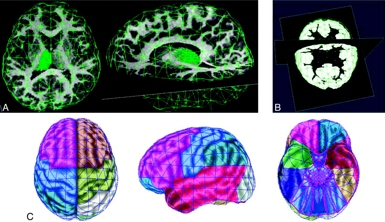
- Fig 1.
Images showing the seed and target definition for fiber tracking. A, Regions of interest were drawn on 7 consecutive planes of each subject's FA images to outline the thalamus. These representative images of 1 subject show the 3D seed region of the left thalamus loaded in the native FSPGR space. B, Representative image of the target layer (white color) in one of the subjects. The target mask contained the layer between the cortical surface and the 2-mm shrunken white matter mask. FTEs were obtained by extending the surface elements 30 mm deeper into the brain to select the underlying portion of the target mask. C, Superior, lateral, and inferior views of the ICBM152 template brain surface with the 8 cortical target regions on each side encompassing sets of surface elements. The cortical regions were defined by using main sulci as references. Those surface elements that extended a landmark sulcus were assigned to the region to which their largest part belonged. Importantly, the territory outlined with magenta (on the inferior surface) was not analyzed.
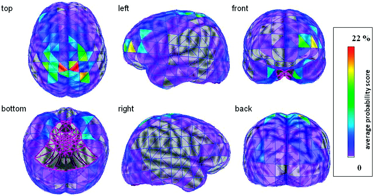
- Fig 2.
Average thalamocortical connectivity map of the 15 subjects visualized on the 3D brain surface of the ICBM152 template brain. The thalamocortical connectivity scores of each homotopic surface element were averaged among the 15 subjects, and the surface elements of the template brain were color-coded based on these average values. Notably, only the ipsilateral thalamic connections of each hemisphere are shown. It can be appreciated that the bilateral sensorimotor cortical areas have the strongest thalamic connectivity and that certain cortical regions (eg, prefrontal, inferior temporal) show profound hemispheric asymmetries.
- Fig 3.
Bar graph showing the thalamocortical connectivity scores (mean ± SE) for the cortex (sum of the analyzed 8 regions) and each individual cortical region. Significant (P < .01) and reproducible (parameter-independent) left>right hemispheric asymmetries are indicated with asterisk.
- Fig 4.
Scatterplot showing a significant positive correlation between age and thalamic connectivity scores of the left and right prefrontal cortex (P = .001 and P = .002, respectively).


In this issue


Jump to section
Related Articles
Cited By...
- The Infraslow Fluctuation of Sigma Power During Sleep in Young Individuals with Schizophrenia
- Age-associated alterations in thalamocortical structural connectivity in psychosis-spectrum youths
- Microstructural development from 9-14 years: evidence from the ABCD Study
- Spatially heterogeneous microstructural development within subcortical regions from 9-13 years