
Article Figures & Data
Figures
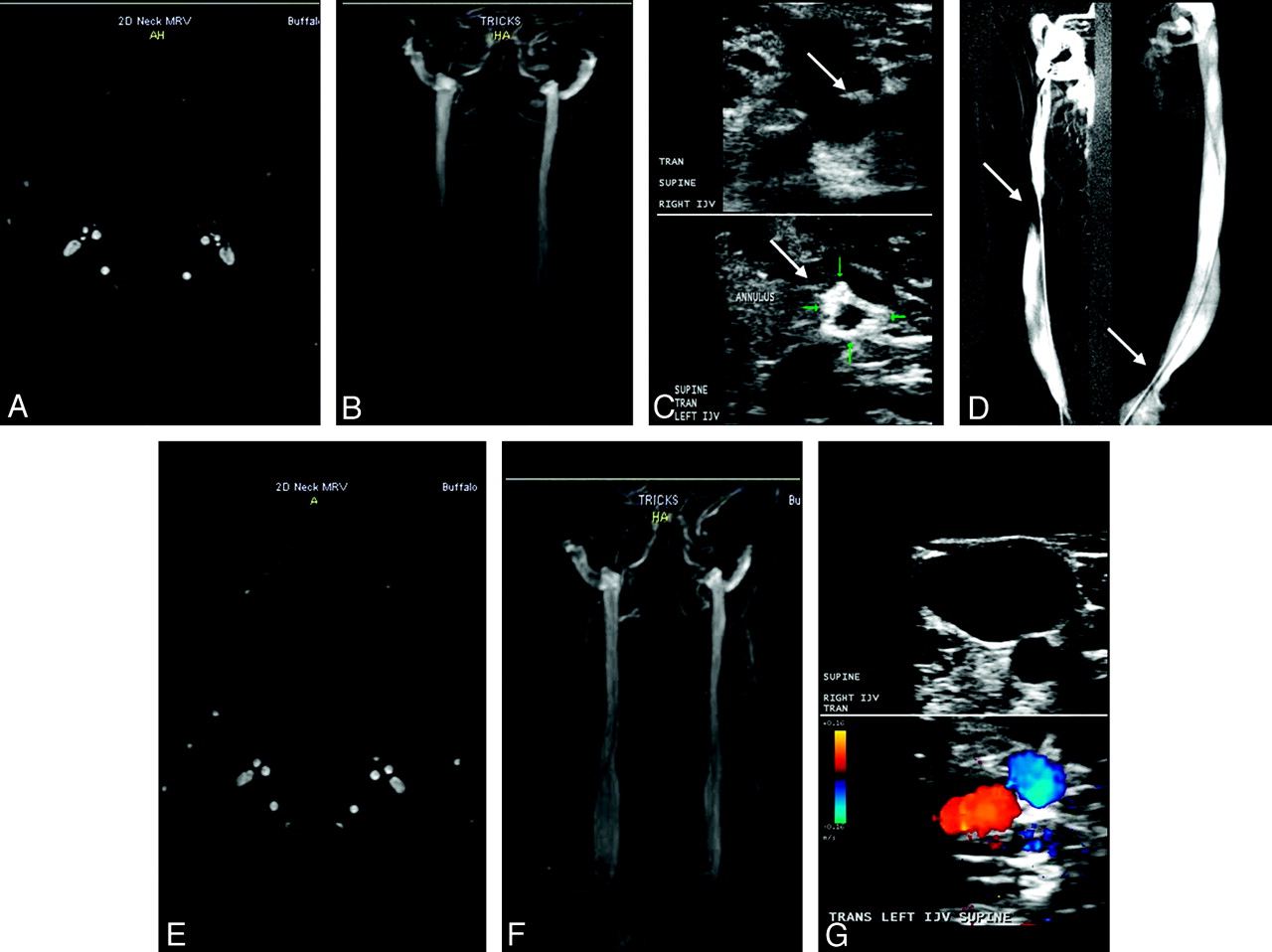
- Fig 1.
Patient with MS (a 43-year-old woman) shows normal examination findings on TOF (A) and TRICKS (B) of both IJVs pretreatment. C, Doppler sonography shows the presence of a septum (arrow) in the right IJV (upper image) and an annulus (arrow) in the left IJV (lower image). D−F, Catheter venography (D) confirms the presence of a septum (arrow) in the right IJV and an annulus (arrow) in the left IJV. The posttreatment 6-month follow-up shows normal examination findings on TOF (E), TRICKS (F), and Doppler sonography (G).
- Fig 2.
Patient with MS (a 44-year-old man) has normal examination findings on TOF (A) and TRICKS (B) of both IJVs pretreatment. C, Doppler sonography shows the presence of stenoses in the right IJV (arrow, upper image) and in the left IJV (arrow, lower image). D, Catheter venography confirms the presence of stenosis (arrows) in the right IJV and in the left IJV. E–G, The posttreatment 6-month follow-up shows normal examination findings on TOF (E) and TRICKS (F), but no change in the stenosis (arrows) on Doppler sonography (G) in the right IJV (upper image) and in the left IJV (lower image).
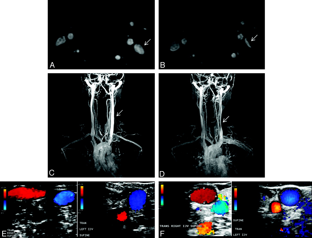
- Fig 3.
Variability between the baseline (A, C, and E, TOF, TRICKS, and Doppler sonography, respectively) and follow-up (B, D, and F, TOF, TRICKS, and Doppler sonography, respectively) examinations in a 42-year-old healthy female control. Flattening of the left IJV (arrows) at follow-up is noted on the TOF (B) and TRICKS (D), whereas Doppler sonography shows normal examination findings like those at baseline (F).
- Fig 4.
Variability between the baseline (A, C, and E, TOF, TRICKS, and Doppler sonography, respectively) and follow-up (B, D, and F, TOF, TRICKS and Doppler sonography, respectively) examinations in a 39-year-old healthy male control. Flattening of the right IJV (arrows) present at the baseline (A and C) examination is not present at follow-up (B and D). Doppler sonography examination shows normal findings at baseline (E) and follow-up (F).


Tables
- Table 1:
MRV, Doppler sonography, and selective venography findings in patients with MS at baseline and 6-month follow-upa
Patients with MS 2D-TOF 3D-TRICKS DS CV RIJV LIJV RIJV LIJV RIJV LIJV RIJV LIJV 1–baseline Normal Normal Normal Abnormal Abnormal Abnormal Normal Abnormal 1–6 mo Normal Normal Normal Normal Normal Normal – – 2–baseline Normal Normal Abnormal Abnormal Abnormal Abnormal Abnormal Abnormal 2–6 mo Abnormal Abnormal Abnormal Abnormal Abnormal Abnormal – – 3–baseline Normal Normal Normal Normal Abnormal Abnormal Abnormal Abnormal 3- 6 mo Normal Normal Normal Normal Normal Abnormal – – 4–baseline Normal Normal Normal Normal Normal Abnormal Normal Abnormal 4–6 mo Normal Normal Abnormal Abnormal Abnormal Normal – – 5–baseline Normal Normal Normal Normal Abnormal Normal Abnormal Normal 5–6 mo Normal Normal Normal Normal Abnormal Normal – – 6–baseline Normal Normal Normal Normal Abnormal Abnormal Abnormal Abnormal 6–6 mo Normal Normal Normal Normal Abnormal Abnormal – – 7–baseline Normal Abnormal Normal Abnormal Normal Abnormal Normal Abnormal 7–6 mo Normal Abnormal Abnormal Abnormal Normal Normal – – 8–baseline Abnormal Abnormal Abnormal Normal Abnormal Abnormal Abnormal Abnormal 8–6 mo Abnormal Abnormal Abnormal Abnormal Abnormal Abnormal – – 9–baseline Normal Abnormal Normal Normal Abnormal Abnormal Abnormal Abnormal 9–6 mo Abnormal Abnormal Normal Abnormal Abnormal Abnormal – – 10–baseline Normal Normal Normal Normal Abnormal Abnormal Abnormal Abnormal 10- 6 mo Normal Normal Normal Normal Normal Normal – – a Absent and pinpoint IJV flow was considered abnormal on TOF and TRICKS; the presence of at least 1 of the following IJV parameters was considered abnormal on a DS examination: B-mode abnormalities (flaps, septa, web), stenoses, absence of detectable flow, and presence of reflux in both sitting and supine positions. On CV, the presence of stenosis >50% of the IJV diameter or at least 1 of the following anomalies was considered abnormal: annulus, septum/valve malformation, hypoplasia, twisting, membrane, and agenesis.
- Table 2:
MRV and Doppler sonography findings in healthy controls at baseline and 6-month follow-upa
Healthy Controls TOF TRICKS DS RIJV LIJV RIJV LIJV RIJV LIJV 1–baseline Normal Normal Normal Normal Normal Normal 1–6 mo Normal Normal Normal Normal Normal Normal 2–baseline Normal Normal Normal Normal Normal Normal 2–6 mo Normal Abnormal Normal Abnormal Normal Normal 3–baseline Normal Normal Normal Normal Normal Normal 3–6 mo Normal Normal Normal Normal Normal Normal 4–baseline Normal Normal Normal Normal Normal Normal 4–6 mo Normal Normal Normal Normal Normal Normal 5–baseline Abnormal Normal Abnormal Normal Normal Abnormal 5–6 mo Normal Normal Abnormal Normal Abnormal Normal 6–baseline Normal Normal Normal Normal Normal Abnormal 6–6 mo Normal Normal Normal Normal Normal Abnormal a Absent and pinpoint IJV flow was considered abnormal on TOF and TRICKS; the presence of at least 1 of the following IJV parameters was considered abnormal on a DS examination: B-mode abnormalities (flaps, septa, and web), stenoses, absence of detectable flow, and presence of reflux in both sitting and supine positions.
- Table 3:
Sensitivity, specificity, PPV, and NPV of MRV (TOF and TRICKS) and Doppler sonography relative to catheter venography (criterion standard) for detection of IJV anomaliesa
Measure/Side Sensitivity (%) Specificity (%) PPV (%) NPV (%) TOF Right 99 (44–100) 33 (3–51) 45 (12–65) 99 (21–100) Left 99 (21–100) 45 (12–065) 33 (3–51) 99 (44–100) Both 99 (51–100) 33 (10–49) 33 (10–49) 99 (51–100) TRICKS Right 99 (44–100) 44 (8–64) 49 (14–69) 99 (34–100) Left 99 (21–100) 45 (12–65) 33 (3–51) 99 (44–100) Both 99 (51–100) 39 (14–56) 35 (11–52) 99 (57–100) DS Right 79 (21–94) 100 (65–100) 99 (34–100) 90 (53–98) Left 99 (21–100) 100 (70–100) 99 (21–100) 100 (70–100) Both 82 (30–95) 100 (81–100) 99 (44–100) 95 (73–99) a 95% confidence intervals are shown in parentheses for all values.
In this issue


Jump to section
Related Articles
Cited By...
- Jugular Anomalies in Multiple Sclerosis Are Associated with Increased Collateral Venous Flow
- Validity of the diagnostic criteria for chronic cerebrospinal venous insufficiency and association with multiple sclerosis
- Cerebral Veins-Why Functional MR Imaging is Worth the Trouble
- Mystery of Chronic Cerebrospinal Venous Insufficiency: Identical Venographic and Ultrasound Findings in Patients with MS and Controls
- No Association Between Conventional Brain MR Imaging and Chronic Cerebrospinal Venous Insufficiency in Multiple Sclerosis
- Extracranial Venous Drainage Patterns in Patients with Multiple Sclerosis and Healthy Controls
- Intra- and Extraluminal Structural and Functional Venous Anomalies in Multiple Sclerosis, as Evidenced by 2 Noninvasive Imaging Techniques
- Prevalence, sensitivity, and specificity of chronic cerebrospinal venous insufficiency in MS
- Venous drainage in multiple sclerosis: A combined MRI and ultrasound study