Advertisement

Main menu

  • Home
  • Content
    • Current Issue
    • Accepted Manuscripts
    • Article Preview
    • Past Issue Archive
    • Video Articles
    • AJNR Case Collection
    • Case of the Week Archive
    • Case of the Month Archive
    • Classic Case Archive
  • Special Collections
    • AJNR Awards
    • Low-Field MRI
    • Alzheimer Disease
    • ASNR Foundation Special Collection
    • Photon-Counting CT
    • View All
  • Multimedia
    • AJNR Podcasts
    • AJNR SCANtastic
    • Trainee Corner
    • MRI Safety Corner
    • Imaging Protocols
  • For Authors
    • Submit a Manuscript
    • Submit a Video Article
    • Submit an eLetter to the Editor/Response
    • Manuscript Submission Guidelines
    • Statistical Tips
    • Fast Publishing of Accepted Manuscripts
    • Graphical Abstract Preparation
    • Imaging Protocol Submission
    • Author Policies
  • About Us
    • About AJNR
    • Editorial Board
    • Editorial Board Alumni
  • More
    • Become a Reviewer/Academy of Reviewers
    • Subscribers
    • Permissions
    • Alerts
    • Feedback
    • Advertisers
    • ASNR Home

User menu

  • Alerts
  • Log in

Search

  • Advanced search
American Journal of Neuroradiology

American Journal of Neuroradiology

ASHNR American Society of Functional Neuroradiology ASHNR American Society of Pediatric Neuroradiology ASSR
  • Alerts
  • Log in

Advanced Search

  • Home
  • Content
    • Current Issue
    • Accepted Manuscripts
    • Article Preview
    • Past Issue Archive
    • Video Articles
    • AJNR Case Collection
    • Case of the Week Archive
    • Case of the Month Archive
    • Classic Case Archive
  • Special Collections
    • AJNR Awards
    • Low-Field MRI
    • Alzheimer Disease
    • ASNR Foundation Special Collection
    • Photon-Counting CT
    • View All
  • Multimedia
    • AJNR Podcasts
    • AJNR SCANtastic
    • Trainee Corner
    • MRI Safety Corner
    • Imaging Protocols
  • For Authors
    • Submit a Manuscript
    • Submit a Video Article
    • Submit an eLetter to the Editor/Response
    • Manuscript Submission Guidelines
    • Statistical Tips
    • Fast Publishing of Accepted Manuscripts
    • Graphical Abstract Preparation
    • Imaging Protocol Submission
    • Author Policies
  • About Us
    • About AJNR
    • Editorial Board
    • Editorial Board Alumni
  • More
    • Become a Reviewer/Academy of Reviewers
    • Subscribers
    • Permissions
    • Alerts
    • Feedback
    • Advertisers
    • ASNR Home
  • Follow AJNR on Twitter
  • Visit AJNR on Facebook
  • Follow AJNR on Instagram
  • Join AJNR on LinkedIn
  • RSS Feeds

AJNR Awards, New Junior Editors, and more. Read the latest AJNR updates

Research ArticleBrainC

Suspicious Neuroimaging Pattern of Thrombotic Microangiopathy

T.N. Ellchuk, L.M. Shah, R.H. Hewlett and A.G. Osborn
American Journal of Neuroradiology April 2011, 32 (4) 734-738; DOI: https://doi.org/10.3174/ajnr.A2351
T.N. Ellchuk
  • Find this author on Google Scholar
  • Find this author on PubMed
  • Search for this author on this site
L.M. Shah
  • Find this author on Google Scholar
  • Find this author on PubMed
  • Search for this author on this site
R.H. Hewlett
  • Find this author on Google Scholar
  • Find this author on PubMed
  • Search for this author on this site
A.G. Osborn
  • Find this author on Google Scholar
  • Find this author on PubMed
  • Search for this author on this site
  • Article
  • Figures & Data
  • Info & Metrics
  • Responses
  • References
  • PDF
Loading

Abstract

SUMMARY: The TMAs are a group of microvascular occlusive disorders characterized by thrombocytopenia and intravascular hemolysis. Literature review reveals a spectrum of neuroimaging findings, including a single case report of multifocal hemorrhagic infarctions. We present a series of 12 patients with TMA demonstrating a similar pattern of multifocal cortical and subcortical hemorrhagic infarctions.

Abbreviations

ADC
apparent diffusion coefficient
AML
acute myelogenous leukemia
CLL
chronic lymphocytic leukemia
CNS
central nervous system
CTV
CT venography
DIC
disseminated intravascular coagulation
DWI
diffusion-weighted imaging
GRE
gradient recalled-echo
HIV
human immunodeficiency virus
HUS
hemolytic uremic syndrome
mHTN
malignant hypertension
MRI
MR imaging
MRV
MR venography
PRES
posterior reversible encephalopathy syndrome
SWI
susceptibility-weighted imaging
TMA
thrombotic microangiopathy
TTP
thrombotic thrombocytopenic purpura
vWF
von Willebrand factor

TMA was defined by Symmers in 19521 as a lesion of arteriole and capillary wall thickening with swelling or detachment of endothelial cells from the basement membrane, accumulation of material in the subendothelial space, and lodging of fibrin-platelet thrombi at the arteriocapillary junction.2 The mechanical injury to erythrocytes results in intravascular hemolysis and schistocytosis. The intraluminal platelet thrombosis results in partial or complete obstruction of the vessel lumina.2

There is a spectrum of microvascular occlusive disorders in which endothelial injury incites platelet aggregation with thrombus formation and vascular occlusion, which includes TTP, HUS, DIC, autoimmune diseases, drug/toxin exposure, and mHTN. These clinically manifest as end-organ ischemia/infarction and/or hemorrhage.3,4 In TTP, brain lesions prevail, while renal lesions predominate in HUS.2 The incidence of TMA varies with the underlying diagnosis. Miller et al5 reported an age-sex standardized incidence of TTP and HUS of 6.5 per million per year in the United States. They found a greater incidence in females.5 This is generalized to systemic disease and not exclusively CNS involvement. Most cases of HUS occur before 20 years of age.5

Literature review reveals a spectrum of TMA neuroimaging findings including parenchymal hematoma, PRES, ischemic infarction, and venous thrombosis, with 1 case report of multifocal hemorrhagic infarctions.3,6,7 It is the latter imaging manifestation on which our case series focuses. We present a series of 12 patients with TMA sharing a similar neuroimaging pattern of multifocal cortical and subcortical hemorrhagic infarctions (Fig 1).

Fig 1.
  • Download figure
  • Open in new tab
  • Download powerpoint
Fig 1.

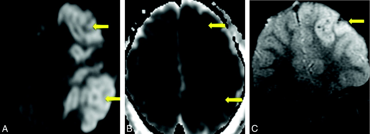
Case 6. A, Axial noncontrast CT scan demonstrates nonspecific peripheral hypoattenuation (arrow). B, Axial T2 MR image demonstrates cortical and subcortical hyperintensity (arrow) without appreciable hemorrhage. C, B = 1000 DWI MR image reveals multifocal cortical and subcortical hyperintensities (arrows). D, The corresponding apparent diffusion coefficient map demonstrates hypointensities in a similar distribution (arrows), consistent with infarction. E and F, GRE MR image reveals subtle blooming from petechial hemorrhage (E, arrow), better demonstrated on the SWI MR image (F, arrow).

Case Series

The cohort of 12 patients was obtained by 2 means. The first group of 7 patients was acquired via an institutional review board−approved retrospective search of the electronic medical records at our institution from 1997 through 2008 for all patients diagnosed with TTP, DIC, and/or mHTN. The patient list was cross-referenced with our imaging data base. Three neuroradiologists reviewed all neuroimaging, and the findings were tabulated. The second group of 5 patients was obtained from a review of our teaching file data base. Archived cases demonstrating multifocal cortical and subcortical hemorrhagic infarctions were included (Fig 2). An in-depth chart review was performed on all 12 patients to confirm the diagnosis of TMA.

Fig 2.
  • Download figure
  • Open in new tab
  • Download powerpoint
Fig 2.

Case 12. A, B = 1000 DWI MR image reveals multifocal cortical and subcortical hyperintensities (arrows). B, The corresponding apparent diffusion coefficient map demonstrates hypointensities in a similar distribution (arrows), consistent with infarction. C, GRE MR image reveals petechial hemorrhage (arrow) in a similar distribution.

The electronic medical record search yielded 623 patients with TMA, 249 (40%) with neuroimaging during admission. Of the 249 patients, 7 (3%) demonstrated the imaging pattern of multifocal cortical and subcortical hemorrhagic infarctions. Review of the teaching file data base yielded an additional 5 cases with the same imaging pattern. Patient demographics, imaging modalities, and clinical information are summarized in the Table.

View this table:
  • View inline
  • View popup

Patient demographics and clinical information

Of the 12 patients with TMA demonstrating the pattern of multifocal cortical and subcortical hemorrhagic infarctions, 8 (67%) had DIC (Fig 3). Three patients (25%) were diagnosed with mHTN. One patient (8%) had a diagnosis of TTP. This distribution of pathology reflects the disease range in the original cohort of 623 patients with TMA.

Fig 3.
  • Download figure
  • Open in new tab
  • Download powerpoint
Fig 3.

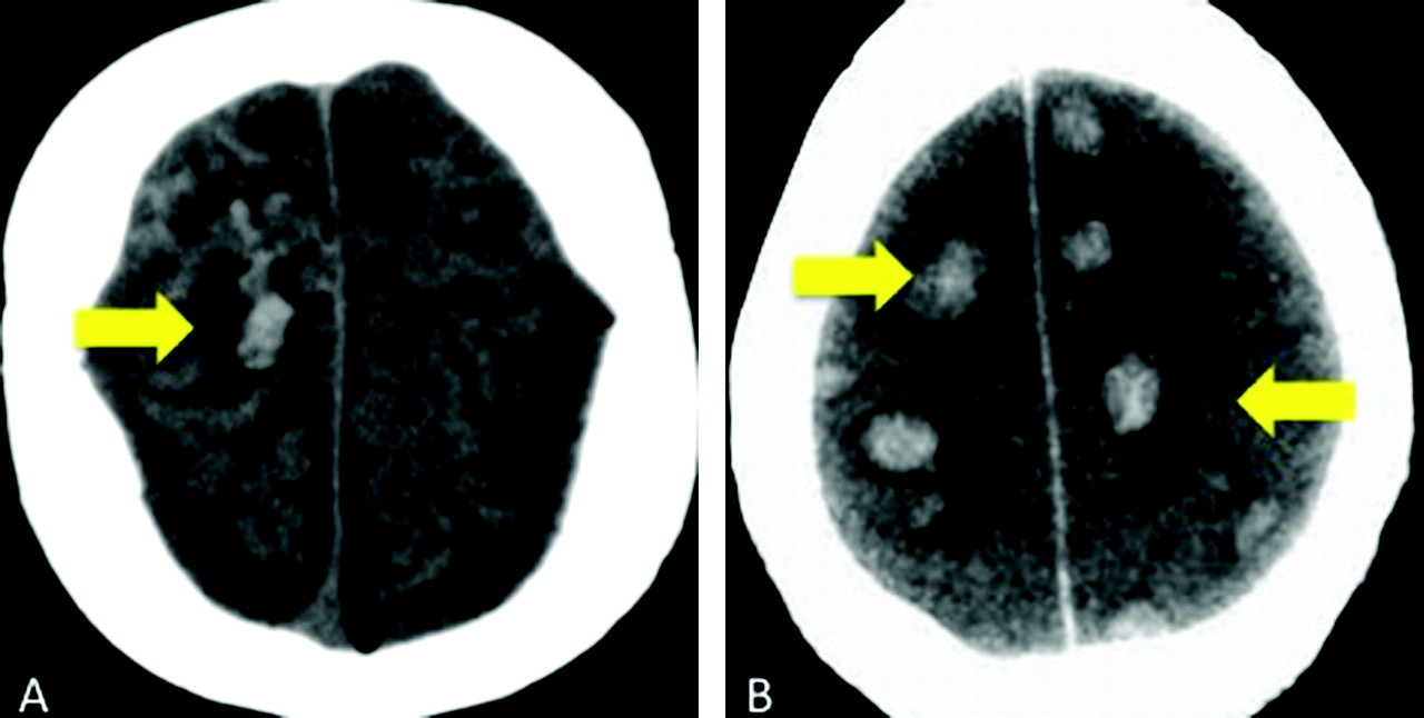
Cases 4 and 8. Axial noncontrast CT scans demonstrate peripheral hemorrhagic infarctions (arrows) in 2 patients with DIC.

Discussion

Injury to endothelial cells is the inciting factor in the sequence of events leading to TMA. The cascade of the microangiopathic process is the following: loss of physiologic thromboresistance, leukocyte adhesion to damaged endothelium, complement consumption, abnormal vWF release and fragmentation, and increased vascular shear stress.2 In TTP, thrombi formation occurs subsequent to the release of multimers of vWF. A specific plasma protease, the ADAMTS13 gene, is responsible for the physiologic degradation of vWF and plays a pathogenic role in a substantial proportion of familial and acute idiopathic cases of TTP.8 A deficiency in ADAMTS13 enzyme levels, along with an inhibitory antibody, is found in most patients with idiopathic TTP. Multiple triggers, such as infection, drugs, cancer, chemotherapy, bone marrow transplantation, and pregnancy, are recognized.9 In HUS, endothelial cell damage is considered the reason for complement and platelet activation leading to thrombus formation. Several complement genes are mutated in HUS.8

Multifocal TMA with intimal-medial dissection by thrombi extending from foci of endothelial damage in small cerebral arteries and arterioles can be secondary to toxins.10,11 A drug as the toxin is a rare cause of TMA.12 Antineoplastic therapy, the most reported agent being mitomycin, has been described as an etiology of renal TMA. The association of HIV and TMA is rare but well known; however, HIV-associated TMA is postulated to have a different pathophysiology than idiopathic TTP.13 Response to infection may generate antibodies that cross-react with platelet antigens. Platelet production may be impaired by infection of megakaryocyte bone marrow−dependent progenitor cells and decreased production of thrombopoietin.14 The classic toxin-induced TMA is HUS,11 in which directed or receptor-mediated verotoxin-induced endothelial injury leads to infarction and hemorrhage. Most studies of CNS involvement with HUS have described basal ganglia lesions.15–19 There are anecdotal reports of territorial infarction and diffuse white matter changes of PRES,18–21 but they are thought to reflect complications rather than specific manifestations of the disease. The pathophysiology of HUS may be, in part, due to blood-brain barrier breakdown and subsequent edema.18 Peripheral cortical involvement has not been described as a predominant imaging pattern in HUS.

Although there is a common pathophysiology in TMAs of microvascular occlusion due to endothelial injury and thrombus formation, the clinical findings can be varied, depending on the underlying disease. These patients may present clinically with altered mental status, seizure, or neurologic deficits. The suspicious neuroimaging of multifocal cortical and subcortical hemorrhagic infarctions in correlation with the underlying disease and laboratory findings enables the diagnosis of TMA. Neurologic manifestation of HUS may be due to hypoxia21 secondary to metabolic derangements, such as hyponatremia, azotemia, hydration disorders, or due to hypertension.11 As stated earlier, the incidence of TMA varies with the underlying diagnosis. Reviewing cases of TMA in our electronic data base for those with this particular imaging pattern showed a greater incidence in males with the most common diagnosis being DIC.

A systematic review of the literature was performed with a focus on those articles discussing TMA involving the CNS. In our retrospective review, DIC (82%) was the most common TMA.4 DIC is associated with many comorbidities including infection, neoplasm, vascular abnormalities, obstetrical and neonatal complications, massive tissue injury, and drug reactions. Clinically, DIC is typified by thrombosis and/or hemorrhage at multiple sites. Characteristic laboratory findings include thrombocytopenia, decreased fibrinogen, increased prothrombin time, and abnormalities in tests for fibrinolysis. In 1980, Buonanno et al7 described a series of 9 patients with DIC in whom venous thrombosis predominated as the most common imaging abnormality. The 2 patients with cross-sectional imaging had lobar hemorrhages.The second most common TMA in our retrospective review was mHTN. Patients with mHTN present with elevated blood pressure and papilledema, often with retinal hemorrhage and exudates.22 The most common reported neuroimaging findings are PRES, parenchymal hemorrhage, and infarctions. Garewal et al3 recently reported a case of mHTN presenting with extensive bilateral peripheral infarctions and hemorrhage with pathologic confirmation of a thrombotic microangiopathy. The least common TMA in our retrospective review, TTP, is characterized clinically by thrombocytopenia, microangiopathic hemolytic anemia, neurologic symptoms, fever, and renal dysfunction. This pentad of signs and symptoms is rarely present in its entirety, and often the triad of thrombocytopenia, elevated lactate dehydrogenase, and schistocytosis is sufficient to suggest the diagnosis.4 In a review of 12 patients with TTP, Bakshi et al6 described 3 imaging patterns: frank hematoma, ischemic infarctions, and reversible bilateral cerebral edema (similar to PRES). In a case report, D'Aprile et al23 described reversible multifocal gray matter edema. Gruber et al24 presented a case report of multiple small areas of cortical infarction. TTP may manifest as multiple punctate T2 hyperintensities in the cerebral white matter.25 Garewal et al3 described ischemic infarctions in the cortical and subcortical regions and PRES as the most characteristic neuroimaging finding in TTP.

There is possible parallel and/or overlap in the pathophysiology of TMA and PRES. One proposed mechanism of PRES is that severe hypertension leads to failed autoregulation and subsequent hyperperfusion with endothelial injury and vasogenic edema.25 The other hypothesized etiology is vasoconstriction and hypoperfusion resulting in brain ischemia and subsequent vasogenic edema.26 There are numerous pathologies, such as preeclampsia/eclampsia, allogenic bone marrow or organ transplantation, autoimmune disorders, and chemotherapy, recognized to produce the characteristic CT/MR imaging focal of regions of symmetric hemispheric edema, predominantly involving the parietal and occipital lobes.27 The basic pattern of PRES resembles watershed zones, with cortex and subcortical and deep white matter involvement to varying degrees. Histopathologic evidence of acute and chronic vessel injury has been described in postmortem studies, including intimal thickening, segmental vessel narrowing, intimal dissection, and organized thrombi.28 This supports a pathologic process similar to that of TMA. Minute hemorrhages as well as focal hematoma and sulcal hemorrhage can be seen with PRES.29 Excluding patients with allogenic bone marrow transplant, we noted no correlation between blood pressure and hemorrhage in PRES.29 Furthermore, Hefzy et al29 found no statistical significance in the coagulation state between hemorrhagic and nonhemorrhagic patients with PRES. The multifocal hemorrhages may be due to postischemic reperfusion injury.30 This differs in comparison with the subjects in our case series who had coagulopathy (ie, DIC and TTP) or elevated blood pressure (ie, mHTN).

Due to the retrospective nature of this work, there are several caveats. First, our cases have this distinct pattern of peripheral cortical and subcortical hemorrhagic infarction, and the diagnosis of TMA was confirmed with laboratory and clinical data. Biopsy was not feasible in these patients with coagulopathy. The small number of patients and the method of data acquisition (electronic data base and teaching files) preclude ascertaining a definite incidence of TMA in our case series. Minute hemorrhages can be absent on initial CT examinations.29 GRE and SWI sequences were not performed on every patient; therefore, subtle minute hemorrhages in a cortical and subcortical distribution may not have been detected on CT or conventional MR imaging. In patients with suspected TMA, the initial CT may be negative for hemorrhage; however, MR imaging can more sensitive for intraparenchymal hemorrhage, particularly echo-planar imaging−GRE T2*-weighted sequences.31 This increased MR imaging sensitivity for parenchymal hemorrhage is depicted in Fig 1. Even more advanced SWI sequences appear to be 3–6 times more sensitive than conventional T2*-weighted GRE sequences in detecting subtle minute hemorrhages.32,33 It is feasible, therefore, that patients with the particular imaging pattern described herein may not have been included. Last, our distribution of underlying disease is a reflection of our adult patient population. HUS is predominantly a childhood disease and was not seen in our referral base.

Conclusions

Despite a common underlying pathophysiology, TMAs demonstrate distinct clinical and laboratory features. Thus, the varied neuroimaging findings documented in the literature are not unexpected. Our series of 12 patients expands the imaging spectrum to include multifocal cortical and subcortical hemorrhagic infarctions. It is this pattern of peripheral cortical and subcortical hemorrhagic infarctions we describe that may prompt further investigation into the clinical diagnosis and alert the clinical service to the possibility of TMA.

References

  1. 1.↵
    1. Symmers WS
    . Thrombotic microangiopathic haemolytic anemia (thrombotic angiopathy). Br Med J 1952;2:897–903
    FREE Full Text
  2. 2.↵
    1. Ruggenenti P,
    2. Noris M,
    3. Remuzzi G
    . Thrombotic microangiopathy, hemolytic uremic syndrome, and thrombotic thrombocytopenic purpura. Kidney Int 2001;60:831–46
    CrossRefPubMed
  3. 3.↵
    1. Garewal M,
    2. Ward C,
    3. Yahya S,
    4. et al
    . MRI changes in thrombotic microangiopathy secondary to malignant hypertension. J Neuroimaging 2007;17:178–80
    CrossRefPubMed
  4. 4.↵
    1. Moake JL
    . Thrombotic microangiopathies. N Engl J Med 2002;347:8:589–600
    CrossRefPubMed
  5. 5.↵
    1. Miller DP,
    2. Kaye JA,
    3. Shea K,
    4. et al
    . Incidence of thrombotic thrombocytopenic purpura/hemolytic uremic syndrome. Epidemiology 2004;15:208–15
    CrossRefPubMed
  6. 6.↵
    1. Bakshi R,
    2. Shaikh ZA,
    3. Bates VE,
    4. et al
    . Thrombotic thrombocytopenic purpura: brain CT and MRI findings in 12 patients. Neurology 1999;52:1285
    Abstract/FREE Full Text
  7. 7.↵
    1. Buonanno FS,
    2. Cooper MR,
    3. Moody DM,
    4. et al
    . Neuroradiologic aspects of cerebral disseminated intravascular coagulation. AJNR Am J Neuroradiol 1980;1:245–50
    Abstract/FREE Full Text
  8. 8.↵
    1. Zipfel PF,
    2. Heinen S,
    3. Skerka C
    . Thrombotic microangiopathies: new insights and new challenges. Curr Opin Nephrol Hypertens 2010;19:372–78
    CrossRefPubMed
  9. 9.↵
    1. Elliott MA,
    2. Nichols WL
    . Thrombotic thrombocytopenic purpura and hemolytic uremic syndrome. Mayo Clin Proc 2001;76:1154–62
    CrossRefPubMed
  10. 10.↵
    1. Malbranque S,
    2. Piercecchi-Marti MD,
    3. Thomas L,
    4. et al
    . Fatal diffuse thrombotic microangiopathy after a bite by the “Fer-de-Lance” pit viper (Bothrops lanceolatus) of Martinique. Am J Trop Med Hyg 2008;78:856–61
    Abstract/FREE Full Text
  11. 11.↵
    1. Steinborn M,
    2. Leiz S,
    3. Rüdisser K,
    4. et al
    . CT and MRI in haemolytic uraemic syndrome with central nervous system involvement: distribution of lesions and prognostic value of imaging findings. Pediatr Radiol 2004;34:805–10.Epub 2004 Jul 29
    PubMed
  12. 12.↵
    1. Pisoni R,
    2. Ruggenenti P,
    3. Remuzzi G
    . Drug-induced thrombotic microangiopathy: incidence, prevention and management. Drug Saf 2001;24:491–501
    CrossRefPubMed
  13. 13.↵
    1. Park YA,
    2. Hay SN,
    3. Brecher ME
    . ADAMTS13 activity levels in patients with human immunodeficiency virus-associated thrombotic microangiopathy and profound CD4 deficiency. J Clin Apher 2009;24:32–36
    CrossRefPubMed
  14. 14.↵
    1. Cines DB,
    2. Liebman H,
    3. Stasi R
    . Pathobiology of secondary immune thrombocytopenia. Semin Hematol 2009;46 (1 suppl 2):S2–14
    CrossRefPubMed
  15. 15.↵
    1. DiMario FJ Jr.,
    2. Brönte-Stewart H,
    3. Sherbotie J,
    4. et al
    . Lacunar infarction of the basal ganglia as a complication of hemolytic-uremic syndrome: MRI and clinical correlations. Clin Pediatr (Phila) 1987;26:586–90
    Abstract/FREE Full Text
  16. 16.↵
    1. Barnett ND,
    2. Kaplan AM,
    3. Bernes SM,
    4. et al
    . Hemolytic uremic syndrome with particular involvement of basal ganglia and favorable outcome. Pediatr Neurol 1995;12:155–58
    CrossRefPubMed
  17. 17.↵
    1. Schmidt S,
    2. Gudinchet F,
    3. Meagher-Villemure K,
    4. et al
    . Brain involvement in haemolytic-uraemic syndrome: MRI features of coagulative necrosis. Neuroradiology 2001;43:581–85
    CrossRefPubMed
  18. 18.↵
    1. Nakamura H,
    2. Takaba H,
    3. Inoue T,
    4. et al
    . MRI findings of hemolytic uremic syndrome with encephalopathy: widespread symmetrical distribution. J Neuroimaging 2003;13:75–78
    PubMed
  19. 19.↵
    1. Theobald I,
    2. Kuwertz-Bröking E,
    3. Schiborr M,
    4. et al
    . Central nervous system involvement in hemolytic uremic syndrome (HUS): a retrospective analysis of cerebral CT and MRI studies. Clin Nephrol 2001;56:S3–8
    PubMed
  20. 20.↵
    1. Ogura H,
    2. Takaoka M,
    3. Kishi M,
    4. et al
    . Reversible MR findings of hemolytic uremic syndrome with mild encephalopathy. AJNR Am J Neuroradiol 1998;19:1144–45
    Abstract
  21. 21.↵
    1. Signorini E,
    2. Lucchi S,
    3. Mastrangelo M,
    4. et al
    . Central nervous system involvement in a child with hemolytic uremic syndrome. Pediatr Nephrol 2000;14:990–92
    CrossRefPubMed
  22. 22.↵
    1. Egan JA,
    2. Bandarenko N,
    3. Hay SN,
    4. et al
    . Differentiating thrombotic microangiopathies induced by severe hypertension from anemia and thrombocytopenia seen in thrombotic thrombocytopenia purpura. J Clin Apher 2004;19:125–29
    CrossRefPubMed
  23. 23.↵
    1. D'Aprile P,
    2. Farchi G,
    3. Pagliarulo R,
    4. et al
    . Thrombotic thrombocytopenic purpura: MR demonstration of reversible brain abnormalities. AJNR Am J Neuroradiol 1994;15:19–20
    Abstract/FREE Full Text
  24. 24.↵
    1. Gruber O,
    2. Wittig I,
    3. Wiggins CJ,
    4. et al
    . Thrombotic thrombocytopenic purpura: MRI demonstration of persistent small cerebral infarcts after clinical recovery. Neuroradiology 2000;42:616–18
    CrossRefPubMed
  25. 25.↵
    1. Tardy B,
    2. Page Y,
    3. Convers P,
    4. et al
    . Thrombotic thrombocytopenic purpura: MR findings. AJNR Am J Neuroradiol 1993;14:489–90
    Abstract/FREE Full Text
  26. 26.↵
    1. Bartynski WS
    . Posterior reversible encephalopathy syndrome. Part 2. Controversies surrounding pathophysiology of vasogenic edema. AJNR Am J Neuroradiol.2008;29:1043–49
    Abstract/FREE Full Text
  27. 27.↵
    1. Bartynski WS
    . Posterior reversible encephalopathy syndrome. Part 1. Fundamental imaging and clinical features. AJNR Am J Neuroradiol 2008;29:1036–42
    Abstract/FREE Full Text
  28. 28.↵
    1. Koide T,
    2. Yamada M,
    3. Takahashi T,
    4. et al
    . Cyclosporine A-associated fatal central nervous system angiopathy in a bone marrow transplant recipient: an autopsy case. Acta Neuropathol 2000;99:680–84
    CrossRefPubMed
  29. 29.↵
    1. Hefzy HM,
    2. Bartynski WS,
    3. Boardman JF,
    4. et al
    . Hemorrhage in posterior reversible encephalopathy syndrome: imaging and clinical features. AJNR Am Neuroradiol 2009;30:1371–79
    Abstract/FREE Full Text
  30. 30.↵
    1. Doss-Esper CE,
    2. Singhal AB,
    3. Smith MS,
    4. et al
    . Reversible posterior leukoencephalopathy, cerebral vasoconstriction, and strokes after intravenous immune globulin therapy in Guillain-Barre syndrome. J Neuroimaging 2005;15:188–92
    CrossRefPubMed
  31. 31.↵
    1. Arnould MC,
    2. Grandin CB,
    3. Peeters A,
    4. et al
    . Comparison of CT and three MR sequences for detecting and categorizing early (48 hours) hemorrhagic transformation in hyperacute ischemic stroke. AJNR Am J Neuroradiol 2004;25:939–44
    Abstract/FREE Full Text
  32. 32.↵
    1. Haacke EM,
    2. Mittal S,
    3. Wu Z,
    4. et al
    . Susceptibility-weighted imaging: technical aspects and clinical applications, part 1. AJNR Am J Neuroradiol 2009;30:19–30.Epub 2008 Nov 27
    Abstract/FREE Full Text
  33. 33.↵
    1. Mittal S,
    2. Wu Z,
    3. Neelavalli J,
    4. et al
    . Susceptibility-weighted imaging: technical aspects and clinical applications, part 2. AJNR Am J Neuroradiol 2009;30:232–52.Epub 2009 Jan 8
    Abstract/FREE Full Text
  • Received May 25, 2010.
  • Accepted after revision August 25, 2010.
  • Copyright © American Society of Neuroradiology
View Abstract
PreviousNext
Back to top

In this issue

American Journal of Neuroradiology: 32 (4)
American Journal of Neuroradiology
Vol. 32, Issue 4
1 Apr 2011
  • Table of Contents
  • Index by author
Advertisement
Print
Download PDF
Email Article

Thank you for your interest in spreading the word on American Journal of Neuroradiology.

NOTE: We only request your email address so that the person you are recommending the page to knows that you wanted them to see it, and that it is not junk mail. We do not capture any email address.

Enter multiple addresses on separate lines or separate them with commas.
Suspicious Neuroimaging Pattern of Thrombotic Microangiopathy
(Your Name) has sent you a message from American Journal of Neuroradiology
(Your Name) thought you would like to see the American Journal of Neuroradiology web site.
CAPTCHA
This question is for testing whether or not you are a human visitor and to prevent automated spam submissions.
Cite this article
T.N. Ellchuk, L.M. Shah, R.H. Hewlett, A.G. Osborn
Suspicious Neuroimaging Pattern of Thrombotic Microangiopathy
American Journal of Neuroradiology Apr 2011, 32 (4) 734-738; DOI: 10.3174/ajnr.A2351

Citation Manager Formats

  • BibTeX
  • Bookends
  • EasyBib
  • EndNote (tagged)
  • EndNote 8 (xml)
  • Medlars
  • Mendeley
  • Papers
  • RefWorks Tagged
  • Ref Manager
  • RIS
  • Zotero
0 Responses
Respond to this article
Share
Bookmark this article
Suspicious Neuroimaging Pattern of Thrombotic Microangiopathy
T.N. Ellchuk, L.M. Shah, R.H. Hewlett, A.G. Osborn
American Journal of Neuroradiology Apr 2011, 32 (4) 734-738; DOI: 10.3174/ajnr.A2351
del.icio.us logo Twitter logo Facebook logo Mendeley logo
  • Tweet Widget
  • Facebook Like
  • Google Plus One
Purchase

Jump to section

  • Article
    • Abstract
    • Abbreviations
    • Case Series
    • Discussion
    • Conclusions
    • References
  • Figures & Data
  • Info & Metrics
  • Responses
  • References
  • PDF

Related Articles

  • No related articles found.
  • PubMed
  • Google Scholar

Cited By...

  • MRI Brain Findings in 126 Patients with COVID-19: Initial Observations from a Descriptive Literature Review
  • Cerebral Venous Thrombosis Associated with COVID-19
  • Retinal and intracranial hemorrhage in acute leukemic disseminated intravascular coagulation
  • Crossref (26)
  • Google Scholar

This article has been cited by the following articles in journals that are participating in Crossref Cited-by Linking.

  • Cerebrovascular disease in patients with COVID-19: neuroimaging, histological and clinical description
    Francisco Hernández-Fernández, Hernán Sandoval Valencia, Rosa Angélica Barbella-Aponte, Rosa Collado-Jiménez, Óscar Ayo-Martín, Cristina Barrena, Juan David Molina-Nuevo, Jorge García-García, Elena Lozano-Setién, Cristian Alcahut-Rodriguez, Álvaro Martínez-Martín, Antonio Sánchez-López, Tomás Segura
    Brain 2020 143 10
  • Cerebral Venous Thrombosis Associated with COVID-19
    D.D. Cavalcanti, E. Raz, M. Shapiro, S. Dehkharghani, S. Yaghi, K. Lillemoe, E. Nossek, J. Torres, R. Jain, H.A. Riina, A. Radmanesh, P.K. Nelson
    American Journal of Neuroradiology 2020 41 8
  • MRI Brain Findings in 126 Patients with COVID-19: Initial Observations from a Descriptive Literature Review
    E. Gulko, M.L. Oleksk, W. Gomes, S. Ali, H. Mehta, P. Overby, F. Al-Mufti, A. Rozenshtein
    American Journal of Neuroradiology 2020 41 12
  • The Spectrum of Neuroimaging Findings on CT and MRI in Adults With COVID-19
    Gul Moonis, Christopher G. Filippi, Claudia F. E. Kirsch, Suyash Mohan, Evan G. Stein, Joshua A. Hirsch, Amit Mahajan
    American Journal of Roentgenology 2021 217 4
  • Cerebral amyloid angiopathy: Review of clinico‐radiological features and mimics
    Rohit Sharma, Stephanie Dearaugo, Bernard Infeld, Richard O'Sullivan, Richard P Gerraty
    Journal of Medical Imaging and Radiation Oncology 2018 62 4
  • Cerebral MRI findings predict the risk of cognitive impairment in thrombotic thrombocytopenic purpura
    Ferras Alwan, Dina Mahdi, Sarrah Tayabali, Lisa Cipolotti, Grace Lakey, Harpreet Hyare, Marie Scully
    British Journal of Haematology 2020 191 5
  • Pediatric Neurology Part II
    Susanne Benseler, Daniela Pohl
    2013 112
  • Cerebral Magnetic Resonance Imaging Findings in Adults with Haemolytic Uraemic Syndrome Following an Infection with Escherichia coli, Subtype O104:H4
    U. Löbel, B. Eckert, O. Simova, M. Meier-Cillien, S. Kluge, C. Gerloff, J. Röther, T. Magnus, J. Fiehler
    Clinical Neuroradiology 2014 24 2
  • Cerebral Microhemorrhage: A Frequent Magnetic Resonance Imaging Finding in Pediatric Patients after Cardiopulmonary Bypass
    Paggie P C Kim, Benjamin W Nasman, Erica L Kinne, Udochukwu E Oyoyo, Daniel K Kido, J P Jacobson
    Journal of Clinical Imaging Science 2017 7
  • Childhood Primary Angiitis of the Central Nervous System With Metachronous Hemorrhagic Infarcts: A Postmortem Study With Clinicopathologic Correlation
    Christos D. Katsetos, Erica Poletto, Kathryn E. Kasmire, Diana Walleigh, Indira Kumar, Judy Mae Pascasio, Agustin Legido, Donald P. Goldsmith
    Seminars in Pediatric Neurology 2014 21 2

More in this TOC Section

  • Fast Contrast-Enhanced 4D MRA and 4D Flow MRI Using Constrained Reconstruction (HYPRFlow): Potential Applications for Brain Arteriovenous Malformations
  • Multimodal CT Provides Improved Performance for Lacunar Infarct Detection
  • Optimal MRI Sequence for Identifying Occlusion Location in Acute Stroke: Which Value of Time-Resolved Contrast-Enhanced MRA?
Show more Brain

Similar Articles

Advertisement

Indexed Content

  • Current Issue
  • Accepted Manuscripts
  • Article Preview
  • Past Issues
  • Editorials
  • Editor's Choice
  • Fellows' Journal Club
  • Letters to the Editor
  • Video Articles

Cases

  • Case Collection
  • Archive - Case of the Week
  • Archive - Case of the Month
  • Archive - Classic Case

More from AJNR

  • Trainee Corner
  • Imaging Protocols
  • MRI Safety Corner
  • Book Reviews

Multimedia

  • AJNR Podcasts
  • AJNR Scantastics

Resources

  • Turnaround Time
  • Submit a Manuscript
  • Submit a Video Article
  • Submit an eLetter to the Editor/Response
  • Manuscript Submission Guidelines
  • Statistical Tips
  • Fast Publishing of Accepted Manuscripts
  • Graphical Abstract Preparation
  • Imaging Protocol Submission
  • Evidence-Based Medicine Level Guide
  • Publishing Checklists
  • Author Policies
  • Become a Reviewer/Academy of Reviewers
  • News and Updates

About Us

  • About AJNR
  • Editorial Board
  • Editorial Board Alumni
  • Alerts
  • Permissions
  • Not an AJNR Subscriber? Join Now
  • Advertise with Us
  • Librarian Resources
  • Feedback
  • Terms and Conditions
  • AJNR Editorial Board Alumni

American Society of Neuroradiology

  • Not an ASNR Member? Join Now

© 2025 by the American Society of Neuroradiology All rights, including for text and data mining, AI training, and similar technologies, are reserved.
Print ISSN: 0195-6108 Online ISSN: 1936-959X

Powered by HighWire