Advertisement

Main menu

  • Home
  • Content
    • Current Issue
    • Accepted Manuscripts
    • Article Preview
    • Past Issue Archive
    • Video Articles
    • AJNR Case Collection
    • Case of the Week Archive
    • Case of the Month Archive
    • Classic Case Archive
  • Special Collections
    • AJNR Awards
    • Low-Field MRI
    • Alzheimer Disease
    • ASNR Foundation Special Collection
    • Photon-Counting CT
    • View All
  • Multimedia
    • AJNR Podcasts
    • AJNR SCANtastic
    • Trainee Corner
    • MRI Safety Corner
    • Imaging Protocols
  • For Authors
    • Submit a Manuscript
    • Submit a Video Article
    • Submit an eLetter to the Editor/Response
    • Manuscript Submission Guidelines
    • Statistical Tips
    • Fast Publishing of Accepted Manuscripts
    • Graphical Abstract Preparation
    • Imaging Protocol Submission
    • Author Policies
  • About Us
    • About AJNR
    • Editorial Board
    • Editorial Board Alumni
  • More
    • Become a Reviewer/Academy of Reviewers
    • Subscribers
    • Permissions
    • Alerts
    • Feedback
    • Advertisers
    • ASNR Home

User menu

  • Alerts
  • Log in

Search

  • Advanced search
American Journal of Neuroradiology

American Journal of Neuroradiology

ASHNR American Society of Functional Neuroradiology ASHNR American Society of Pediatric Neuroradiology ASSR
  • Alerts
  • Log in

Advanced Search

  • Home
  • Content
    • Current Issue
    • Accepted Manuscripts
    • Article Preview
    • Past Issue Archive
    • Video Articles
    • AJNR Case Collection
    • Case of the Week Archive
    • Case of the Month Archive
    • Classic Case Archive
  • Special Collections
    • AJNR Awards
    • Low-Field MRI
    • Alzheimer Disease
    • ASNR Foundation Special Collection
    • Photon-Counting CT
    • View All
  • Multimedia
    • AJNR Podcasts
    • AJNR SCANtastic
    • Trainee Corner
    • MRI Safety Corner
    • Imaging Protocols
  • For Authors
    • Submit a Manuscript
    • Submit a Video Article
    • Submit an eLetter to the Editor/Response
    • Manuscript Submission Guidelines
    • Statistical Tips
    • Fast Publishing of Accepted Manuscripts
    • Graphical Abstract Preparation
    • Imaging Protocol Submission
    • Author Policies
  • About Us
    • About AJNR
    • Editorial Board
    • Editorial Board Alumni
  • More
    • Become a Reviewer/Academy of Reviewers
    • Subscribers
    • Permissions
    • Alerts
    • Feedback
    • Advertisers
    • ASNR Home
  • Follow AJNR on Twitter
  • Visit AJNR on Facebook
  • Follow AJNR on Instagram
  • Join AJNR on LinkedIn
  • RSS Feeds

AJNR Awards, New Junior Editors, and more. Read the latest AJNR updates

Research ArticleHead and Neck

Identification of the Nervus Intermedius Using 3T MR Imaging

H.P. Burmeister, P.A. Baltzer, M. Dietzel, I. Krumbein, T. Bitter, A. Schrott-Fischer, O. Guntinas-Lichius and W.A. Kaiser
American Journal of Neuroradiology March 2011, 32 (3) 460-464; DOI: https://doi.org/10.3174/ajnr.A2338
H.P. Burmeister
  • Find this author on Google Scholar
  • Find this author on PubMed
  • Search for this author on this site
P.A. Baltzer
  • Find this author on Google Scholar
  • Find this author on PubMed
  • Search for this author on this site
M. Dietzel
  • Find this author on Google Scholar
  • Find this author on PubMed
  • Search for this author on this site
I. Krumbein
  • Find this author on Google Scholar
  • Find this author on PubMed
  • Search for this author on this site
T. Bitter
  • Find this author on Google Scholar
  • Find this author on PubMed
  • Search for this author on this site
A. Schrott-Fischer
  • Find this author on Google Scholar
  • Find this author on PubMed
  • Search for this author on this site
O. Guntinas-Lichius
  • Find this author on Google Scholar
  • Find this author on PubMed
  • Search for this author on this site
W.A. Kaiser
  • Find this author on Google Scholar
  • Find this author on PubMed
  • Search for this author on this site
  • Article
  • Figures & Data
  • Supplemental
  • Info & Metrics
  • Responses
  • References
  • PDF
Loading

Article Figures & Data

Figures

  • Tables
  • Fig 1.
    • Download figure
    • Open in new tab
    • Download powerpoint
    Fig 1.

    Para-axial CISS sequence image (TR, 12.18 ms; TE, 6.09 ms; flip angle, 50°) of the CPA at 3T depicting the infrequent finding of an approximate symmetric course of the NI: 1) nervus facialis; 2) nervus intermedius; 3) nervus vestibulocochlearis; 4) loop of the anterior inferior cerebellar artery between the origin of 1 and 2; 5) loop of the anterior inferior cerebellar artery dorsal of the nervus intermedius; 6) lateral semicircular canal; and 7) brain stem.

  • Fig 2.
    • Download figure
    • Open in new tab
    • Download powerpoint
    Fig 2.

    Parasagittal reformatted CISS sequence images (1–6) (TR, 12.18 ms; TE, 6.09 ms; flip angle, 50°) of the CPA and IAC at 3T depicting the course of the NI and additional reference images on the right: 1) NI originates (white arrows) anterior to the nervus vestibularis superior from the brain stem, 2) NI in the middle third of the CPA between the nervus facialis (anterior) and vestibularis superior (posterior), 3) NI in the distal third of the CPA between the nervus facialis (anterior) and vestibularis superior (posterior), (4) loop of the anterior inferior cerebellar artery in the proximal IAC slightly elevating the NI, (5) NI in the middle third of the IAC between the nervus facialis (anterior) and vestibularis superior (posterior), and 6) NI in the distal third of the IAC attached to the nervus facialis (anterior). Reference pictures: upper row, para-axial CISS sequence image with dotted reference lines for images; 1–6, lower row, schematic drawing of the order of nerves (clockwise direction) in the IAC.

  • Fig 3.
    • Download figure
    • Open in new tab
    • Download powerpoint
    Fig 3.

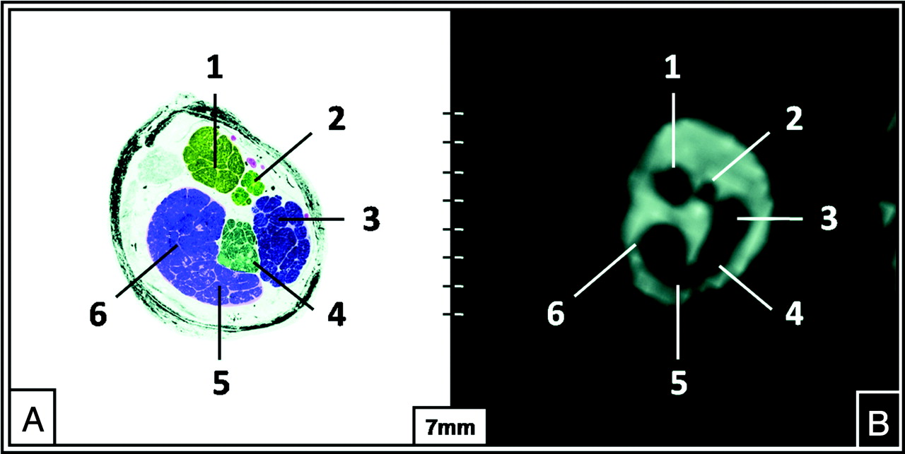
    The intermediate position of the NI in the middle third of the IAC (TR, 12.18 ms; TE, 6.09 ms; flip angle, 50°). Semi-thin anatomic section of the left IAC (A) and a correlating parasagittal in vivo CISS sequence image (B) (TR, 12.18 ms; TE, 6.09 ms) at 3T depicting the nervus facialis (1), nervus intermedius (2), nervus vestibularis superior (3), nervus vestibularis inferior (4), hook of the nervus cochlearis (5), and the nervus cochlearis (6).

Tables

  • Figures
    • View popup
    Table 1:

    Cross-tabulations of image-quality ratingsa

    Image QualityObserver BTotal% A
    012
    Observer A
        010011.9
        121221629.6
        205323768.5
    Total3173454100
    % B5.531.563.0100
    • a 0 indicates insufficient; 1, adequate; 2, excellent.

    • View popup
    Table 2:

    Cross-tabulations of identifiability ratingsa

    IdentifiabilityObserver BTotal% A
    012
    Observer A
        022102342.6
        11921222.2
        202171935.2
    Total23121954100
    % B42.622.235.2100
    • a 0 indicates none; 1, adequate; 2, precise.

PreviousNext
Back to top

In this issue

American Journal of Neuroradiology: 32 (3)
American Journal of Neuroradiology
Vol. 32, Issue 3
1 Mar 2011
  • Table of Contents
  • Index by author
Advertisement
Print
Download PDF
Email Article

Thank you for your interest in spreading the word on American Journal of Neuroradiology.

NOTE: We only request your email address so that the person you are recommending the page to knows that you wanted them to see it, and that it is not junk mail. We do not capture any email address.

Enter multiple addresses on separate lines or separate them with commas.
Identification of the Nervus Intermedius Using 3T MR Imaging
(Your Name) has sent you a message from American Journal of Neuroradiology
(Your Name) thought you would like to see the American Journal of Neuroradiology web site.
CAPTCHA
This question is for testing whether or not you are a human visitor and to prevent automated spam submissions.
Cite this article
H.P. Burmeister, P.A. Baltzer, M. Dietzel, I. Krumbein, T. Bitter, A. Schrott-Fischer, O. Guntinas-Lichius, W.A. Kaiser
Identification of the Nervus Intermedius Using 3T MR Imaging
American Journal of Neuroradiology Mar 2011, 32 (3) 460-464; DOI: 10.3174/ajnr.A2338

Citation Manager Formats

  • BibTeX
  • Bookends
  • EasyBib
  • EndNote (tagged)
  • EndNote 8 (xml)
  • Medlars
  • Mendeley
  • Papers
  • RefWorks Tagged
  • Ref Manager
  • RIS
  • Zotero
0 Responses
Respond to this article
Share
Bookmark this article
Identification of the Nervus Intermedius Using 3T MR Imaging
H.P. Burmeister, P.A. Baltzer, M. Dietzel, I. Krumbein, T. Bitter, A. Schrott-Fischer, O. Guntinas-Lichius, W.A. Kaiser
American Journal of Neuroradiology Mar 2011, 32 (3) 460-464; DOI: 10.3174/ajnr.A2338
del.icio.us logo Twitter logo Facebook logo Mendeley logo
  • Tweet Widget
  • Facebook Like
  • Google Plus One
Purchase

Jump to section

  • Article
    • Abstract
    • Abbreviations
    • Materials and Methods
    • Results
    • Discussion
    • Conclusions
    • Footnotes
    • References
  • Figures & Data
  • Supplemental
  • Info & Metrics
  • Responses
  • References
  • PDF

Related Articles

  • PubMed
  • Google Scholar

Cited By...

  • The Functions of the Nervus Intermedius
  • Reply:
  • Crossref (31)
  • Google Scholar

This article has been cited by the following articles in journals that are participating in Crossref Cited-by Linking.

  • The Nervus Intermedius: A Review of Its Anatomy, Function, Pathology, and Role in Neurosurgery
    R. Shane Tubbs, Dominik T. Steck, Martin M. Mortazavi, Aaron A. Cohen-Gadol
    World Neurosurgery 2013 79 5-6
  • MRI of the Internal Auditory Canal, Labyrinth, and Middle Ear: How We Do It
    John C. Benson, Matthew L. Carlson, John I. Lane
    Radiology 2020 297 2
  • The Functions of the Nervus Intermedius
    A. Alfieri, J. Fleischhammer, J. Prell
    American Journal of Neuroradiology 2011 32 7
  • The nervus intermedius as a variable landmark and critical structure in cerebellopontine angle surgery: an anatomical study and classification
    Alex Alfieri, Julius Fleischhammer, Elmar Peschke, Christian Strauss
    Acta Neurochirurgica 2012 154 7
  • The central myelin–peripheral myelin transitional zone of the nervus intermedius and its implications for microsurgery in the cerebellopontine angle
    Alex Alfieri, Julius Fleischhammer, Christian Strauss, Elmar Peschke
    Clinical Anatomy 2012 25 7
  • Temporal Bone Anatomy
    John C. Benson, John I. Lane
    Neuroimaging Clinics of North America 2022 32 4
  • The Facial Nerve: Anatomy and Pathology
    Ana Carolina Ottaiano, Gustavo Dalul Gomez, Tomás de Andrade Lourenção Freddi
    Seminars in Ultrasound, CT and MRI 2023 44 2
  • A Case of Nervus Intermedius Neuralgia
    Keisuke Onoda, Ai Kawaguchi, Yoshinori Takaya, Yuzo Saito, Hisashi Ishikawa, Takeshi Uno, Michiyuki Miyamoto, Kenichi Oyama, Akira Matsuno
    World Neurosurgery 2020 137
  • Clinical implications of nervus intermedius variants in patients with geniculate neuralgia: Let anatomy be the guide
    William E. Clifton, Sanjeet Grewal, Larry Lundy, William P. Cheshire, R. Shane Tubbs, Robert E. Wharen
    Clinical Anatomy 2020 33 7
  • Imaging Anatomy and Pathology of the Intracranial and Intratemporal Facial Nerve
    Burce Ozgen Mocan
    Neuroimaging Clinics of North America 2021 31 4

More in this TOC Section

  • Parathyroid Lesions: Characterization with Dual-Phase Arterial and Venous Enhanced CT of the Neck
  • Correlation of Apparent Diffusion Coefficient at 3T with Prognostic Parameters of Retinoblastoma
  • Efficacy of Diffusion-Weighted Imaging for the Differentiation between Lymphomas and Carcinomas of the Nasopharynx and Oropharynx: Correlations of Apparent Diffusion Coefficients and Histologic Features
Show more HEAD AND NECK

Similar Articles

Advertisement

Indexed Content

  • Current Issue
  • Accepted Manuscripts
  • Article Preview
  • Past Issues
  • Editorials
  • Editor's Choice
  • Fellows' Journal Club
  • Letters to the Editor
  • Video Articles

Cases

  • Case Collection
  • Archive - Case of the Week
  • Archive - Case of the Month
  • Archive - Classic Case

More from AJNR

  • Trainee Corner
  • Imaging Protocols
  • MRI Safety Corner
  • Book Reviews

Multimedia

  • AJNR Podcasts
  • AJNR Scantastics

Resources

  • Turnaround Time
  • Submit a Manuscript
  • Submit a Video Article
  • Submit an eLetter to the Editor/Response
  • Manuscript Submission Guidelines
  • Statistical Tips
  • Fast Publishing of Accepted Manuscripts
  • Graphical Abstract Preparation
  • Imaging Protocol Submission
  • Evidence-Based Medicine Level Guide
  • Publishing Checklists
  • Author Policies
  • Become a Reviewer/Academy of Reviewers
  • News and Updates

About Us

  • About AJNR
  • Editorial Board
  • Editorial Board Alumni
  • Alerts
  • Permissions
  • Not an AJNR Subscriber? Join Now
  • Advertise with Us
  • Librarian Resources
  • Feedback
  • Terms and Conditions
  • AJNR Editorial Board Alumni

American Society of Neuroradiology

  • Not an ASNR Member? Join Now

© 2025 by the American Society of Neuroradiology All rights, including for text and data mining, AI training, and similar technologies, are reserved.
Print ISSN: 0195-6108 Online ISSN: 1936-959X

Powered by HighWire