Advertisement

Main menu

  • Home
  • Content
    • Current Issue
    • Accepted Manuscripts
    • Article Preview
    • Past Issue Archive
    • Video Articles
    • AJNR Case Collection
    • Case of the Week Archive
    • Case of the Month Archive
    • Classic Case Archive
  • Special Collections
    • AJNR Awards
    • Low-Field MRI
    • Alzheimer Disease
    • ASNR Foundation Special Collection
    • Photon-Counting CT
    • View All
  • Multimedia
    • AJNR Podcasts
    • AJNR SCANtastic
    • Trainee Corner
    • MRI Safety Corner
    • Imaging Protocols
  • For Authors
    • Submit a Manuscript
    • Submit a Video Article
    • Submit an eLetter to the Editor/Response
    • Manuscript Submission Guidelines
    • Statistical Tips
    • Fast Publishing of Accepted Manuscripts
    • Graphical Abstract Preparation
    • Imaging Protocol Submission
    • Author Policies
  • About Us
    • About AJNR
    • Editorial Board
    • Editorial Board Alumni
  • More
    • Become a Reviewer/Academy of Reviewers
    • Subscribers
    • Permissions
    • Alerts
    • Feedback
    • Advertisers
    • ASNR Home

User menu

  • Alerts
  • Log in

Search

  • Advanced search
American Journal of Neuroradiology

American Journal of Neuroradiology

ASHNR American Society of Functional Neuroradiology ASHNR American Society of Pediatric Neuroradiology ASSR
  • Alerts
  • Log in

Advanced Search

  • Home
  • Content
    • Current Issue
    • Accepted Manuscripts
    • Article Preview
    • Past Issue Archive
    • Video Articles
    • AJNR Case Collection
    • Case of the Week Archive
    • Case of the Month Archive
    • Classic Case Archive
  • Special Collections
    • AJNR Awards
    • Low-Field MRI
    • Alzheimer Disease
    • ASNR Foundation Special Collection
    • Photon-Counting CT
    • View All
  • Multimedia
    • AJNR Podcasts
    • AJNR SCANtastic
    • Trainee Corner
    • MRI Safety Corner
    • Imaging Protocols
  • For Authors
    • Submit a Manuscript
    • Submit a Video Article
    • Submit an eLetter to the Editor/Response
    • Manuscript Submission Guidelines
    • Statistical Tips
    • Fast Publishing of Accepted Manuscripts
    • Graphical Abstract Preparation
    • Imaging Protocol Submission
    • Author Policies
  • About Us
    • About AJNR
    • Editorial Board
    • Editorial Board Alumni
  • More
    • Become a Reviewer/Academy of Reviewers
    • Subscribers
    • Permissions
    • Alerts
    • Feedback
    • Advertisers
    • ASNR Home
  • Follow AJNR on Twitter
  • Visit AJNR on Facebook
  • Follow AJNR on Instagram
  • Join AJNR on LinkedIn
  • RSS Feeds

AJNR Awards, New Junior Editors, and more. Read the latest AJNR updates

Research ArticleHead and Neck

CT Angiography as a Screening Tool for Dural Arteriovenous Fistula in Patients with Pulsatile Tinnitus: Feasibility and Test Characteristics

J. Narvid, H.M. Do, N.H. Blevins and N.J. Fischbein
American Journal of Neuroradiology March 2011, 32 (3) 446-453; DOI: https://doi.org/10.3174/ajnr.A2328
J. Narvid
  • Find this author on Google Scholar
  • Find this author on PubMed
  • Search for this author on this site
H.M. Do
  • Find this author on Google Scholar
  • Find this author on PubMed
  • Search for this author on this site
N.H. Blevins
  • Find this author on Google Scholar
  • Find this author on PubMed
  • Search for this author on this site
N.J. Fischbein
  • Find this author on Google Scholar
  • Find this author on PubMed
  • Search for this author on this site
  • Article
  • Figures & Data
  • Info & Metrics
  • Responses
  • References
  • PDF
Loading

Article Figures & Data

Figures

  • Tables
  • Fig 1.
    • Download figure
    • Open in new tab
    • Download powerpoint
    Fig 1.

    A, Axial 3-mm-slab MIP from CTA. A clearly abnormal “ring” of arterial feeders is seen around the right distal cervical carotid artery at the C1–2 level (white arrow) in a patient with a right sigmoid DAVF (patient 1). B, Large transosseous venous channels (white arrows) are present on the right in patient 1. This is an uncommon but specific finding for DAVF. These channels do not track to the occipitomastoid suture, a structure from which they must be distinguished in some cases by viewing serial sections. C, Markedly asymmetric venous collaterals along the right occipital bone, both intracranially (white arrow) and extracranially (white arrowhead), as well as asymmetrically enlarged and numerous veins throughout the infratemporal fossa (patient 2). This patient also had large transosseous collaterals (not shown). D, DSA, arterial phase, right external carotid injection in a lateral projection in patient 2, shows arterial feeders from the markedly enlarged occipital artery (short black arrow), as well as from the middle meningeal artery (black arrowheads), the correlates of the arterial feeders shown on the CTA in C, with a sigmoid DAVF (long black arrow).
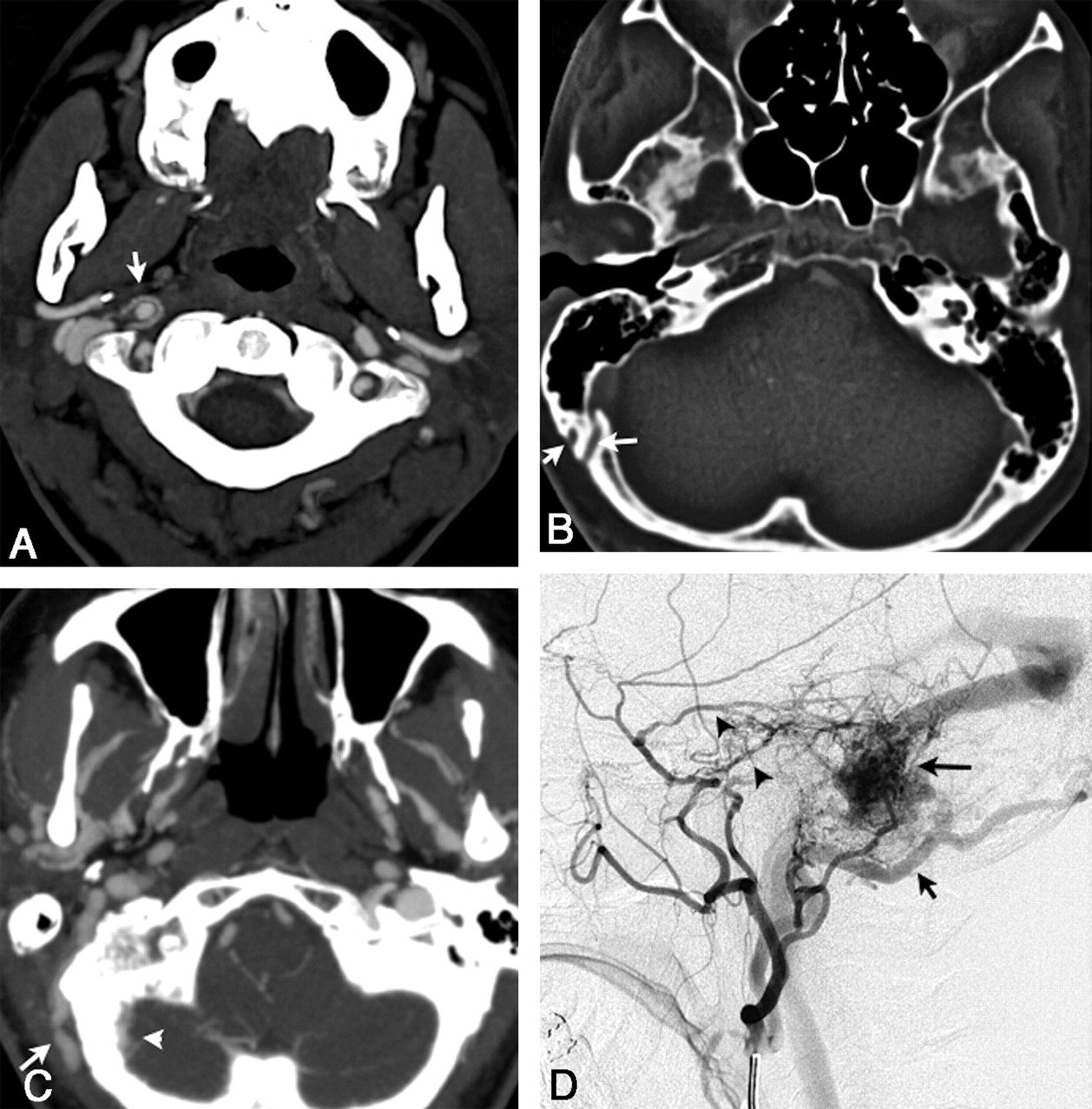
  • Fig 2.
    • Download figure
    • Open in new tab
    • Download powerpoint
    Fig 2.

    A, Axial 3-mm-slab MIP from CTA in patient 3 with a right sigmoid DAVF exhibits a markedly enlarged R occipital artery at the C1–2 level (large white arrow). There are also abnormally prominent small vessels surrounding the right vertebral and distal internal carotid arteries (white arrowheads). B, DSA, arterial phase, right external carotid injection in a lateral projection, demonstrates a markedly enlarged occipital artery (long black arrows), the angiographic correlate of the CTA in A. Transosseous branches (black arrowheads) of the occipital artery and middle meningeal artery feeders (white arrowheads) contribute to this sigmoid dural fistula. C, Axial 3-mm-slab MIP. The right sigmoid sinus is markedly enlarged and has a very irregular medial margin (arrow), an example of the shaggy sinus sign (patient 2). D, Another shaggy sinus sign involving the right transverse sinus. (The shaggy sinus only occurred in patients and was not seen in the control population.) E, DSA, right external carotid injection in a lateral projection, shows a typical DAVF with abnormal early opacification of the proximal occluded sigmoid sinus with retrograde flow through the transverse sinus to the contralateral dural sinuses. Arterial feeders arise from the middle meningeal artery (white arrowhead) and posterior auricular artery (black arrow) in patient 2. Angiographically, this sinus also appears shaggy (black arrowheads).

  • Fig 3.
    • Download figure
    • Open in new tab
    • Download powerpoint
    Fig 3.

    A, Coronal 3-mm-slab MIP from CTA in patient 3 with a right transverse sinus DAVF, predominantly supplied by right occipital, middle meningeal, and marginal tentorial branches; some supply from the left internal carotid is also seen, as described below. This case illustrates the “asymmetric jugular attenuation” sign, which is easily appreciated when the R and L IJVs are compared. The attenuation of the R IJV is 402 HU, and the L IJV is 318 HU. B, DSA, late arterial phase, left internal carotid injection in an anteroposterior projection in same patient as in A. Fistula supply from small left internal carotid artery dural and tentorial branches (black arrows) contributes to early opacification of the right sigmoid sinus and IJV (note the catheter near the jugular bulb). Due to the arteriovenous shunt surgery and drainage pattern, iodinated contrast preferentially fills the right jugular vein (black arrowheads) and leads to an asymmetric right-sided increase in CT attenuation, which is seen in A.

  • Fig 4.
    • Download figure
    • Open in new tab
    • Download powerpoint
    Fig 4.

    A, Axial 3-mm-slab MIP from CTA in patient 7 illustrates a cluster of abnormally prominent, tortuous, and numerous veins overlying the right temporal lobe (black arrows). These are dilated cortical venous efferents, which are thought to confer increased risk of hemorrhage. B, DSA, right external carotid injection, venous phase, in a lateral projection in the same patient as in A, demonstrates retrograde cortical or leptomeningeal draining veins (black arrows) in the right temporal region in this Cognard IIa+b fistula. Note multiple arterial feeders arising from the occipital artery (white arrows).

Tables

  • Figures
    • View popup
    Table 1:

    Clinical and angiographic characteristics of patients (PT and confirmed DAVF)

    PatientAge (yr)/SexArterial SupplyVenous DrainageStenosesCortical VenousCognard Stage
    146/FR Occ/R Vert/R APAMarginalNoneNoIIa
    246/FR PCA/R Vert/R Occ/R MM/R APA/L OccSigmoidR JugularNoIIa
    379/FR MM/R Occ/R MHTOccipitalR JugularNoI
    455/MR Occ/R MHT/R MMSigmoidR JugularNoIIa
    543/MR Occ/R PAur/R MHTMarginalNoneNoI
    652/FL Occ/L VertMarginalNoneNoIIa
    740/FR MM/R APA/R OccSigmoidR SigmoidYesIIa+b
    • View popup
    Table 2:

    Clinical characteristics of controls (PT without DAVF)

    PatientAge (yr)/SexSymptomsCTA Findings
    A53/FR PTSigmoid sinus diverticulum
    B30/FR PTNo dx
    C53/FR PTTransverse sinus stenosis
    D48/FL PTNo dx
    E39/FR PTNo dx
    F33/ML PTNo dx
    G75/MR PTNo dx
    • View popup
    Table 3:

    Test characteristics of CTA findings

    CTA FindingSensitivity (%)95% CISpecificity (%)95% CIPPV (%)95% CINPV (%)95% CI
    Asymmetrically increased arterial feeders8642–9910052–10010056–1008867–88
    Asymmetrically increased venous collaterals4211–797130–956017–935638–70
    Transcalvarial channels295–708642–996712–985543–62
    Shaggy sinus or tentorium4211–7910056–10010031–1006455–64
    Prominent cortical venous drainage295–707130–95509–905035–63
PreviousNext
Back to top

In this issue

American Journal of Neuroradiology: 32 (3)
American Journal of Neuroradiology
Vol. 32, Issue 3
1 Mar 2011
  • Table of Contents
  • Index by author
Advertisement
Print
Download PDF
Email Article

Thank you for your interest in spreading the word on American Journal of Neuroradiology.

NOTE: We only request your email address so that the person you are recommending the page to knows that you wanted them to see it, and that it is not junk mail. We do not capture any email address.

Enter multiple addresses on separate lines or separate them with commas.
CT Angiography as a Screening Tool for Dural Arteriovenous Fistula in Patients with Pulsatile Tinnitus: Feasibility and Test Characteristics
(Your Name) has sent you a message from American Journal of Neuroradiology
(Your Name) thought you would like to see the American Journal of Neuroradiology web site.
CAPTCHA
This question is for testing whether or not you are a human visitor and to prevent automated spam submissions.
Cite this article
J. Narvid, H.M. Do, N.H. Blevins, N.J. Fischbein
CT Angiography as a Screening Tool for Dural Arteriovenous Fistula in Patients with Pulsatile Tinnitus: Feasibility and Test Characteristics
American Journal of Neuroradiology Mar 2011, 32 (3) 446-453; DOI: 10.3174/ajnr.A2328

Citation Manager Formats

  • BibTeX
  • Bookends
  • EasyBib
  • EndNote (tagged)
  • EndNote 8 (xml)
  • Medlars
  • Mendeley
  • Papers
  • RefWorks Tagged
  • Ref Manager
  • RIS
  • Zotero
0 Responses
Respond to this article
Share
Bookmark this article
CT Angiography as a Screening Tool for Dural Arteriovenous Fistula in Patients with Pulsatile Tinnitus: Feasibility and Test Characteristics
J. Narvid, H.M. Do, N.H. Blevins, N.J. Fischbein
American Journal of Neuroradiology Mar 2011, 32 (3) 446-453; DOI: 10.3174/ajnr.A2328
del.icio.us logo Twitter logo Facebook logo Mendeley logo
  • Tweet Widget
  • Facebook Like
  • Google Plus One
Purchase

Jump to section

  • Article
    • Abstract
    • Abbreviations
    • Materials and Methods
    • Results
    • Discussion
    • Conclusions
    • References
  • Figures & Data
  • Info & Metrics
  • Responses
  • References
  • PDF

Related Articles

  • No related articles found.
  • PubMed
  • Google Scholar

Cited By...

  • Non-invasive imaging modalities for diagnosing pulsatile tinnitus: a comprehensive review and recommended imaging algorithm
  • Clinical Reasoning: A 68-year-old man with rapid cognitive decline
  • Carotid CTA at the Lowest Tube Voltage (70 kV) in Comparison with Automated Tube Voltage Adaption
  • The application of susceptibility-weighted MRI in pre-interventional evaluation of intracranial dural arteriovenous fistulas
  • Response to Letter Regarding Article, "Intracranial Dural Arteriovenous Fistulae: Clinical Presentation and Management Strategies"
  • Intracranial Dural Arteriovenous Fistulae: Clinical Presentation and Management Strategies
  • Carotid CTA: Radiation Exposure and Image Quality with the Use of Attenuation-Based, Automated Kilovolt Selection
  • Crossref (83)
  • Google Scholar

This article has been cited by the following articles in journals that are participating in Crossref Cited-by Linking.

  • Intracranial Dural Arteriovenous Fistulae
    Timothy R. Miller, Dheeraj Gandhi
    Stroke 2015 46 7
  • ACR Appropriateness Criteria ® Cerebrovascular Disease
    Michael B. Salmela, Shabnam Mortazavi, Bharathi D. Jagadeesan, Daniel F. Broderick, Judah Burns, Tejaswini K. Deshmukh, H. Benjamin Harvey, Jenny Hoang, Christopher H. Hunt, Tabassum A. Kennedy, Alexander A. Khalessi, William Mack, Nandini D. Patel, Joel S. Perlmutter, Bruno Policeni, Jason W. Schroeder, Gavin Setzen, Matthew T. Whitehead, Rebecca S. Cornelius, Amanda S. Corey
    Journal of the American College of Radiology 2017 14 5
  • Evaluation and treatment of pulsatile tinnitus associated with sigmoid sinus wall anomalies
    David J. Eisenman, Prashant Raghavan, Ronna Hertzano, Robert Morales
    The Laryngoscope 2018 128 S2
  • Pulse-Synchronous Tinnitus and Sigmoid Sinus Wall Anomalies
    Rebecca S. Harvey, Ronna Hertzano, Shalom E. Kelman, David J. Eisenman
    Otology & Neurotology 2014 35 1
  • Incidence of Vascular Anomalies and Variants Associated with Unilateral Venous Pulsatile Tinnitus in 242 Patients Based on Dual-phase Contrast-enhanced Computed Tomography
    Cheng Dong, Peng-Fei Zhao, Ji-Gang Yang, Zhao-Hui Liu, Zhen-Chang Wang
    Chinese Medical Journal 2015 128 5
  • State of the Art: Venous Causes of Pulsatile Tinnitus and Diagnostic Considerations Guiding Endovascular Therapy
    Mohamad Abdalkader, Thanh N. Nguyen, Alexander M. Norbash, Eytan Raz, Maksim Shapiro, Stéphanie Lenck, Waleed Brinjikji, Peter Weber, Osamu Sakai
    Radiology 2021 300 1
  • Sigmoid sinus diverticulum and pulsatile tinnitus: analysis of CT scans from 15 cases
    Zhaohui Liu, Chengfang Chen, Zhenchang Wang, Shusheng Gong, Junfang Xian, Yongzhe Wang, Xihong Liang, Xiaobo Ma, Yi Li
    Acta Radiologica 2013 54 7
  • Dural Arteriovenous Fistulae
    Yafell Serulle, Timothy R. Miller, Dheeraj Gandhi
    Neuroimaging Clinics of North America 2016 26 2
  • Endovascular treatment of two concomitant causes of pulsatile tinnitus: sigmoid sinus stenosis and ipsilateral jugular bulb diverticulum. Case report and literature review
    Francesco Signorelli, Kalid Mahla, Francis Turjman
    Acta Neurochirurgica 2012 154 1
  • Carotid CTA: Radiation Exposure and Image Quality with the Use of Attenuation-Based, Automated Kilovolt Selection
    A. Eller, W. Wuest, M. Kramer, M. May, A. Schmid, M. Uder, M. M. Lell
    American Journal of Neuroradiology 2014 35 2

More in this TOC Section

  • Correlation of Apparent Diffusion Coefficient at 3T with Prognostic Parameters of Retinoblastoma
  • Parathyroid Lesions: Characterization with Dual-Phase Arterial and Venous Enhanced CT of the Neck
  • Comparison of Contrast Effect on the Cochlear Perilymph after Intratympanic and Intravenous Gadolinium Injection
Show more HEAD AND NECK

Similar Articles

Advertisement

Indexed Content

  • Current Issue
  • Accepted Manuscripts
  • Article Preview
  • Past Issues
  • Editorials
  • Editor's Choice
  • Fellows' Journal Club
  • Letters to the Editor
  • Video Articles

Cases

  • Case Collection
  • Archive - Case of the Week
  • Archive - Case of the Month
  • Archive - Classic Case

More from AJNR

  • Trainee Corner
  • Imaging Protocols
  • MRI Safety Corner
  • Book Reviews

Multimedia

  • AJNR Podcasts
  • AJNR Scantastics

Resources

  • Turnaround Time
  • Submit a Manuscript
  • Submit a Video Article
  • Submit an eLetter to the Editor/Response
  • Manuscript Submission Guidelines
  • Statistical Tips
  • Fast Publishing of Accepted Manuscripts
  • Graphical Abstract Preparation
  • Imaging Protocol Submission
  • Evidence-Based Medicine Level Guide
  • Publishing Checklists
  • Author Policies
  • Become a Reviewer/Academy of Reviewers
  • News and Updates

About Us

  • About AJNR
  • Editorial Board
  • Editorial Board Alumni
  • Alerts
  • Permissions
  • Not an AJNR Subscriber? Join Now
  • Advertise with Us
  • Librarian Resources
  • Feedback
  • Terms and Conditions
  • AJNR Editorial Board Alumni

American Society of Neuroradiology

  • Not an ASNR Member? Join Now

© 2025 by the American Society of Neuroradiology All rights, including for text and data mining, AI training, and similar technologies, are reserved.
Print ISSN: 0195-6108 Online ISSN: 1936-959X

Powered by HighWire