Advertisement

Main menu

  • Home
  • Content
    • Current Issue
    • Accepted Manuscripts
    • Article Preview
    • Past Issue Archive
    • Video Articles
    • AJNR Case Collection
    • Case of the Week Archive
    • Case of the Month Archive
    • Classic Case Archive
  • Special Collections
    • AJNR Awards
    • Low-Field MRI
    • Alzheimer Disease
    • ASNR Foundation Special Collection
    • Photon-Counting CT
    • View All
  • Multimedia
    • AJNR Podcasts
    • AJNR SCANtastic
    • Trainee Corner
    • MRI Safety Corner
    • Imaging Protocols
  • For Authors
    • Submit a Manuscript
    • Submit a Video Article
    • Submit an eLetter to the Editor/Response
    • Manuscript Submission Guidelines
    • Statistical Tips
    • Fast Publishing of Accepted Manuscripts
    • Graphical Abstract Preparation
    • Imaging Protocol Submission
    • Author Policies
  • About Us
    • About AJNR
    • Editorial Board
    • Editorial Board Alumni
  • More
    • Become a Reviewer/Academy of Reviewers
    • Subscribers
    • Permissions
    • Alerts
    • Feedback
    • Advertisers
    • ASNR Home

User menu

  • Alerts
  • Log in

Search

  • Advanced search
American Journal of Neuroradiology

American Journal of Neuroradiology

ASHNR American Society of Functional Neuroradiology ASHNR American Society of Pediatric Neuroradiology ASSR
  • Alerts
  • Log in

Advanced Search

  • Home
  • Content
    • Current Issue
    • Accepted Manuscripts
    • Article Preview
    • Past Issue Archive
    • Video Articles
    • AJNR Case Collection
    • Case of the Week Archive
    • Case of the Month Archive
    • Classic Case Archive
  • Special Collections
    • AJNR Awards
    • Low-Field MRI
    • Alzheimer Disease
    • ASNR Foundation Special Collection
    • Photon-Counting CT
    • View All
  • Multimedia
    • AJNR Podcasts
    • AJNR SCANtastic
    • Trainee Corner
    • MRI Safety Corner
    • Imaging Protocols
  • For Authors
    • Submit a Manuscript
    • Submit a Video Article
    • Submit an eLetter to the Editor/Response
    • Manuscript Submission Guidelines
    • Statistical Tips
    • Fast Publishing of Accepted Manuscripts
    • Graphical Abstract Preparation
    • Imaging Protocol Submission
    • Author Policies
  • About Us
    • About AJNR
    • Editorial Board
    • Editorial Board Alumni
  • More
    • Become a Reviewer/Academy of Reviewers
    • Subscribers
    • Permissions
    • Alerts
    • Feedback
    • Advertisers
    • ASNR Home
  • Follow AJNR on Twitter
  • Visit AJNR on Facebook
  • Follow AJNR on Instagram
  • Join AJNR on LinkedIn
  • RSS Feeds

AJNR Awards, New Junior Editors, and more. Read the latest AJNR updates

Research ArticleBrainC

MR Imaging-Guided Intravenous Thrombolysis in Posterior Cerebral Artery Stroke

A. Förster, A. Gass, R. Kern, M.E. Wolf, M.G. Hennerici and K. Szabo
American Journal of Neuroradiology February 2011, 32 (2) 419-421; DOI: https://doi.org/10.3174/ajnr.A2300
A. Förster
  • Find this author on Google Scholar
  • Find this author on PubMed
  • Search for this author on this site
A. Gass
  • Find this author on Google Scholar
  • Find this author on PubMed
  • Search for this author on this site
R. Kern
  • Find this author on Google Scholar
  • Find this author on PubMed
  • Search for this author on this site
M.E. Wolf
  • Find this author on Google Scholar
  • Find this author on PubMed
  • Search for this author on this site
M.G. Hennerici
  • Find this author on Google Scholar
  • Find this author on PubMed
  • Search for this author on this site
K. Szabo
  • Find this author on Google Scholar
  • Find this author on PubMed
  • Search for this author on this site
  • Article
  • Figures & Data
  • Info & Metrics
  • Responses
  • References
  • PDF
Loading

Abstract

SUMMARY: PCA stroke was under-represented in or excluded from the clinical trials examining thrombolysis based on the PWI-DWI mismatch concept. We present 6 patients with PCA stroke treated with thrombolysis in an extended time window by using MR imaging criteria. Symptoms included aphasia, sensorimotor hemiparesis, hemineglect, and homonymous hemianopia. Initial MR imaging demonstrated circumscribed ischemic lesions in the thalamus or hippocampus; MR angiography showed PCA occlusion with corresponding hypoperfusion. Follow-up MR imaging showed partial/complete recanalization in 4 patients with minor infarction growth, while in 1 patient, PCA occlusion persisted, resulting in a large PCA infarction. Three patients improved within 2 hours; at discharge, homonymous hemianopia had resolved in 3 patients. At 3-month follow-up, 4 patients had an mRS score of 0 or 1. These results support the approach to treat patients with PCA stroke with thrombolysis based on the mismatch concept. Because rehabilitation options for hemianopia are limited, thrombolysis may enhance the chance of a favorable outcome.

Abbreviations

A-S-C-O
A for atherosclerosis, S for small vessel disease, C for cardiac source, O for other cause
DWI
diffusion-weighted imaging
FLAIR
fluid-attenuated inversion recovery
MRA
MR angiography
mRS
modified Rankin Scale
NIHSS
National Institutes of Health Stroke Scale
OTT
onset to treatment time
PCA
posterior cerebral artery
PWI
perfusion-weighted imaging
rtPA
recombinant tissue plasminogen activator
TTP
time to peak

Posterior circulation stroke accounts for approximately 15% of all acute ischemic strokes and may cause permanent hemianopia, hemiparesis, or neuropsychological deficits, which may result in considerable permanent disability.1 Nevertheless, the large clinical trials investigating acute therapy with rtPA in acute ischemic stroke focused on anterior circulation stroke or even excluded patients with posterior circulation stroke from participation.2–4 The same holds true for clinical trials examining MR imaging−guided thrombolysis based on the PWI-DWI mismatch concept.5,6 We report a case series of 6 patients with acute ischemic stroke in the PCA territory treated with intravenous thrombolysis in an extended time window based on the DWI-PWI-mismatch concept.

Materials and Methods

From a prospectively collected stroke unit registry, we identified 5948 patients with acute ischemic stroke admitted between January 2004 and December 2009. Altogether, 818 (13.8%) patients were treated with rtPA; among these, 71 (8.7%) patients received intravenous thrombolysis (0.9-mg/kg rtPA; maximum, 90 mg; with 10% of the dose given as a bolus followed by an infusion lasting 60 minutes) in an extended time window guided by acute stroke MR imaging based on the DWI-PWI mismatch concept. Treatment was performed on an individual off-label-use basis with patient informed consent documented according to ethics committee approval. Six of the 71 (8.5%) patients had acute ischemic stroke in the PCA territory. All clinical data and technical investigations were collected and documented according to a standardized acute stroke care protocol.

Initial and follow-up MR imaging was performed on a 1.5T system (Magnetom Sonata; Siemens, Erlangen, Germany). A standardized protocol was used in all patients: 1) transverse, coronal, and sagittal localizing sequences followed by transverse oblique contiguous images aligned with the inferior borders of the corpus callosum (applied on sequences 2–6), 2) T2-weighted images, 3) T1-weighted images, 4) DWI, 5) FLAIR, and 6) PWI following the first pass of contrast bolus through the brain (20-mL gadoterate meglumine, Dotarem; Guerbet, Aulney-sous-Bois, France) injected manually through a large-gauge venous cannula into the antecubital vein; and 7) 3D time-of-flight MRA sequences of the circle of Willis and the neck arteries in the coronal direction.

The size of the acute ischemic lesion was measured on DWI. The size of perfusion deficit was measured on TTP maps by manually delineating the region of interest with commercially available image analysis software, nordicICE (Nordic Imaging Lab, Bergen, Norway). Probable stroke cause was classified according to the A-S-C-O (phenotypic) classification of stroke described recently.7

Results

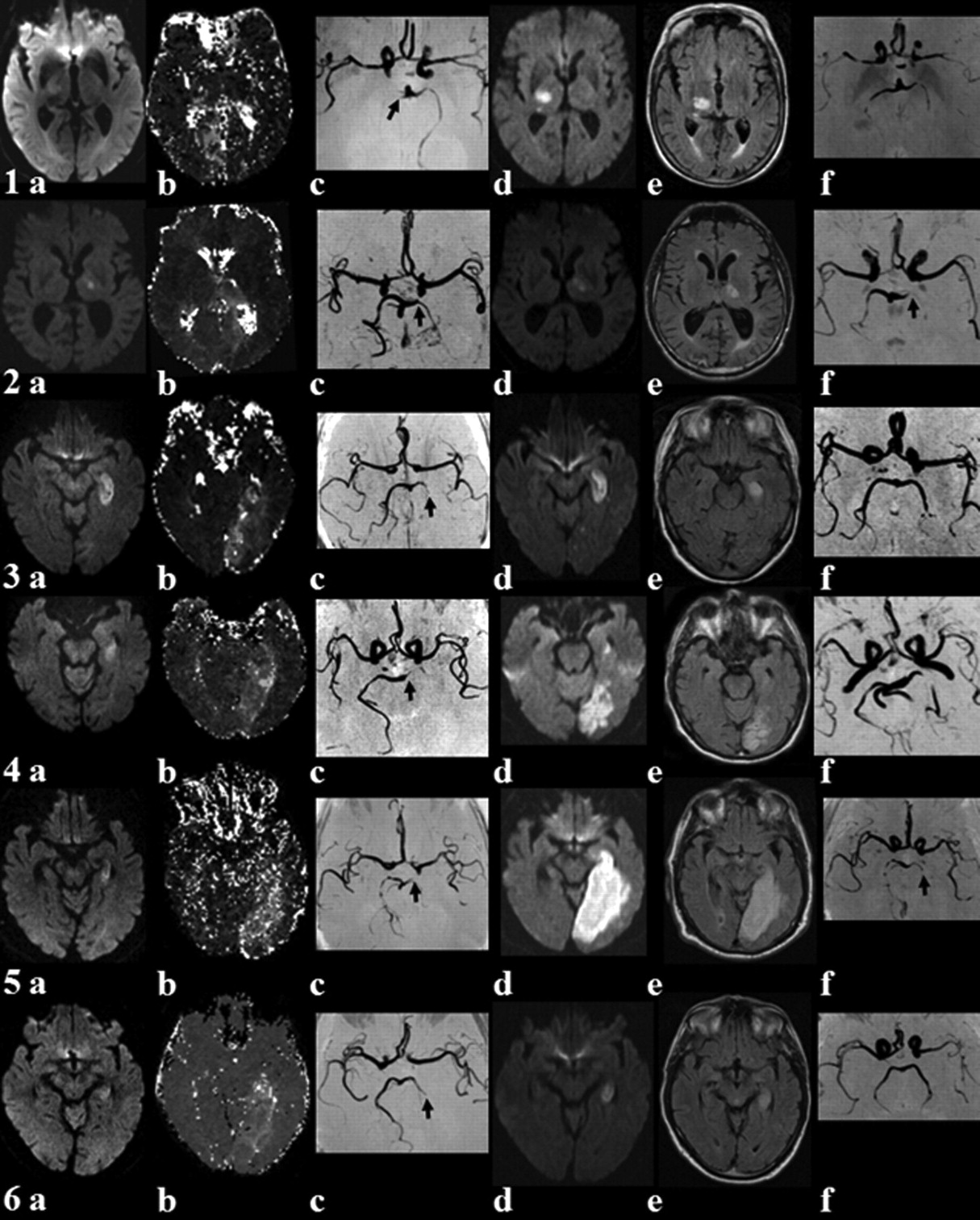
The Table shows the baseline and MR imaging characteristics of the 6 patients (4 men, 2 women). On initial DWI, 1 patient had thalamic infarction (patient 2), while in the other 5 cases, the hippocampus was affected. MRA showed occlusion of the right or left PCA and correspondingly hypoperfusion in the right or left PCA territory (Fig 1). The time from symptom onset to treatment varied from 185 to 390 minutes.

View this table:
  • View inline
  • View popup

Baseline data, clinical outcome, A-S-C-O subtype, and MR imaging characteristics

Fig 1.
  • Download figure
  • Open in new tab
  • Download powerpoint
Fig 1.

MR imaging in posterior circulation stroke before (a−c) and after (d−f) intravenous thrombolysis. Initial MR imaging shows the ischemic lesion on DWI (a), hypoperfusion on PWI (TTP map) (b), and vessel pathology (black arrow) on MRA (c). Follow-up MR imaging lesion on DWI (d) and T2 FLAIR imaging (e), as well as recanalization or persistent vessel pathology (black arrow) on MRA (f).

Four patients improved significantly within 2 hours after therapy, and at discharge, homonymous hemianopia had resolved completely in 3 patients (patients 3, 4, and 6). The condition of patient 5 deteriorated with conjugate horizontal gaze palsy and right sensorimotor hemiparesis. In patient 2, neither significant improvement nor deterioration of clinical symptoms was observed. Follow-up MR imaging was performed after 1–6 days and showed complete or partial recanalization in 4 patients. DWI demonstrated acute ischemic stroke in the thalamus only in patient 2; in the thalamus and hippocampus in patients 1 and 6; in the thalamus, hippocampus, and occipital lobe in patient 3; and in the thalamus, hippocampus, splenium, and occipital lobe in patient 4. A remarkable growth of infarct size was detected in patient 5, with nearly complete PCA territory infarction and persisting PCA occlusion.

At 3-month follow-up 4 patients had an mRS score of 0 or 1, 1 patient had an mRS score of 3, and 1 patient had an mRS score of 5. Only 1 minor complication (gum bleeding) occurred after thrombolysis.

Discussion

Due to strict study protocols focusing on anterior circulation stroke—mostly in the middle cerebral artery territory—there are limited data on thrombolysis in PCA stroke. PCA ischemia may present with a nondebilitating neurologic syndrome. Consequently patients might take longer to consult a physician, who in turn might hesitate to recommend thrombolysis, especially because PCA stroke symptoms are not well represented by the NIHSS.8 However, because rehabilitation options for visual field defects are limited,9 acute PCA stroke should be treated with the same regimen and emphasis as anterior circulation stroke.

We demonstrated that MR imaging identifies constellations in PCA stroke very comparable with those in anterior circulation stroke. A mismatch between an early DWI and PWI lesion can be seen, and the DWI lesion may increase in size (patient 5) unless early recanalization is achieved (patients 1, 3, 4, and 6). Natural history studies of PCA stroke indicate that tissue vulnerability and the treatment window in the PCA territory are comparable with anterior circulation stroke and that early recanalization may facilitate a good outcome. MR imaging, including MRA, DWI, and PWI, provides a diagnosis of the vascular lesion and perfusion deficit, which allows estimating the risk for growth of an ischemic lesion.

References

  1. 1.↵
    1. Ng YS,
    2. Stein J,
    3. Salles SS,
    4. et al
    . Clinical characteristics and rehabilitation outcomes of patients with posterior cerebral artery stroke. Arch Phys Med Rehabil 2005;86:2138–43
    CrossRefPubMed
  2. 2.↵
    1. De Keyser J,
    2. Gdovinova Z,
    3. Uyttenboogaart M,
    4. et al
    . Intravenous alteplase for stroke: beyond the guidelines and in particular clinical situations. Stroke 2007;38:2612–18. Epub 2007 Jul 26
    Abstract/FREE Full Text
  3. 3.↵
    1. Hacke W,
    2. Kaste M,
    3. Fieschi C,
    4. et al
    . Intravenous thrombolysis with recombinant tissue plasminogen activator for acute hemispheric stroke: the European Cooperative Acute Stroke Study (ECASS). JAMA 1995;274:1017–25
    CrossRefPubMed
  4. 4.↵
    1. Hacke W,
    2. Kaste M,
    3. Fieschi C,
    4. et al
    . Randomised double-blind placebo-controlled trial of thrombolytic therapy with intravenous alteplase in acute ischaemic stroke (ECASS II): Second European-Australasian Acute Stroke Study Investigators. Lancet 1998;352:1245–51
    CrossRefPubMed
  5. 5.↵
    1. Röther J,
    2. Schellinger PD,
    3. Gass A,
    4. et al
    . Effect of intravenous thrombolysis on MRI parameters and functional outcome in acute stroke <6 hours. Stroke 2002;33:2438–45
    Abstract/FREE Full Text
  6. 6.↵
    1. Albers GW,
    2. Thijs VN,
    3. Wechsler L,
    4. et al.
    , for the DEFUSE Investigators. Magnetic resonance imaging profiles predict clinical response to early reperfusion: the diffusion and perfusion imaging evaluation for understanding stroke evolution (DEFUSE) study. Ann Neurol 2006;60:508–17
    CrossRefPubMed
  7. 7.↵
    1. Amarenco P,
    2. Bogousslavsky J,
    3. Caplan LR,
    4. et al
    . New approach to stroke subtyping: the A-S-C-O (phenotypic) classification of stroke. Cerebrovasc Dis 2009;27:502–08
    CrossRefPubMed
  8. 8.↵
    1. Sato S,
    2. Toyoda K,
    3. Uehara T,
    4. et al
    . Baseline NIH Stroke Scale score predicting outcome in anterior and posterior circulation strokes. Neurology 2008;70(24 pt 2):2371–77. Epub 2008 Apr 23
    Abstract/FREE Full Text
  9. 9.↵
    1. Schofield TM,
    2. Leff AP
    . Rehabilitation of hemianopia. Curr Opin Neurol 2009;22:36–40
    CrossRefPubMed
  • Received June 26, 2010.
  • Accepted after revision July 22, 2010.
  • Copyright © American Society of Neuroradiology
View Abstract
PreviousNext
Back to top

In this issue

American Journal of Neuroradiology: 32 (2)
American Journal of Neuroradiology
Vol. 32, Issue 2
1 Feb 2011
  • Table of Contents
  • Index by author
Advertisement
Print
Download PDF
Email Article

Thank you for your interest in spreading the word on American Journal of Neuroradiology.

NOTE: We only request your email address so that the person you are recommending the page to knows that you wanted them to see it, and that it is not junk mail. We do not capture any email address.

Enter multiple addresses on separate lines or separate them with commas.
MR Imaging-Guided Intravenous Thrombolysis in Posterior Cerebral Artery Stroke
(Your Name) has sent you a message from American Journal of Neuroradiology
(Your Name) thought you would like to see the American Journal of Neuroradiology web site.
CAPTCHA
This question is for testing whether or not you are a human visitor and to prevent automated spam submissions.
Cite this article
A. Förster, A. Gass, R. Kern, M.E. Wolf, M.G. Hennerici, K. Szabo
MR Imaging-Guided Intravenous Thrombolysis in Posterior Cerebral Artery Stroke
American Journal of Neuroradiology Feb 2011, 32 (2) 419-421; DOI: 10.3174/ajnr.A2300

Citation Manager Formats

  • BibTeX
  • Bookends
  • EasyBib
  • EndNote (tagged)
  • EndNote 8 (xml)
  • Medlars
  • Mendeley
  • Papers
  • RefWorks Tagged
  • Ref Manager
  • RIS
  • Zotero
0 Responses
Respond to this article
Share
Bookmark this article
MR Imaging-Guided Intravenous Thrombolysis in Posterior Cerebral Artery Stroke
A. Förster, A. Gass, R. Kern, M.E. Wolf, M.G. Hennerici, K. Szabo
American Journal of Neuroradiology Feb 2011, 32 (2) 419-421; DOI: 10.3174/ajnr.A2300
del.icio.us logo Twitter logo Facebook logo Mendeley logo
  • Tweet Widget
  • Facebook Like
  • Google Plus One
Purchase

Jump to section

  • Article
    • Abstract
    • Abbreviations
    • Materials and Methods
    • Results
    • Discussion
    • References
  • Figures & Data
  • Info & Metrics
  • Responses
  • References
  • PDF

Related Articles

  • No related articles found.
  • PubMed
  • Google Scholar

Cited By...

  • Mechanical thrombectomy versus intravenous alteplase alone in acute isolated posterior cerebral artery occlusion: a systematic review
  • Pretreatment Advanced Imaging in Patients with Stroke Treated with IV Thrombolysis: Evaluation of a Multihospital Data Base
  • Crossref (13)
  • Google Scholar

This article has been cited by the following articles in journals that are participating in Crossref Cited-by Linking.

  • Endovascular therapy versus no endovascular therapy in patients receiving best medical management for acute isolated occlusion of the posterior cerebral artery: A systematic review andmeta‐analysis
    Anne Berberich, Stephanos Finitsis, Davide Strambo, Patrik Michel, Christian Herweh, Lukas Meyer, Uta Hanning, Daniel Strbian, Mohamad Abdalkader, Raul G. Nogueira, Volker Puetz, Daniel P. O. Kaiser, Marta Olive‐Gadea, Marc Ribo, Isabel Fragata, João Pedro Marto, Michele Romoli, Peter A. Ringleb, Thanh N. Nguyen, Simon Nagel
    European Journal of Neurology 2022 29 9
  • Susceptibility Vessel Sign for Intra-arterial Thrombus in Acute Posterior Cerebral Artery Infarction
    Min-Gyu Park, Chang Hyo Yoon, Seung Kug Baik, Kyung-Pil Park
    Journal of Stroke and Cerebrovascular Diseases 2015 24 6
  • Vestibular migraine: the chameleon in vestibular disease
    Minping Li, Xue Xu, Weiwei Qi, Yingyin Liang, Yongxin Huang, Haiwei Huang
    Neurological Sciences 2021 42 5
  • Mechanical thrombectomy versus intravenous alteplase alone in acute isolated posterior cerebral artery occlusion: a systematic review
    Andre Monteiro, Slah Khan, Muhammad Waqas, Rimal H Dossani, Nicco Ruggiero, Nehaal M Siddiqi, Ammad A Baig, Hamid H Rai, Justin M Cappuzzo, Elad I Levy, Adnan H Siddiqui
    Journal of NeuroInterventional Surgery 2022 14 6
  • Recent Advances in Magnetic Resonance Imaging in Posterior Circulation Stroke: Implications for Diagnosis and Prognosis
    Alex Förster, Martin Griebe, Achim Gass, Michael G. Hennerici, Kristina Szabo
    Current Treatment Options in Cardiovascular Medicine 2011 13 3
  • Advanced Neuroimaging to Guide Acute Stroke Therapy
    Gurpreet Singh Sandhu, Jeffrey L. Sunshine
    Current Cardiology Reports 2012 14 6
  • FLAIR vascular hyperintensities and dynamic 4D angiograms for the estimation of collateral blood flow in posterior circulation occlusion
    Alex Förster, Holger Wenz, Hans Ulrich Kerl, Mansour Al-Zghloul, Sonia Habich, Christoph Groden
    Neuroradiology 2014 56 9
  • Pretreatment Advanced Imaging in Patients with Stroke Treated with IV Thrombolysis: Evaluation of a Multihospital Data Base
    J.S. McDonald, J. Fan, D.F. Kallmes, H.J. Cloft
    American Journal of Neuroradiology 2014 35 3
  • Artery of Percheron Infarction
    Salman S. Ikramuddin, John A. Coburn, Solmaz Ramezani, Christopher Streib
    Neurology Clinical Practice 2024 14 2
  • Intravenous thrombolysis in ischemic stroke patients with isolated homonymous hemianopia
    D. Strbian, L. Soinne, T. Sairanen, F. Scheperjans, O. Salonen, M. Palomäki, M. Kaste, T. Tatlisumak
    Acta Neurologica Scandinavica 2012 126 4

More in this TOC Section

  • Multimodal CT Provides Improved Performance for Lacunar Infarct Detection
  • Optimal MRI Sequence for Identifying Occlusion Location in Acute Stroke: Which Value of Time-Resolved Contrast-Enhanced MRA?
  • Evaluating the Effects of White Matter Multiple Sclerosis Lesions on the Volume Estimation of 6 Brain Tissue Segmentation Methods
Show more Brain

Similar Articles

Advertisement

Indexed Content

  • Current Issue
  • Accepted Manuscripts
  • Article Preview
  • Past Issues
  • Editorials
  • Editor's Choice
  • Fellows' Journal Club
  • Letters to the Editor
  • Video Articles

Cases

  • Case Collection
  • Archive - Case of the Week
  • Archive - Case of the Month
  • Archive - Classic Case

More from AJNR

  • Trainee Corner
  • Imaging Protocols
  • MRI Safety Corner
  • Book Reviews

Multimedia

  • AJNR Podcasts
  • AJNR Scantastics

Resources

  • Turnaround Time
  • Submit a Manuscript
  • Submit a Video Article
  • Submit an eLetter to the Editor/Response
  • Manuscript Submission Guidelines
  • Statistical Tips
  • Fast Publishing of Accepted Manuscripts
  • Graphical Abstract Preparation
  • Imaging Protocol Submission
  • Evidence-Based Medicine Level Guide
  • Publishing Checklists
  • Author Policies
  • Become a Reviewer/Academy of Reviewers
  • News and Updates

About Us

  • About AJNR
  • Editorial Board
  • Editorial Board Alumni
  • Alerts
  • Permissions
  • Not an AJNR Subscriber? Join Now
  • Advertise with Us
  • Librarian Resources
  • Feedback
  • Terms and Conditions
  • AJNR Editorial Board Alumni

American Society of Neuroradiology

  • Not an ASNR Member? Join Now

© 2025 by the American Society of Neuroradiology All rights, including for text and data mining, AI training, and similar technologies, are reserved.
Print ISSN: 0195-6108 Online ISSN: 1936-959X

Powered by HighWire