
Article Figures & Data
Figures
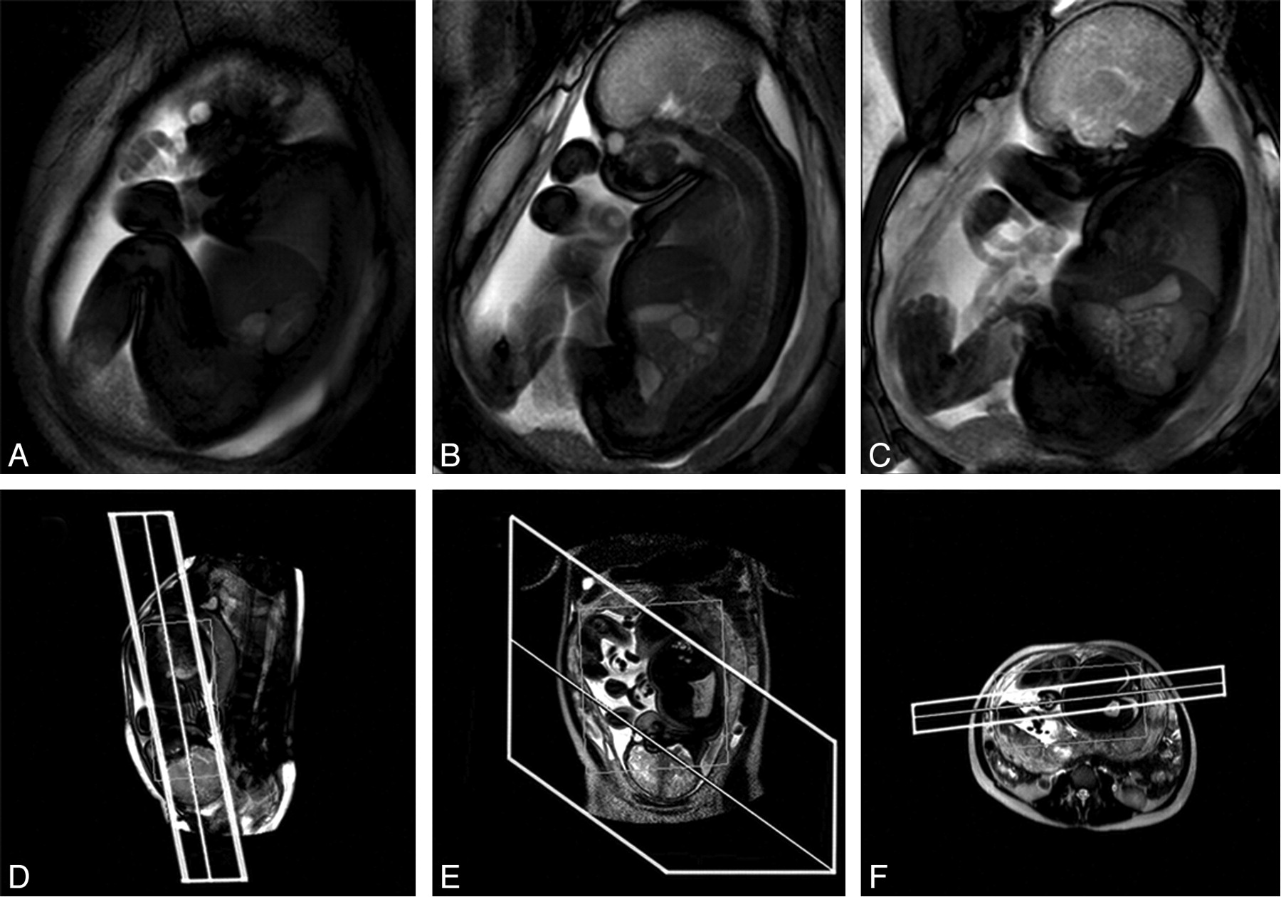
- Fig 1.
Example of a cine acquisition at GA 36 weeks. A–C, Successive frames from a multisection cine image sequence (section thickness, 30 mm). D–F, Piloting procedure used to set up the scan. Upper and lower limbs, trunk, and head are all visible within the FOV. D, Fetal localizer scan, which is used to generate sagittal-oriented cine data; E and F, Maternal pilots in orthogonal planes used to ensure that the prescribed sections have FOVs that cover the full extent of maternal tissue.
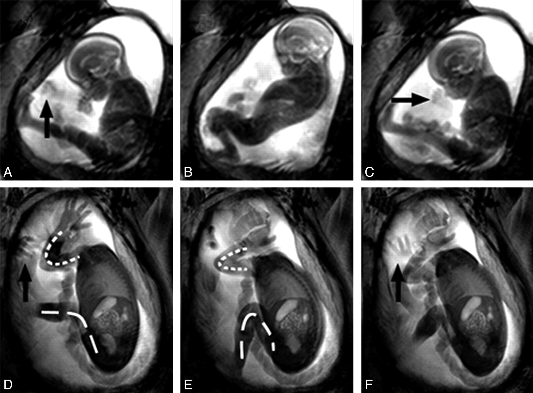
- Fig 2.
Examples of the fetal movement repertoire. A–C, Time points 0, 4, and 8 seconds of cine data from a GA 22 weeks' fetus. D–F, A GA 30 weeks' fetus at time points 0, 6, and 10 seconds. The younger fetus is engaging in GM, demonstrating a complete flexion at its knees and extension in its trunk, followed by trunk relaxation and knee extension. D–F, The older fetus also shows GM consisting of flexion at the knee joint (dashed line) as well as at the elbow (dotted line). Arrows indicate hands, 1 of which is rotating in the older fetus. The overall variability in amplitude of movements that older fetuses perform is significantly reduced and appears associated with uterine restrictions giving a cramped appearance to movements. This is in contrast to younger fetuses, that tend to make full use of their surrounding space.
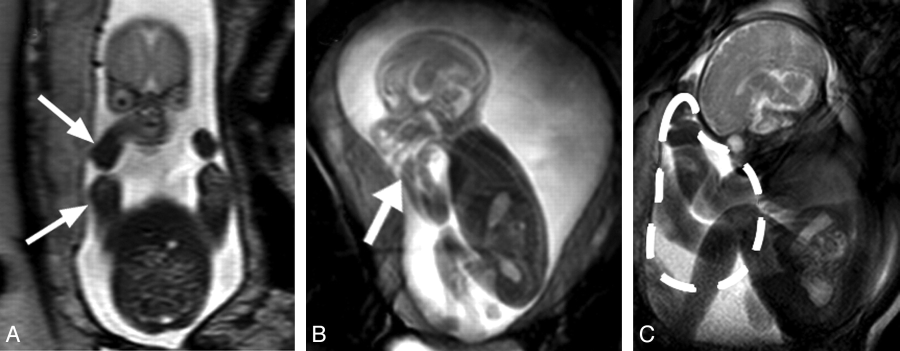
- Fig 3.
Facilitation of flexed posture by the uterine wall. A, T2-weighted coronal section through a GA 22 weeks' fetus. The uterine wall is in close proximity to the fetus's lateral aspects (arrows). B, Still image from cine data of the same fetus. The fetal arm is flexed and located adjacent to the fetal head (thick arrow). C, Image of a GA 35 weeks' fetus demonstrates the redistribution of amniotic fluid volume near term, anterior to the thorax of the fetus (dashed line), due to its posture and the uterine limits; this collection of amniotic fluid may facilitate upper limb movements and trunk extensions.
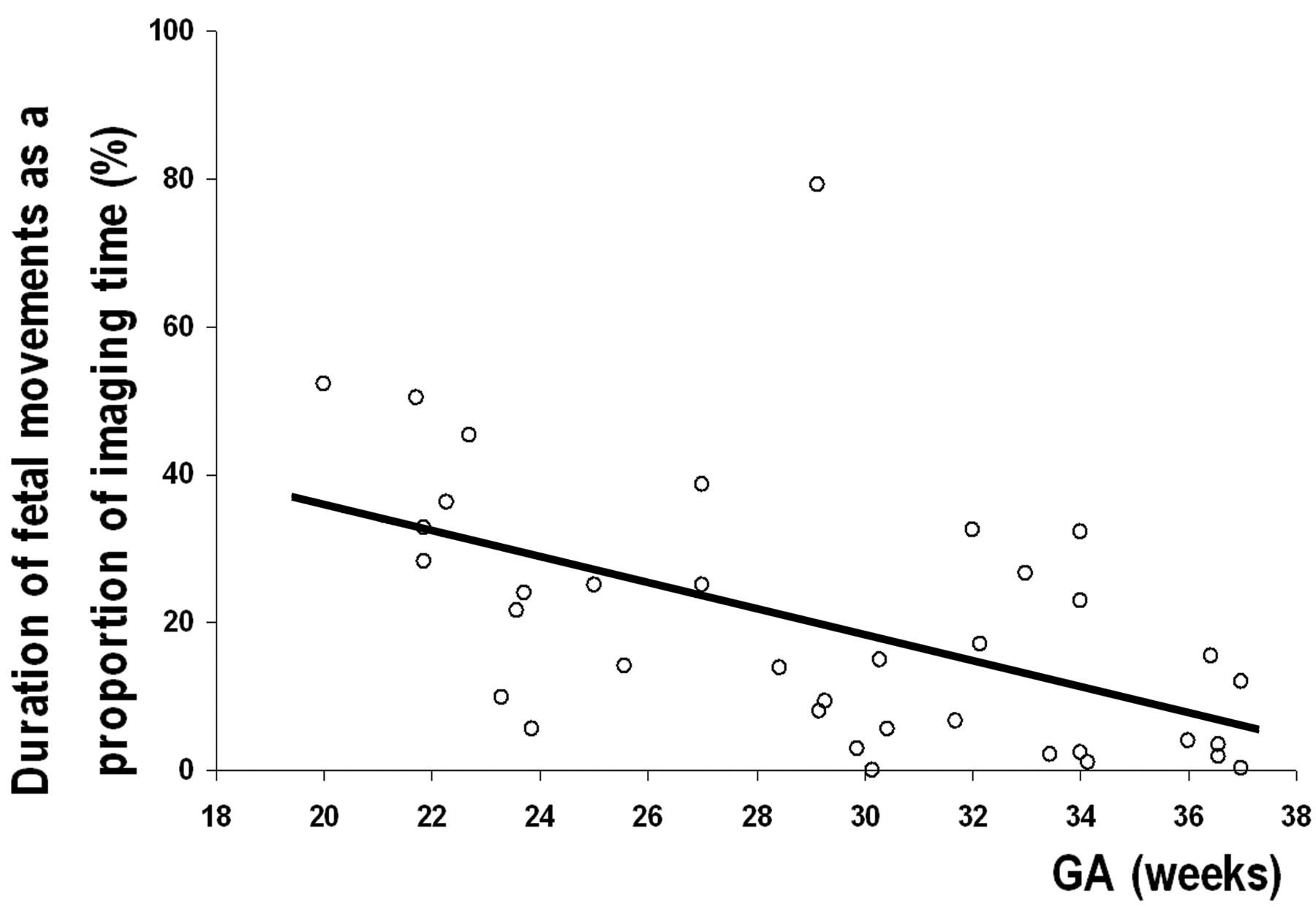
- Fig 4.
Graph shows that the proportion of imaging time spent moving by all fetuses decreases significantly with increasing GA (r = −0.514, P = .001).
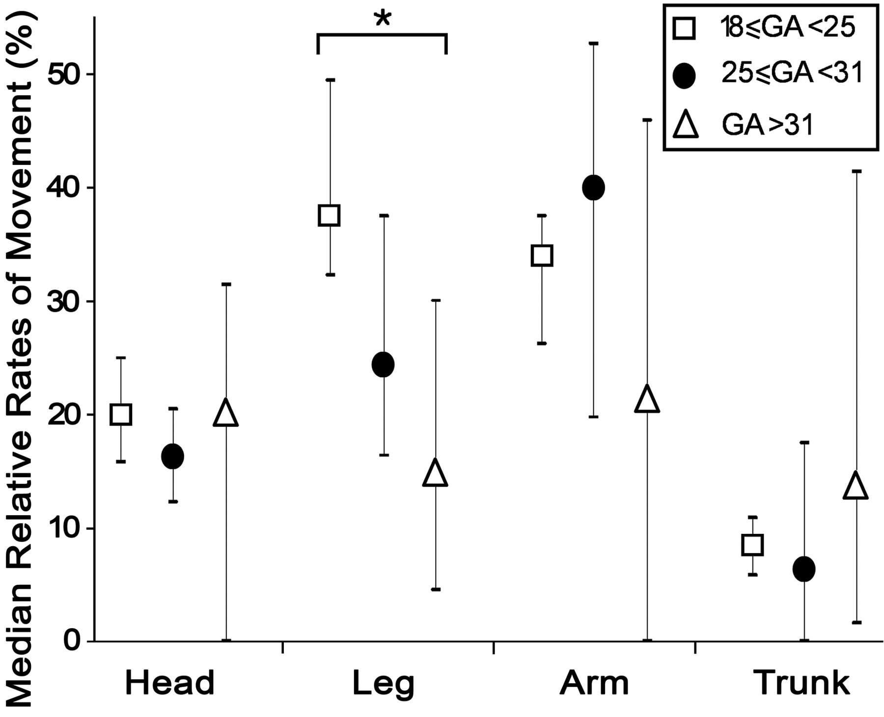
- Fig 5.
Graph shows the median frequency of movements in fetuses grouped by age (± interquartile range). There is a significant decrease in the rate of leg movements between fetuses younger than GA 25 weeks and those older than GA 31 weeks (P = .015). There are no statistically significant differences in the median rates of head, arm, or trunk movements between age groups (P = .746, P = .310, and P = .397, respectively).
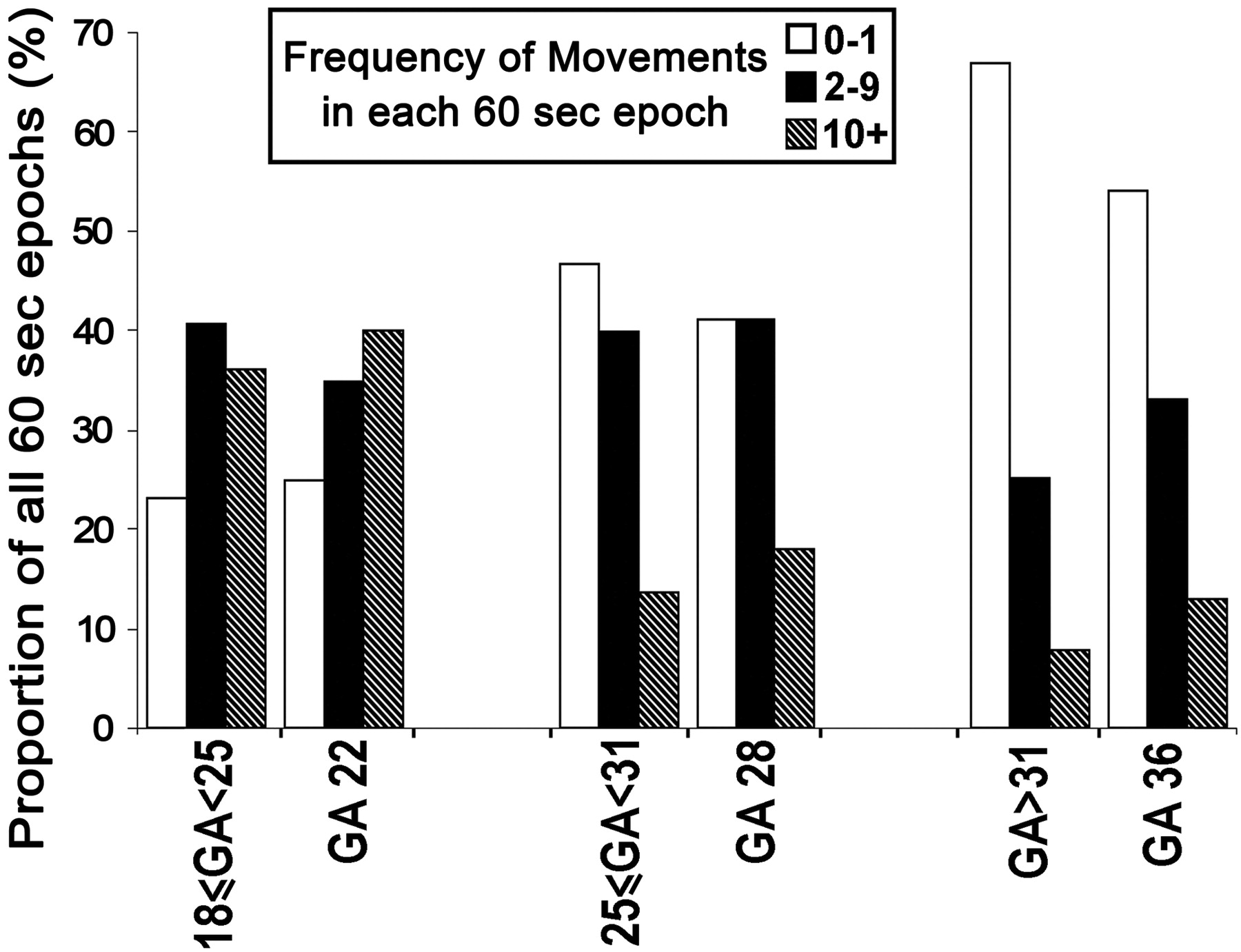
- Fig 6.
Graph shows overall activity levels in long-duration cine acquisitions compared with pooled shorter duration data from the corresponding age groups. The results show that there are similar proportions of 60-second epochs that contain each level of motility between each age group and the corresponding long acquisition.
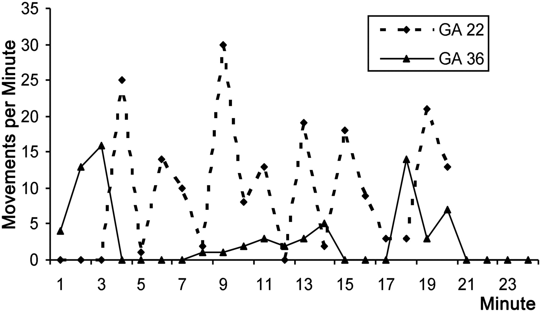
- Fig 7.
Graph shows activity levels during 20 minutes. Data from the long cine acquisitions from the fetuses at GA 22 (dashed line) and GA 36 (solid line) are shown. There are notable differences in the maximum frequency of movements. Also the behavior of the GA 36 weeks' fetus appears more ordered with more clearly defined periods of activity and inactivity than the GA 22 weeks' fetus.
- Fig 8.
Graph of movement frequency in a subset of 24 healthy fetuses shows a significant reduction as the proportion of uterine volume that the fetus occupies increases (r = −0.703, P = .0001).


