
Article Figures & Data
Figures
- Fig 1.
The defined mask based on the AAL template includes the bilateral primary sensorimotor cortex and supplementary motor area. The numbers below the images refer to the z coordinates in the TT space. R indicates right; L, left; Z, the z coordinates.
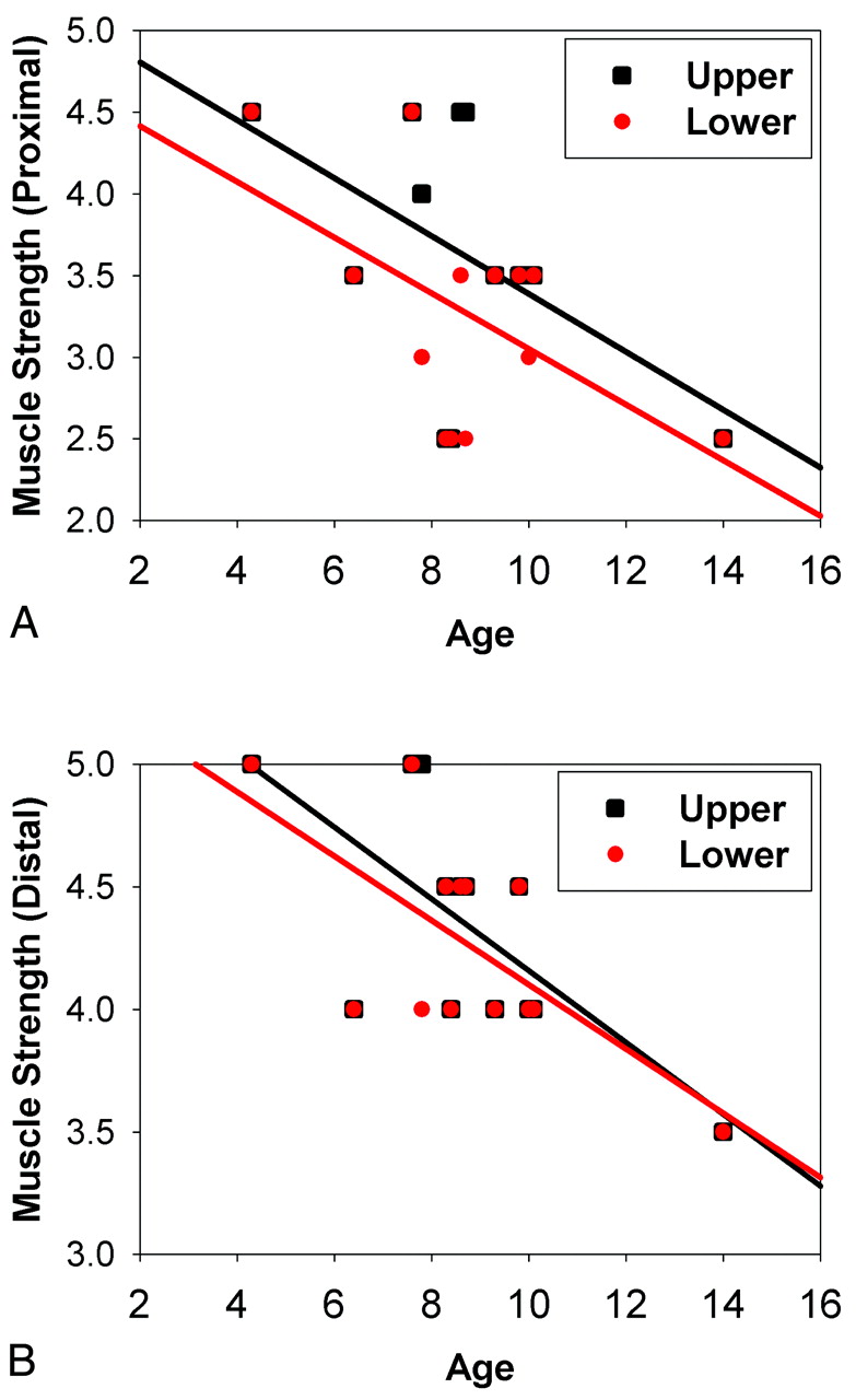
- Fig 2.
Correlation between age and muscle strength in patients with DMD. A, Proximal limb. B, Distal limb. Black squares and regression lines indicate the correlations between age and the upper limb, while red dots and regression lines indicate the correlation between age and the lower limb.
- Fig 3.
The brain areas of deceased ReHo (A) and decreased GMC (B) in boys with DMD compared with controls. The numbers below the images refer to the z coordinates in the TT space. R indicates right; L, left; Z, the z coordinates.
- Fig 4.
Correlation between ReHo at the left PSMC (BA 4) and muscle strength of the distal upper limb (DUL) with age as covariance. The x- and y-axes are the residuals of the original ReHo and muscle strength of the DUL, respectively, with age regressed out.


Tables
Muscle Strength r P value Proximal upper limb 3.6 ± 0.8 −0.517 .070 Distal upper limb 4.3 ± 0.5 −0.693 .009 Proximal lower limb 3.3 ± 0.7 −0.548 .052 Distal lower limb 4.3 ± 0.4 −0.670 .012 - Table 2:
Brain areas of regional homogeneity and gray matter concentration difference between 2 groups in the defined motor cortex maska
Volume (mm3) Area Hemisphere BA TT Coordinates Peak T Value X Y Z Decreased ReHo in DMD 2214 PSMC L 4 −43 −25 53 −2.73 PSMC L 5 −22 −49 59 −2.81 1404 SMA R 6 11 −19 50 −3.33 459 PSMC R 4 47 −19 47 −2.61 Decreased GMC in DMD 675 PSMC L 4 −43 −13 32 −4.40 Note:—L indicates left; R, right; BA = Brodmann area.
↵a P < .05.
- Table 3:
The correlation between MR imaging measures and the muscle strength in patients with DMD
Area ReHo Value GMC Value L-PSMC (BA 4) L-PSMC (BA 5) R-SMA (BA 6) R-PSMC (BA 4) L-PSMC (BA 4) Proximal upper limb r −0.347 −0.388 0.353 0.260 −0.550 P 0.360 0.302 0.352 0.500 0.125 Distal upper limb r −0.784 −0.235 0.482 −0.407 −0.626 P 0.012 0.543 0.189 0.277 0.071 Proximal lower limb r −0.105 0.208 −0.530 0.012 0.248 P 0.789 0.592 0.142 0.976 0.520 Distal lower limb r −0.428 −0.435 0.094 −0.271 −0.520 P 0.250 0.242 0.810 0.481 0.151 Note:—L indicates left; R, right; BA, Brodmann area.


