
Article Figures & Data
Figures
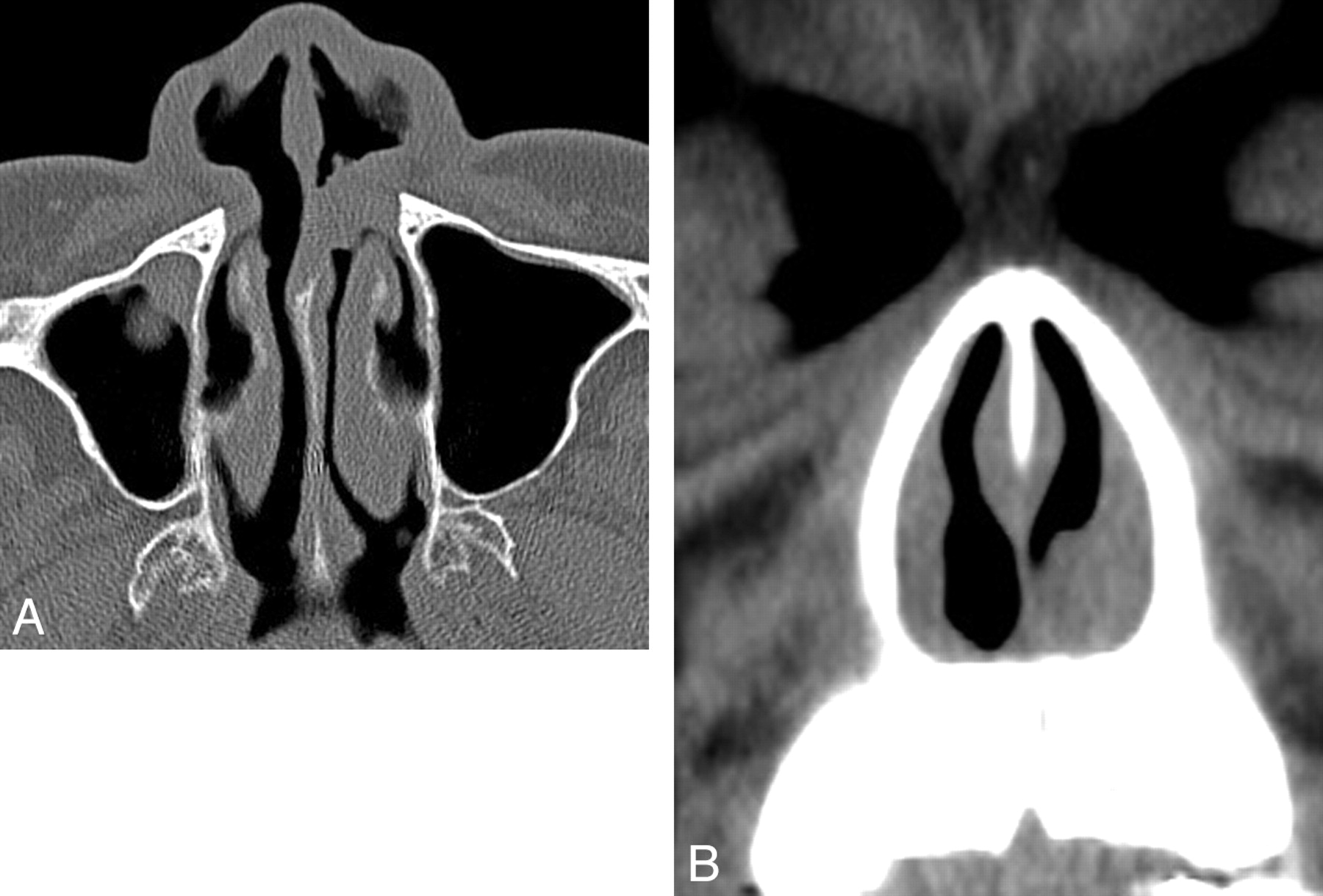
- Fig 1.
Case 5. Axial CT image shows symmetrical thickening of the septum with lateral nasal walls bilaterally.
- Fig 2.
Case 6. Axial (A) and coronal (B) CT images show an oval-shaped, well-defined, and isointense soft-tissue mass arising from left lateral nasal wall and extending into the nasal septum.
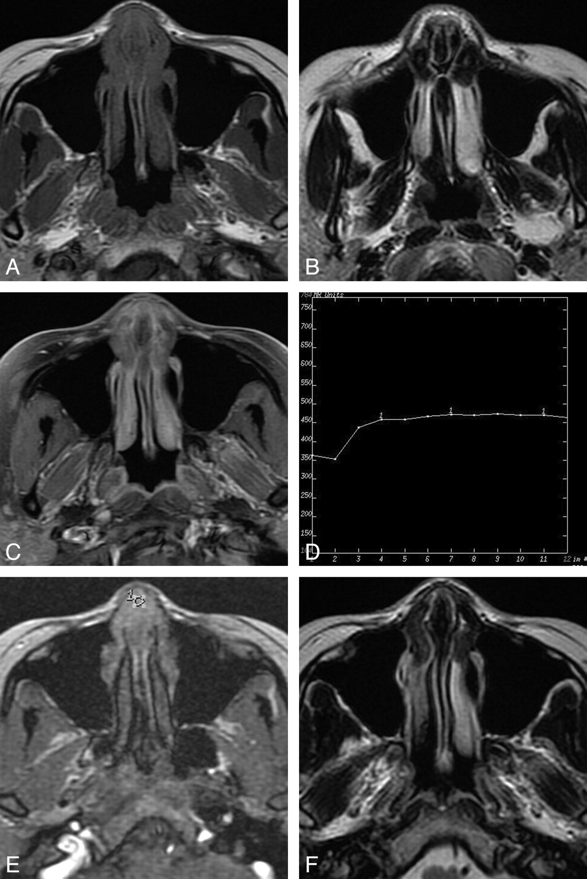
- Fig 3.
Case 1. A, Axial T1-weighted image shows symmetrical thickening of the septum and lateral nasal walls with isointense signal intensity. B, The lesion has hypointense signal intensity on the axial T2-weighted image. C, Axial contrast-enhanced T1-weighted image with fat saturation shows moderate inhomogeneous enhancement of the lesion. D, Corresponding axial DCE MR image depicts the rapidly enhancing and slow washout pattern (Type II). E, The round cursors mark the region of interest selected for signal intensity measurement at dynamic MR imaging. F, Axial T2-weighted image shows a recurrent lesion in the primary site.
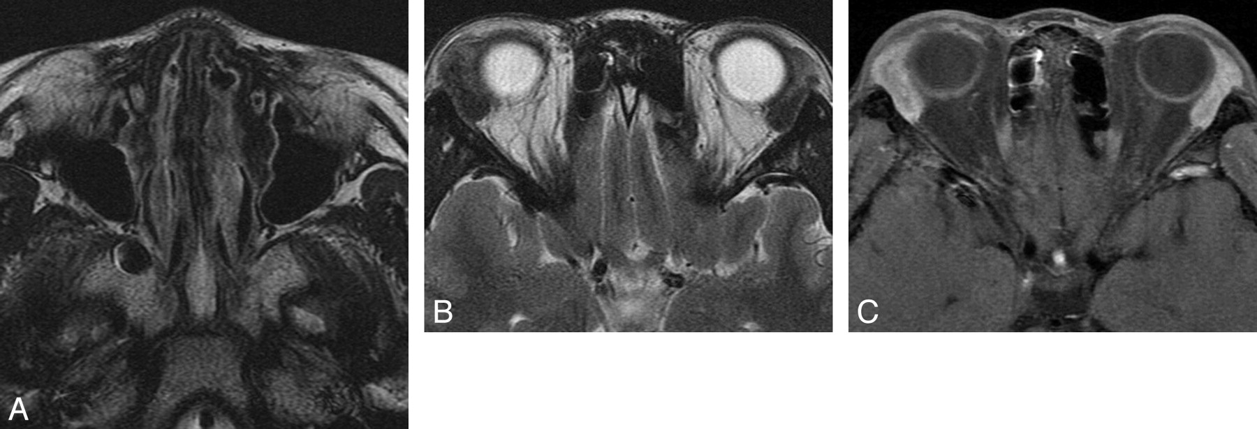
- Fig 4.
Case 2. A, Axial T2-weighted image shows diffuse thickening of the septum and lateral nasal walls with hypointense signal intensity. B, Axial T2-weighted image shows bilateral lacrimal gland enlargement, appearing as hypointense signal intensity. C, Axial contrast-enhanced T1-weighted image with fat saturation shows moderate homogeneous enhancement of bilateral lacrimal glands.


Tables
Nasal cavity eosinophilic angiocentric fibrosis: CT and MR imaging characteristics
Patient No. Sex/ Age (years) Location Lesion Size (mm) Unenhanced CT MR Imaging Follow-Up (years) T1WI T2WI Contrast 1 F/26 Nasal septum 33 Isodense Isointense Hypointense Moderate 2 2 M/16 Nasal septum 52 Isodense Isointense Hypointense Moderate N 3 F/62 Right lateral nasal wall 21 Isodense Isointense Isointense Moderate 4 4 M/28 Nasal septum 26 Isodense N N N 3 5 F/24 Nasal septum 35 Isodense N N N 5 6 M/73 Left lateral nasal wall 12 Isodense N N N 0.5 Note:—Lesion size indicates maximum diameter. CT density and MR imaging signal intensity are compared with cerebral gray matter. N indicates none.
Five patients were followed up for 0.5–5 years following surgery. Patient 1 recurred after 2 years (Fig 3F); the other 5 patients showed no evidence of recurrence. After the second surgery, a follow-up of approximately 1 year for patient 1, there was no clinical evidence of recurrence.


