Advertisement
  • Main menu
  • User menu
  • Search
  • English ▼
    • English
    • Afrikaans
    • Albanian
    • Amharic
    • Arabic
    • Armenian
    • Azerbaijani
    • Basque
    • Belarusian
    • Bengali
    • Bosnian
    • Bulgarian
    • Catalan
    • Cebuano
    • Chichewa
    • Chinese (Simplified)
    • Chinese (Traditional)
    • Corsican
    • Croatian
    • Czech
    • Danish
    • Dutch
    • Esperanto
    • Estonian
    • Filipino
    • Finnish
    • French
    • Frisian
    • Galician
    • Georgian
    • German
    • Greek
    • Gujarati
    • Haitian Creole
    • Hausa
    • Hawaiian
    • Hebrew
    • Hindi
    • Hmong
    • Hungarian
    • Icelandic
    • Igbo
    • Indonesian
    • Irish
    • Italian
    • Japanese
    • Javanese
    • Kannada
    • Kazakh
    • Khmer
    • Korean
    • Kurdish (Kurmanji)
    • Kyrgyz
    • Lao
    • Latin
    • Latvian
    • Lithuanian
    • Luxembourgish
    • Macedonian
    • Malagasy
    • Malay
    • Malayalam
    • Maltese
    • Maori
    • Marathi
    • Mongolian
    • Myanmar (Burmese)
    • Nepali
    • Norwegian
    • Pashto
    • Persian
    • Polish
    • Portuguese
    • Punjabi
    • Romanian
    • Russian
    • Samoan
    • Scottish Gaelic
    • Serbian
    • Sesotho
    • Shona
    • Sindhi
    • Sinhala
    • Slovak
    • Slovenian
    • Somali
    • Spanish
    • Sudanese
    • Swahili
    • Swedish
    • Tajik
    • Tamil
    • Telugu
    • Thai
    • Turkish
    • Ukrainian
    • Urdu
    • Uzbek
    • Vietnamese
    • Welsh
    • Xhosa
    • Yiddish
    • Yoruba
    • Zulu

Main menu

  • Home
  • Content
    • Current Issue
    • Accepted Manuscripts
    • Article Preview
    • Past Issue Archive
    • Video Articles
    • AJNR Case Collection
    • Case of the Week Archive
    • Case of the Month Archive
    • Classic Case Archive
  • Special Collections
    • Advancing NeuroMRI with High-Relaxivity Contrast Agents
    • AJNR Awards
    • Low-Field MRI
    • Alzheimer Disease
    • ASNR Foundation Special Collection
    • Photon-Counting CT
    • View All
  • Multimedia
    • AJNR Podcasts
    • AJNR SCANtastic
    • Trainee Corner
    • MRI Safety Corner
    • Imaging Protocols
  • For Authors
    • Submit a Manuscript
    • Submit a Video Article
    • Submit an eLetter to the Editor/Response
    • Manuscript Submission Guidelines
    • Statistical Tips
    • Fast Publishing of Accepted Manuscripts
    • Graphical Abstract Preparation
    • Imaging Protocol Submission
    • Author Policies
  • About Us
    • About AJNR
    • Editorial Board
    • Editorial Board Alumni
  • More
    • Become a Reviewer/Academy of Reviewers
    • Subscribers
    • Permissions
    • Alerts
    • Feedback
    • Advertisers
    • ASNR Home

User menu

  • Alerts
  • Log in

Search

  • Advanced search
American Journal of Neuroradiology

American Journal of Neuroradiology

ASHNR American Society of Functional Neuroradiology ASHNR American Society of Pediatric Neuroradiology ASSR
  • Alerts
  • Log in

Advanced Search

English ▼
  • English
  • Afrikaans
  • Albanian
  • Amharic
  • Arabic
  • Armenian
  • Azerbaijani
  • Basque
  • Belarusian
  • Bengali
  • Bosnian
  • Bulgarian
  • Catalan
  • Cebuano
  • Chichewa
  • Chinese (Simplified)
  • Chinese (Traditional)
  • Corsican
  • Croatian
  • Czech
  • Danish
  • Dutch
  • Esperanto
  • Estonian
  • Filipino
  • Finnish
  • French
  • Frisian
  • Galician
  • Georgian
  • German
  • Greek
  • Gujarati
  • Haitian Creole
  • Hausa
  • Hawaiian
  • Hebrew
  • Hindi
  • Hmong
  • Hungarian
  • Icelandic
  • Igbo
  • Indonesian
  • Irish
  • Italian
  • Japanese
  • Javanese
  • Kannada
  • Kazakh
  • Khmer
  • Korean
  • Kurdish (Kurmanji)
  • Kyrgyz
  • Lao
  • Latin
  • Latvian
  • Lithuanian
  • Luxembourgish
  • Macedonian
  • Malagasy
  • Malay
  • Malayalam
  • Maltese
  • Maori
  • Marathi
  • Mongolian
  • Myanmar (Burmese)
  • Nepali
  • Norwegian
  • Pashto
  • Persian
  • Polish
  • Portuguese
  • Punjabi
  • Romanian
  • Russian
  • Samoan
  • Scottish Gaelic
  • Serbian
  • Sesotho
  • Shona
  • Sindhi
  • Sinhala
  • Slovak
  • Slovenian
  • Somali
  • Spanish
  • Sudanese
  • Swahili
  • Swedish
  • Tajik
  • Tamil
  • Telugu
  • Thai
  • Turkish
  • Ukrainian
  • Urdu
  • Uzbek
  • Vietnamese
  • Welsh
  • Xhosa
  • Yiddish
  • Yoruba
  • Zulu
  • Home
  • Content
    • Current Issue
    • Accepted Manuscripts
    • Article Preview
    • Past Issue Archive
    • Video Articles
    • AJNR Case Collection
    • Case of the Week Archive
    • Case of the Month Archive
    • Classic Case Archive
  • Special Collections
    • Advancing NeuroMRI with High-Relaxivity Contrast Agents
    • AJNR Awards
    • Low-Field MRI
    • Alzheimer Disease
    • ASNR Foundation Special Collection
    • Photon-Counting CT
    • View All
  • Multimedia
    • AJNR Podcasts
    • AJNR SCANtastic
    • Trainee Corner
    • MRI Safety Corner
    • Imaging Protocols
  • For Authors
    • Submit a Manuscript
    • Submit a Video Article
    • Submit an eLetter to the Editor/Response
    • Manuscript Submission Guidelines
    • Statistical Tips
    • Fast Publishing of Accepted Manuscripts
    • Graphical Abstract Preparation
    • Imaging Protocol Submission
    • Author Policies
  • About Us
    • About AJNR
    • Editorial Board
    • Editorial Board Alumni
  • More
    • Become a Reviewer/Academy of Reviewers
    • Subscribers
    • Permissions
    • Alerts
    • Feedback
    • Advertisers
    • ASNR Home
  • Follow AJNR on Twitter
  • Visit AJNR on Facebook
  • Follow AJNR on Instagram
  • Join AJNR on LinkedIn
  • RSS Feeds

AJNR Awards, New Junior Editors, and more. Read the latest AJNR updates


Improved Turnaround Times | Median time to first decision: 12 days

Research ArticleHead and Neck Imaging

Utility and Interobserver Agreement of Ultrasound Elastography in the Detection of Malignant Thyroid Nodules in Clinical Care

S. Merino, J. Arrazola, A. Cárdenas, M. Mendoza, P. De Miguel, C. Fernández and T. Ganado
American Journal of Neuroradiology December 2011, 32 (11) 2142-2148; DOI: https://doi.org/10.3174/ajnr.A2716
S. Merino
aFrom the Departments of Radiology (S.M., J.A., A.C., T.G.)
  • Find this author on Google Scholar
  • Find this author on PubMed
  • Search for this author on this site
J. Arrazola
aFrom the Departments of Radiology (S.M., J.A., A.C., T.G.)
  • Find this author on Google Scholar
  • Find this author on PubMed
  • Search for this author on this site
A. Cárdenas
aFrom the Departments of Radiology (S.M., J.A., A.C., T.G.)
  • Find this author on Google Scholar
  • Find this author on PubMed
  • Search for this author on this site
M. Mendoza
bPathology (M.M.)
  • Find this author on Google Scholar
  • Find this author on PubMed
  • Search for this author on this site
P. De Miguel
cEndocrinology (P.D.M.)
  • Find this author on Google Scholar
  • Find this author on PubMed
  • Search for this author on this site
C. Fernández
dPreventive Medicine (C.F.), Hospital Clínico San Carlos, Madrid, Spain.
  • Find this author on Google Scholar
  • Find this author on PubMed
  • Search for this author on this site
T. Ganado
aFrom the Departments of Radiology (S.M., J.A., A.C., T.G.)
  • Find this author on Google Scholar
  • Find this author on PubMed
  • Search for this author on this site
  • Article
  • Figures & Data
  • Info & Metrics
  • Responses
  • References
  • PDF
Loading

Article Figures & Data

Figures

  • Tables
  • Fig 1.
    • Download figure
    • Open in new tab
    • Download powerpoint
    Fig 1.

    Flow chart of the study. Three patients were excluded from the sample population because no definite pathologic result was available. A total of 103 patients and 106 nodules were definitively included in the study (analyzed population)

  • Fig 2.
    • Download figure
    • Open in new tab
    • Download powerpoint
    Fig 2.

    Classification of nodules (absolute numbers) depending on suspicion of malignancy or benignity of lesions with US elastography for each radiologist and by consensus.
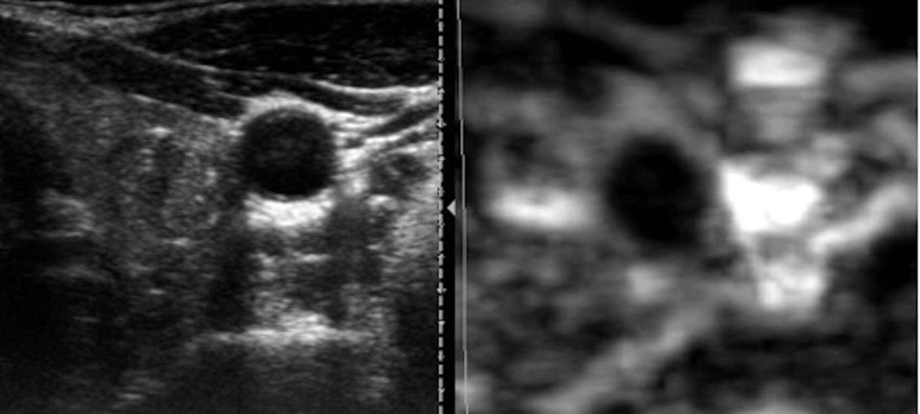
  • Fig 3.
    • Download figure
    • Open in new tab
    • Download powerpoint
    Fig 3.

    A predominantly hypoechoic nodule with microcalcifications is seen in conventional B-mode sonography image. US elastography image shows a homogeneously black nodule representing a predominantly rigid lesion, sized similar to that on B-mode image. This nodule corresponded to a papillary carcinoma on FNAB and surgery.
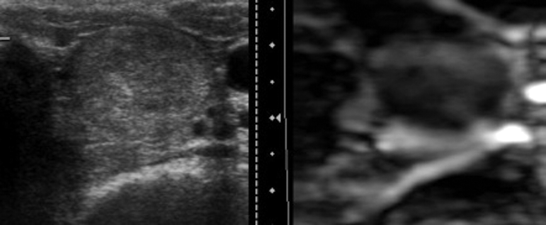
  • Fig 4.
    • Download figure
    • Open in new tab
    • Download powerpoint
    Fig 4.

    B-mode sonographic and US elastographic images of a benign nodule are shown. A well-defined heterogeneous nodule on conventional sonography shows a predominantly soft pattern of stiffness on the US elastogram.

Tables

  • Figures
    • View popup
    Table 1:

    Frequency in absolute numbers and percentage of different variables studied with US elastography combined with nodule location in the thyroid gland

    Total of No. of NodulesNo. of Nodules in Left LobeNo. of Nodules in Right LobeNo. of Nodules in the IsthmusP Value
    Elasticity
        Predominantly rigid16 (15.1%)9 (56.3%)7 (43.7%)0 (0%).436
        Intermediate46 (43.4%)18 (39.1%)26 (56.5%)2 (4.4%)
        Predominantly soft44 (41.5%)22 (50%)19 (43.2%)3 (6.8%)
    Size
        Greater diameter in US elastography than B-mode US15 (14.2%)7 (46.7%)8 (53.3%)0 (0.0%).138
        Same size62 (58.5%)29 (46.8%)32 (51.6%)1 (1.6%)
        Lesser diameter in US elastography than B-mode US29 (27.3%)13 (44.8%)12 (41.4%)4 (13.8%)
    Margins
        Ill-defined or irregular41 (38.7%)20 (48.8%)19 (46.3%)2 (4.9%).905
        Well-defined or regular65 (61.3%)29 (44.6%)33 (50.8%)3 (4.6%)
    • View popup
    Table 2:

    Utility of US elastography: correlation among different variables studied with US elastography and defined by consensus between radiologists and pathologic results (reference standard)

    US Elastography Variables by ConsensusMalignant NodulesBenign NodulesP Value
    Elasticity
        Predominantly rigid (n = 16)6 (37.5%)10 (62.5%).000
        Indeterminate (n = 46)4 (8.7%)42 (91.3%)
        Predominantly soft (n = 44)0 (0%)44 (100%)
    Size
        Greater diameter in US elastography than B-mode image (n = 15)4 (26.7%)11 (73.3%).068
        Same size as B-mode image (n = 62)5 (8.1%)57 (91.9%)
        Lesser diameter in US elastography than B-mode (n = 29)1 (3.4%)28 (96.6%)
    Margins
        Ill-defined or irregular (n = 41)3 (7.3%)38 (92.7%).548
        Well-defined or regular (n = 65)7 (10.8%)58 (89.2%)
    • View popup
    Table 3:

    Utility of classification with US elastography in groups I, II, and III, according to benign or malignant suspicion obtained by consensus: correlation with pathologic results (reference standard)

    Classification of Nodules with US Elastography by ConsensusMalignant NodulesBenign NodulesP Value
    Type I: suspicious for benignity (n = 39)039.001
    Type II: indeterminate (n = 51)546
    Type III: suspicious for malignancy (n = 16)511
PreviousNext
Back to top

In this issue

American Journal of Neuroradiology: 32 (11)
American Journal of Neuroradiology
Vol. 32, Issue 11
1 Dec 2011
  • Table of Contents
  • Index by author
Advertisement
Print
Download PDF
Email Article
Cite this article
0 Responses
Respond to this article
Share
Bookmark this article
Utility and Interobserver Agreement of Ultrasound Elastography in the Detection of Malignant Thyroid Nodules in Clinical Care
S. Merino, J. Arrazola, A. Cárdenas, M. Mendoza, P. De Miguel, C. Fernández, T. Ganado
American Journal of Neuroradiology Dec 2011, 32 (11) 2142-2148; DOI: 10.3174/ajnr.A2716
del.icio.us logo Twitter logo Facebook logo Mendeley logo
  • Tweet Widget
Purchase

Jump to section

  • Article
    • Abstract
    • ABBREVIATIONS
    • Materials and Methods
    • Results
    • Discussion
    • Conclusions
    • References
  • Figures & Data
  • Info & Metrics
  • Responses
  • References
  • PDF

Related Articles

  • No related articles found.
  • PubMed
  • Google Scholar

Cited By...

  • Real-time Ultrasound Elastography for Differentiation of Benign and Malignant Thyroid Nodules: A Meta-analysis
  • Crossref
  • Google Scholar

This article has not yet been cited by articles in journals that are participating in Crossref Cited-by Linking.

More in this TOC Section

  • Diagnostic Performance of US in Neck Node NIRADS 2
  • Hydrops Herniation into the Semicircular Canals
  • ASL Sensitivity for Head and Neck Paraganglioma
Show more HEAD AND NECK IMAGING

Similar Articles

Advertisement

Indexed Content

  • Current Issue
  • Accepted Manuscripts
  • Article Preview
  • Past Issues
  • Editorials
  • Editor's Choice
  • Fellows' Journal Club
  • Letters to the Editor
  • Video Articles

Cases

  • Case Collection
  • Archive - Case of the Week
  • Archive - Case of the Month
  • Archive - Classic Case

More from AJNR

  • Trainee Corner
  • Imaging Protocols
  • MRI Safety Corner
  • Book Reviews

Multimedia

  • AJNR Podcasts
  • AJNR Scantastics

Resources

  • Turnaround Time
  • Submit a Manuscript
  • Submit a Video Article
  • Submit an eLetter to the Editor/Response
  • Manuscript Submission Guidelines
  • Statistical Tips
  • Fast Publishing of Accepted Manuscripts
  • Graphical Abstract Preparation
  • Imaging Protocol Submission
  • Evidence-Based Medicine Level Guide
  • Publishing Checklists
  • Author Policies
  • Become a Reviewer/Academy of Reviewers
  • News and Updates

About Us

  • About AJNR
  • Editorial Board
  • Editorial Board Alumni
  • Alerts
  • Permissions
  • Not an AJNR Subscriber? Join Now
  • Advertise with Us
  • Librarian Resources
  • Feedback
  • Terms and Conditions
  • AJNR Editorial Board Alumni

American Society of Neuroradiology

  • Not an ASNR Member? Join Now

© 2025 by the American Society of Neuroradiology All rights, including for text and data mining, AI training, and similar technologies, are reserved.
Print ISSN: 0195-6108 Online ISSN: 1936-959X

Powered by HighWire
Email this Article

Thank you for your interest in spreading the word on American Journal of Neuroradiology.

NOTE: We only request your email address so that the person you are recommending the page to knows that you wanted them to see it, and that it is not junk mail. We do not capture any email address.

Enter multiple addresses on separate lines or separate them with commas.
Utility and Interobserver Agreement of Ultrasound Elastography in the Detection of Malignant Thyroid Nodules in Clinical Care
(Your Name) has sent you a message from American Journal of Neuroradiology
(Your Name) thought you would like to see the American Journal of Neuroradiology web site.
CAPTCHA
This question is for testing whether or not you are a human visitor and to prevent automated spam submissions.
Cite this article
S. Merino, J. Arrazola, A. Cárdenas, M. Mendoza, P. De Miguel, C. Fernández, T. Ganado
Utility and Interobserver Agreement of Ultrasound Elastography in the Detection of Malignant Thyroid Nodules in Clinical Care
American Journal of Neuroradiology Dec 2011, 32 (11) 2142-2148; DOI: 10.3174/ajnr.A2716

Citation Manager Formats

  • BibTeX
  • Bookends
  • EasyBib
  • EndNote (tagged)
  • EndNote 8 (xml)
  • Medlars
  • Mendeley
  • Papers
  • RefWorks Tagged
  • Ref Manager
  • RIS
  • Zotero

We use cookies on this site to enhance your user experience. By clicking any link on this page you are giving your consent for us to set cookies.