Advertisement

Main menu

  • Home
  • Content
    • Current Issue
    • Accepted Manuscripts
    • Article Preview
    • Past Issue Archive
    • Video Articles
    • AJNR Case Collection
    • Case of the Week Archive
    • Case of the Month Archive
    • Classic Case Archive
  • Special Collections
    • AJNR Awards
    • Low-Field MRI
    • Alzheimer Disease
    • ASNR Foundation Special Collection
    • Photon-Counting CT
    • View All
  • Multimedia
    • AJNR Podcasts
    • AJNR SCANtastic
    • Trainee Corner
    • MRI Safety Corner
    • Imaging Protocols
  • For Authors
    • Submit a Manuscript
    • Submit a Video Article
    • Submit an eLetter to the Editor/Response
    • Manuscript Submission Guidelines
    • Statistical Tips
    • Fast Publishing of Accepted Manuscripts
    • Graphical Abstract Preparation
    • Imaging Protocol Submission
    • Author Policies
  • About Us
    • About AJNR
    • Editorial Board
    • Editorial Board Alumni
  • More
    • Become a Reviewer/Academy of Reviewers
    • Subscribers
    • Permissions
    • Alerts
    • Feedback
    • Advertisers
    • ASNR Home

User menu

  • Alerts
  • Log in

Search

  • Advanced search
American Journal of Neuroradiology

American Journal of Neuroradiology

ASHNR American Society of Functional Neuroradiology ASHNR American Society of Pediatric Neuroradiology ASSR
  • Alerts
  • Log in

Advanced Search

  • Home
  • Content
    • Current Issue
    • Accepted Manuscripts
    • Article Preview
    • Past Issue Archive
    • Video Articles
    • AJNR Case Collection
    • Case of the Week Archive
    • Case of the Month Archive
    • Classic Case Archive
  • Special Collections
    • AJNR Awards
    • Low-Field MRI
    • Alzheimer Disease
    • ASNR Foundation Special Collection
    • Photon-Counting CT
    • View All
  • Multimedia
    • AJNR Podcasts
    • AJNR SCANtastic
    • Trainee Corner
    • MRI Safety Corner
    • Imaging Protocols
  • For Authors
    • Submit a Manuscript
    • Submit a Video Article
    • Submit an eLetter to the Editor/Response
    • Manuscript Submission Guidelines
    • Statistical Tips
    • Fast Publishing of Accepted Manuscripts
    • Graphical Abstract Preparation
    • Imaging Protocol Submission
    • Author Policies
  • About Us
    • About AJNR
    • Editorial Board
    • Editorial Board Alumni
  • More
    • Become a Reviewer/Academy of Reviewers
    • Subscribers
    • Permissions
    • Alerts
    • Feedback
    • Advertisers
    • ASNR Home
  • Follow AJNR on Twitter
  • Visit AJNR on Facebook
  • Follow AJNR on Instagram
  • Join AJNR on LinkedIn
  • RSS Feeds

AJNR Awards, New Junior Editors, and more. Read the latest AJNR updates

Research ArticleBrain

Structural Brain Abnormalities in Patients with Parkinson Disease: A Comparative Voxel-Based Analysis Using T1-Weighted MR Imaging and Magnetization Transfer Imaging

K. Morgen, G. Sammer, L. Weber, B. Aslan, C. Müller, G.F. Bachmann, D. Sandmann, M. Oechsner, D. Vaitl, M. Kaps and I. Reuter
American Journal of Neuroradiology December 2011, 32 (11) 2080-2086; DOI: https://doi.org/10.3174/ajnr.A2837
K. Morgen
aFrom the Central Institute of Mental Health (K.M.), Mannheim, Germany
bDepartment of Neurology (K.M., L.W., B.A., I.R., M.K.), University of Giessen, Giessen, Germany
cBender Institute of Neuroimaging (K.M., G.S., D.V.), Giessen, Germany
  • Find this author on Google Scholar
  • Find this author on PubMed
  • Search for this author on this site
G. Sammer
cBender Institute of Neuroimaging (K.M., G.S., D.V.), Giessen, Germany
dCognitive Neuroscience Lab at the Center for Psychiatry (G.S., B.A.), Giessen, Germany
  • Find this author on Google Scholar
  • Find this author on PubMed
  • Search for this author on this site
L. Weber
bDepartment of Neurology (K.M., L.W., B.A., I.R., M.K.), University of Giessen, Giessen, Germany
  • Find this author on Google Scholar
  • Find this author on PubMed
  • Search for this author on this site
B. Aslan
bDepartment of Neurology (K.M., L.W., B.A., I.R., M.K.), University of Giessen, Giessen, Germany
dCognitive Neuroscience Lab at the Center for Psychiatry (G.S., B.A.), Giessen, Germany
  • Find this author on Google Scholar
  • Find this author on PubMed
  • Search for this author on this site
C. Müller
eDepartment of Radiology (C.M., G.F.B.), Kerckhoff-Klinik, Bad Nauheim, Germany
  • Find this author on Google Scholar
  • Find this author on PubMed
  • Search for this author on this site
G.F. Bachmann
eDepartment of Radiology (C.M., G.F.B.), Kerckhoff-Klinik, Bad Nauheim, Germany
  • Find this author on Google Scholar
  • Find this author on PubMed
  • Search for this author on this site
D. Sandmann
fSoemmerring-Institut (D.S., I.R.), Bad Nauheim, Germany
  • Find this author on Google Scholar
  • Find this author on PubMed
  • Search for this author on this site
M. Oechsner
gParkinson-Klinik (M.O.), Bad Nauheim, Germany.
  • Find this author on Google Scholar
  • Find this author on PubMed
  • Search for this author on this site
D. Vaitl
cBender Institute of Neuroimaging (K.M., G.S., D.V.), Giessen, Germany
  • Find this author on Google Scholar
  • Find this author on PubMed
  • Search for this author on this site
M. Kaps
bDepartment of Neurology (K.M., L.W., B.A., I.R., M.K.), University of Giessen, Giessen, Germany
  • Find this author on Google Scholar
  • Find this author on PubMed
  • Search for this author on this site
I. Reuter
bDepartment of Neurology (K.M., L.W., B.A., I.R., M.K.), University of Giessen, Giessen, Germany
fSoemmerring-Institut (D.S., I.R.), Bad Nauheim, Germany
  • Find this author on Google Scholar
  • Find this author on PubMed
  • Search for this author on this site
  • Article
  • Figures & Data
  • Supplemental
  • Info & Metrics
  • Responses
  • References
  • PDF
Loading

Article Figures & Data

Figures

  • Fig 1.
    • Download figure
    • Open in new tab
    • Download powerpoint
    Fig 1.

    Results are presented at P < .001 whole-brain uncorrected for GM images and P < .01 for midbrain images; significant effects are marked with arrows and labeled. A, The comparison of control subjects and patients shows reduced tissue integrity only on MTR images in the left olfactory cortex bordering the amygdala (A3). B and C, MTR images also show effects in bilateral SNc (C3), whereas midbrain volume images indicate atrophy only in the left SNc (C1), and density images show no effect (C2). All GM images reveal an effect in the left parahippocampal gyrus (B1–3).
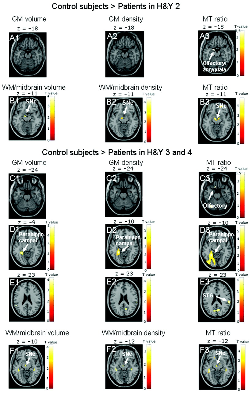
  • Fig 2.
    • Download figure
    • Open in new tab
    • Download powerpoint
    Fig 2.

    Results are presented at P < .001 whole-brain uncorrected, for GM images, and P < .01, for midbrain images; significant effects are marked with arrows and labeled. A, The comparison of control subjects and patients in H&Y stage 2 indicates reduced tissue integrity only on MTR images in the left olfactory cortex bordering on the amygdala (A3). B, MTR images also show effects in the bilateral SNc (B3), whereas midbrain volume and density images indicate tissue damage only in the left SNc (B1–2). C−F, When control subjects are compared with patients in H&Y stages 3 and 4, only MTR images show effects in the left olfactory cortex (C3) and right STG (E3). All images reveal tissue damage in the left parahippocampal gyrus (D1–3) and left SNc (F1–3).

  • Fig 3.
    • Download figure
    • Open in new tab
    • Download powerpoint
    Fig 3.

    Results are presented at P < .001 whole-brain uncorrected; significant effects are marked with arrows and labeled. A, The comparison of patients in H&Y stages 1 and 2 shows a significant effect in the subcallosal area on MTR images (A3), but not on GM volume or density maps (A1, A2). B and C, When patients in H&Y 3 and 4 are compared with patients in H&Y stage 2, only MTR images (C3) indicate a reduction in tissue integrity in the right STG. Both MTR images (B3) and GM volume maps (B1) show a progression of pathology in the left olfactory cortex. There are no significant differences in GM density between patients in H&Y stages 3 and 4 and patients in H&Y stage 2 (B2 and C2).

  • Fig 4.
    • Download figure
    • Open in new tab
    • Download powerpoint
    Fig 4.

    Results are presented at P < .001 whole-brain uncorrected; significant effects are marked with arrows and labeled. A and B, Only MTR images show a correlation with the UPDRS in the right STG (A3) and adjacent left ILF (B3). Neither GM volume maps (A1 and B1) nor GM density maps (A2 and B2) based on T1-weighted MRI indicate correlations with UPDRS.

PreviousNext
Back to top

In this issue

American Journal of Neuroradiology: 32 (11)
American Journal of Neuroradiology
Vol. 32, Issue 11
1 Dec 2011
  • Table of Contents
  • Index by author
Advertisement
Print
Download PDF
Email Article

Thank you for your interest in spreading the word on American Journal of Neuroradiology.

NOTE: We only request your email address so that the person you are recommending the page to knows that you wanted them to see it, and that it is not junk mail. We do not capture any email address.

Enter multiple addresses on separate lines or separate them with commas.
Structural Brain Abnormalities in Patients with Parkinson Disease: A Comparative Voxel-Based Analysis Using T1-Weighted MR Imaging and Magnetization Transfer Imaging
(Your Name) has sent you a message from American Journal of Neuroradiology
(Your Name) thought you would like to see the American Journal of Neuroradiology web site.
CAPTCHA
This question is for testing whether or not you are a human visitor and to prevent automated spam submissions.
Cite this article
K. Morgen, G. Sammer, L. Weber, B. Aslan, C. Müller, G.F. Bachmann, D. Sandmann, M. Oechsner, D. Vaitl, M. Kaps, I. Reuter
Structural Brain Abnormalities in Patients with Parkinson Disease: A Comparative Voxel-Based Analysis Using T1-Weighted MR Imaging and Magnetization Transfer Imaging
American Journal of Neuroradiology Dec 2011, 32 (11) 2080-2086; DOI: 10.3174/ajnr.A2837

Citation Manager Formats

  • BibTeX
  • Bookends
  • EasyBib
  • EndNote (tagged)
  • EndNote 8 (xml)
  • Medlars
  • Mendeley
  • Papers
  • RefWorks Tagged
  • Ref Manager
  • RIS
  • Zotero
0 Responses
Respond to this article
Share
Bookmark this article
Structural Brain Abnormalities in Patients with Parkinson Disease: A Comparative Voxel-Based Analysis Using T1-Weighted MR Imaging and Magnetization Transfer Imaging
K. Morgen, G. Sammer, L. Weber, B. Aslan, C. Müller, G.F. Bachmann, D. Sandmann, M. Oechsner, D. Vaitl, M. Kaps, I. Reuter
American Journal of Neuroradiology Dec 2011, 32 (11) 2080-2086; DOI: 10.3174/ajnr.A2837
del.icio.us logo Twitter logo Facebook logo Mendeley logo
  • Tweet Widget
  • Facebook Like
  • Google Plus One
Purchase

Jump to section

  • Article
    • Abstract
    • ABBREVIATIONS
    • Materials and Methods
    • Results
    • Discussion
    • Conclusions
    • Footnotes
    • References
  • Figures & Data
  • Supplemental
  • Info & Metrics
  • Responses
  • References
  • PDF

Related Articles

  • PubMed
  • Google Scholar

Cited By...

  • A smaller amygdala is associated with anxiety in Parkinsons disease: a combined FreeSurfer--VBM study
  • Individual Detection of Patients with Parkinson Disease using Support Vector Machine Analysis of Diffusion Tensor Imaging Data: Initial Results
  • Crossref
  • Google Scholar

This article has not yet been cited by articles in journals that are participating in Crossref Cited-by Linking.

More in this TOC Section

  • Progression of Microstructural Damage in Spinocerebellar Ataxia Type 2: A Longitudinal DTI Study
  • SWI or T2*: Which MRI Sequence to Use in the Detection of Cerebral Microbleeds? The Karolinska Imaging Dementia Study
  • Statin Therapy Does Not Affect the Radiographic and Clinical Profile of Patients with TIA and Minor Stroke
Show more Brain

Similar Articles

Advertisement

Indexed Content

  • Current Issue
  • Accepted Manuscripts
  • Article Preview
  • Past Issues
  • Editorials
  • Editor's Choice
  • Fellows' Journal Club
  • Letters to the Editor
  • Video Articles

Cases

  • Case Collection
  • Archive - Case of the Week
  • Archive - Case of the Month
  • Archive - Classic Case

More from AJNR

  • Trainee Corner
  • Imaging Protocols
  • MRI Safety Corner
  • Book Reviews

Multimedia

  • AJNR Podcasts
  • AJNR Scantastics

Resources

  • Turnaround Time
  • Submit a Manuscript
  • Submit a Video Article
  • Submit an eLetter to the Editor/Response
  • Manuscript Submission Guidelines
  • Statistical Tips
  • Fast Publishing of Accepted Manuscripts
  • Graphical Abstract Preparation
  • Imaging Protocol Submission
  • Evidence-Based Medicine Level Guide
  • Publishing Checklists
  • Author Policies
  • Become a Reviewer/Academy of Reviewers
  • News and Updates

About Us

  • About AJNR
  • Editorial Board
  • Editorial Board Alumni
  • Alerts
  • Permissions
  • Not an AJNR Subscriber? Join Now
  • Advertise with Us
  • Librarian Resources
  • Feedback
  • Terms and Conditions
  • AJNR Editorial Board Alumni

American Society of Neuroradiology

  • Not an ASNR Member? Join Now

© 2025 by the American Society of Neuroradiology All rights, including for text and data mining, AI training, and similar technologies, are reserved.
Print ISSN: 0195-6108 Online ISSN: 1936-959X

Powered by HighWire