Advertisement

Main menu

  • Home
  • Content
    • Current Issue
    • Accepted Manuscripts
    • Article Preview
    • Past Issue Archive
    • Video Articles
    • AJNR Case Collection
    • Case of the Week Archive
    • Case of the Month Archive
    • Classic Case Archive
  • Special Collections
    • AJNR Awards
    • Low-Field MRI
    • Alzheimer Disease
    • ASNR Foundation Special Collection
    • Photon-Counting CT
    • View All
  • Multimedia
    • AJNR Podcasts
    • AJNR SCANtastic
    • Trainee Corner
    • MRI Safety Corner
    • Imaging Protocols
  • For Authors
    • Submit a Manuscript
    • Submit a Video Article
    • Submit an eLetter to the Editor/Response
    • Manuscript Submission Guidelines
    • Statistical Tips
    • Fast Publishing of Accepted Manuscripts
    • Graphical Abstract Preparation
    • Imaging Protocol Submission
    • Author Policies
  • About Us
    • About AJNR
    • Editorial Board
    • Editorial Board Alumni
  • More
    • Become a Reviewer/Academy of Reviewers
    • Subscribers
    • Permissions
    • Alerts
    • Feedback
    • Advertisers
    • ASNR Home

User menu

  • Alerts
  • Log in

Search

  • Advanced search
American Journal of Neuroradiology

American Journal of Neuroradiology

ASHNR American Society of Functional Neuroradiology ASHNR American Society of Pediatric Neuroradiology ASSR
  • Alerts
  • Log in

Advanced Search

  • Home
  • Content
    • Current Issue
    • Accepted Manuscripts
    • Article Preview
    • Past Issue Archive
    • Video Articles
    • AJNR Case Collection
    • Case of the Week Archive
    • Case of the Month Archive
    • Classic Case Archive
  • Special Collections
    • AJNR Awards
    • Low-Field MRI
    • Alzheimer Disease
    • ASNR Foundation Special Collection
    • Photon-Counting CT
    • View All
  • Multimedia
    • AJNR Podcasts
    • AJNR SCANtastic
    • Trainee Corner
    • MRI Safety Corner
    • Imaging Protocols
  • For Authors
    • Submit a Manuscript
    • Submit a Video Article
    • Submit an eLetter to the Editor/Response
    • Manuscript Submission Guidelines
    • Statistical Tips
    • Fast Publishing of Accepted Manuscripts
    • Graphical Abstract Preparation
    • Imaging Protocol Submission
    • Author Policies
  • About Us
    • About AJNR
    • Editorial Board
    • Editorial Board Alumni
  • More
    • Become a Reviewer/Academy of Reviewers
    • Subscribers
    • Permissions
    • Alerts
    • Feedback
    • Advertisers
    • ASNR Home
  • Follow AJNR on Twitter
  • Visit AJNR on Facebook
  • Follow AJNR on Instagram
  • Join AJNR on LinkedIn
  • RSS Feeds

AJNR Awards, New Junior Editors, and more. Read the latest AJNR updates

Research ArticleBrainF
Open Access

Whole-Brain Perfusion CT Patterns of Brain Arteriovenous Malformations: A Pilot Study in 18 Patients

D.J. Kim and T. Krings
American Journal of Neuroradiology December 2011, 32 (11) 2061-2066; DOI: https://doi.org/10.3174/ajnr.A2659
D.J. Kim
aFrom the Division of Neuroradiology (D.J.K., T.K.), Department of Medical Imaging, Toronto Western Hospital, University of Toronto, Toronto, Ontario, Canada
bDepartment of Radiology (D.J.K.), Yonsei University College of Medicine, Seoul, Korea.
  • Find this author on Google Scholar
  • Find this author on PubMed
  • Search for this author on this site
T. Krings
aFrom the Division of Neuroradiology (D.J.K., T.K.), Department of Medical Imaging, Toronto Western Hospital, University of Toronto, Toronto, Ontario, Canada
  • Find this author on Google Scholar
  • Find this author on PubMed
  • Search for this author on this site
  • Article
  • Figures & Data
  • Info & Metrics
  • Responses
  • References
  • PDF
Loading

Article Figures & Data

Figures

  • Tables
  • Fig 1.
    • Download figure
    • Open in new tab
    • Download powerpoint
    Fig 1.

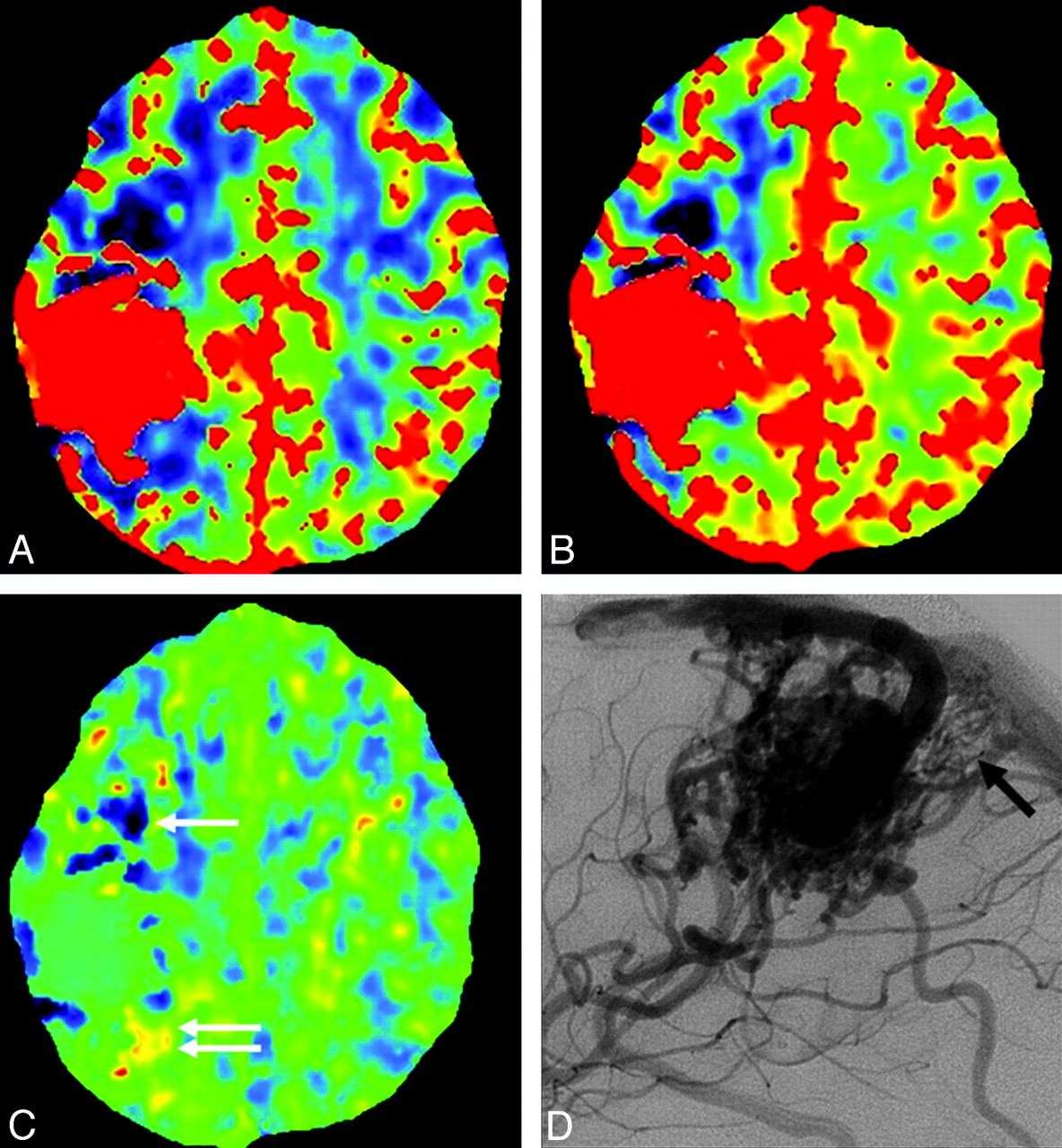
    Case 4. A−C, CBF, CBV, and MTT maps. Decreased CBF and CBV are seen in the anterior and posterior perinidal areas, suggestive of arterial steal. The MTT in the anterior aspect is decreased (pattern 1, white arrow); however, MTT is increased in the posterior aspect (pattern 2, white arrows). D, Lateral view of conventional angiography shows small tortuous vessels posterior to the nidus, suggesting sprouting angiogenesis (black arrow).

  • Fig 2.
    • Download figure
    • Open in new tab
    • Download powerpoint
    Fig 2.

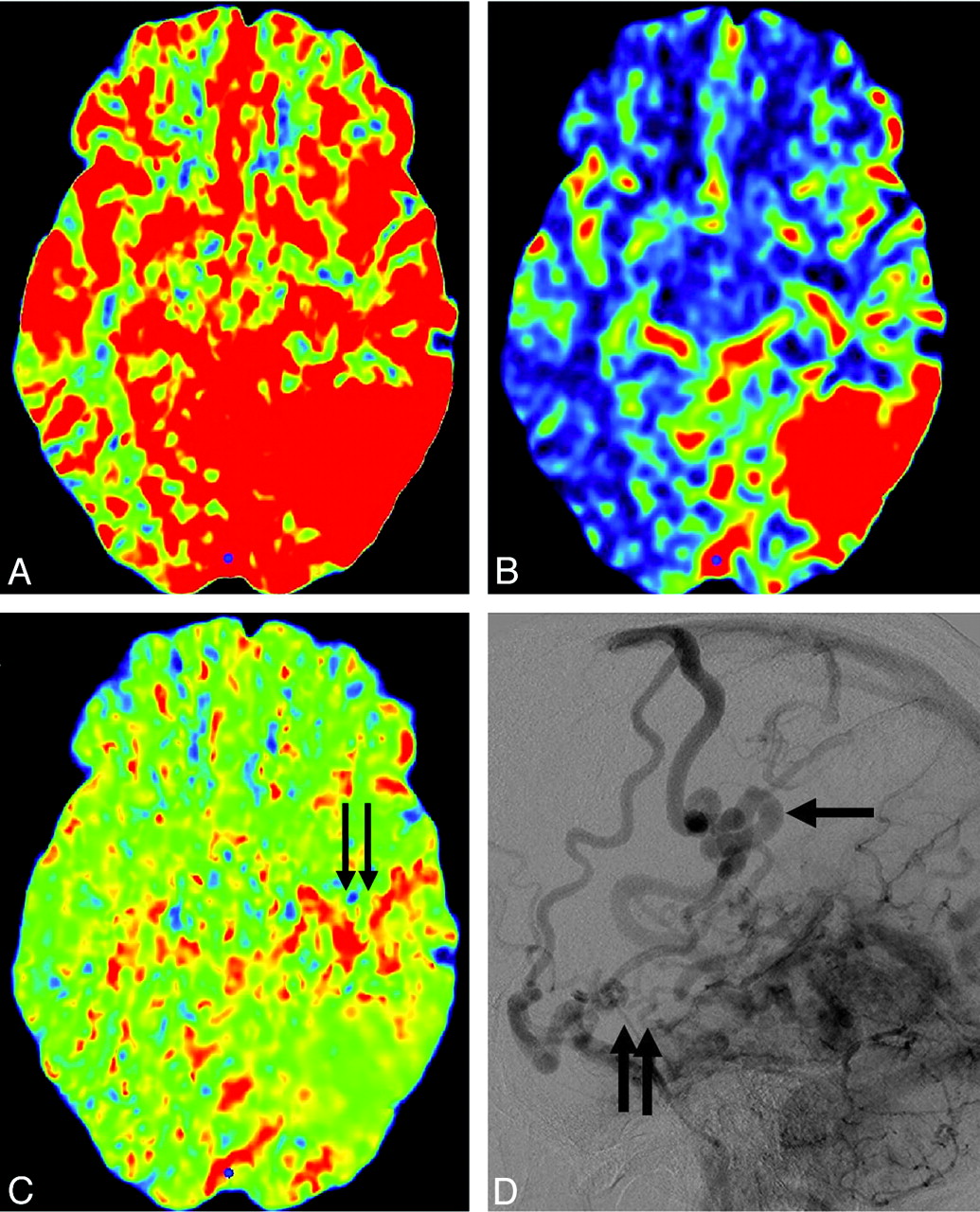
    Case 7. A−C, CBF, CBV, and MTT maps. Increased CBV and MTT are seen anterior to the nidus, which is suggestive of venous congestion (pattern 3, black arrows). D, Delayed phase of lateral conventional angiography shows tortuous engorged pial veins (pseudophlebitic pattern, black arrows) slowly draining anteriorly in addition to the main draining vein (arrow).

  • Fig 3.
    • Download figure
    • Open in new tab
    • Download powerpoint
    Fig 3.

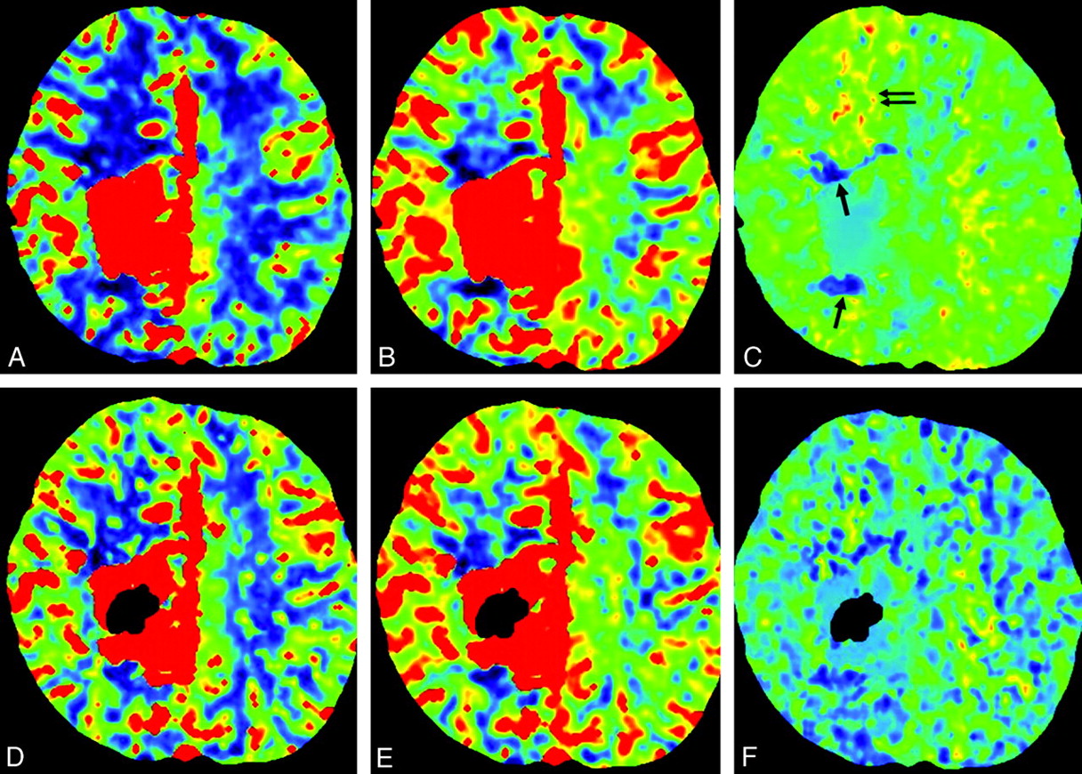
    Case 17. A−C, Perinidal areas of pattern 1 are seen in anterior and posterior aspects of the nidus (arrow). Increased blood volume and increased transit time (pattern 3) in the remote anterior frontal lobe are suggestive of venous congestion (arrows). D−F, Postembolization CBF, CBV, and MTT maps. Glue is shown as dark signal intensity in the nidus. Improvement of pattern 3 is seen in the right frontal lobe. Residual but slight improvement of pattern 1 in the perinidal area is also suggested.

  • Fig 4.
    • Download figure
    • Open in new tab
    • Download powerpoint
    Fig 4.

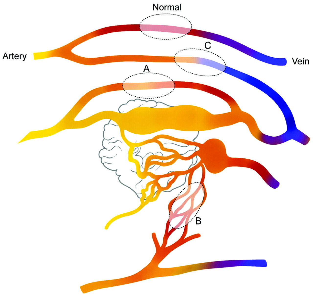
    Schematic diagram of the PCT patterns in patients with brain AVM. Fast shunting of flow is noted in the nidus of the AVM. Blood flow in the normal brain parenchyma is seen (normal). Sump effect from the vascular pedicle supplying the shunt causes functional steal (pattern 1) in the brain adjacent to this pedicle (area A). Areas supplied by indirectly recruited collateral flow to the shunt from adjacent arteries cause ischemic steal (area B, pattern 2). High-pressure flow in the draining veins of the AVM causes venous congestion in remote parts of the brain (area C, pattern 3).

Tables

  • Figures
    • View popup
    Table 1:

    Summary of cases

    CaseSex/Age (yr)SymptomVolume (cm3)LocationDrainagePCT Abnormality Pattern (location)a
    1F/49Seizure23.0Frontal, ltSuperficialPattern 1 (remote and perinidal)
    2F/40None2.7Frontal, rtSuperficialPattern 2 (perinidal)
    3M/47Seizure3.8Frontal, rtSuperficialPattern 1 (perinidal)
    4F/21FND17.2Parietal, rtSuperficialPatterns 1 and 2 (perinidal)
    5F/49FND15.3Occipital, rtSuperficialPattern 3
    6F/61none0.4Frontal, rtSuperficialNone
    7M/53FND24.7Temporo-occipital, ltSuperficial, deepPattern 3
    8F/17Hemorrhage0.7Temporal, rtSuperficialNone
    9M/64Hemorrhage2.1Temporo-parietal, rtSuperficialNone
    10M/62Chemosis14.1Frontal, rtSuperficial, deepPattern 3
    11F/30None2.4Frontal, ltSuperficial, deepPattern 1 (perinidal)
    12F/28Hemorrhage19.1Frontal, rtSuperficial, deepPatterns 1 (perinidal) and 3
    13F/53FND10.2Temporal, rtDeepPattern 2 (remote)
    14M/39FND8.0Occipital, ltSuperficialPattern 2 (perinidal)
    15F/42Seizure0.3Parietal, ltSuperficialPattern 1 (perinidal)
    16M/37Seizure21.2Frontal, rtSuperficial, deepPattern 1 (perinidal)
    17M/23Seizure27.4Parietal, rtSuperficialPatterns 1 (perinidal) and 3
    18F/39Hemorrhage3.1Cerebellum, rtSuperficialNone
    • ↵a Pattern 1 is decreased CBF, CBV, and MTT; pattern 2, decreased CBF and CBV, and increased MTT; pattern 3, increased CBV and MTT.

    • View popup
    Table 2:

    Catheter angiography correlated with PCT findings

    Pattern 1 (n = 8)Pattern 2 (n = 4)Pattern 3 (n = 5)
    Arterial phase
        Neoangiogenesis (n = 15)7 (88%)3 (75%)5 (100%)
        Leptomeningeal recruit (n = 8)4 (50%)2 (50%)4 (80%)
        Transdural recruit (n = 2)01 (25%)1 (20%)
        ≥2 Signs4 (50%)2 (50%)4 (80%)
    Venous phase
        Pseudophlebitic (n = 5)2 (25%)04 (80%)
        Venous reflux (n = 8)4 (50%)1 (25%)5 (100%)
        ≥2 Signs1 (13%)04 (80%)
PreviousNext
Back to top

In this issue

American Journal of Neuroradiology: 32 (11)
American Journal of Neuroradiology
Vol. 32, Issue 11
1 Dec 2011
  • Table of Contents
  • Index by author
Advertisement
Print
Download PDF
Email Article

Thank you for your interest in spreading the word on American Journal of Neuroradiology.

NOTE: We only request your email address so that the person you are recommending the page to knows that you wanted them to see it, and that it is not junk mail. We do not capture any email address.

Enter multiple addresses on separate lines or separate them with commas.
Whole-Brain Perfusion CT Patterns of Brain Arteriovenous Malformations: A Pilot Study in 18 Patients
(Your Name) has sent you a message from American Journal of Neuroradiology
(Your Name) thought you would like to see the American Journal of Neuroradiology web site.
CAPTCHA
This question is for testing whether or not you are a human visitor and to prevent automated spam submissions.
Cite this article
D.J. Kim, T. Krings
Whole-Brain Perfusion CT Patterns of Brain Arteriovenous Malformations: A Pilot Study in 18 Patients
American Journal of Neuroradiology Dec 2011, 32 (11) 2061-2066; DOI: 10.3174/ajnr.A2659

Citation Manager Formats

  • BibTeX
  • Bookends
  • EasyBib
  • EndNote (tagged)
  • EndNote 8 (xml)
  • Medlars
  • Mendeley
  • Papers
  • RefWorks Tagged
  • Ref Manager
  • RIS
  • Zotero
0 Responses
Respond to this article
Share
Bookmark this article
Whole-Brain Perfusion CT Patterns of Brain Arteriovenous Malformations: A Pilot Study in 18 Patients
D.J. Kim, T. Krings
American Journal of Neuroradiology Dec 2011, 32 (11) 2061-2066; DOI: 10.3174/ajnr.A2659
del.icio.us logo Twitter logo Facebook logo Mendeley logo
  • Tweet Widget
  • Facebook Like
  • Google Plus One
Purchase

Jump to section

  • Article
    • Abstract
    • Materials and Methods
    • Results
    • Discussion
    • Conclusions
    • Footnotes
    • References
  • Figures & Data
  • Info & Metrics
  • Responses
  • References
  • PDF

Related Articles

  • No related articles found.
  • PubMed
  • Google Scholar

Cited By...

  • MR characteristics of unruptured intracranial arteriovenous malformations associated with seizure as initial clinical presentation
  • Dural Arteriovenous Fistulas: A Characteristic Pattern of Edema and Enhancement of the Medulla on MRI
  • Feasibility of Flat Panel Detector CT in Perfusion Assessment of Brain Arteriovenous Malformations: Initial Clinical Experience
  • The clinical dilemma of treating transient ischaemic attack-like symptoms in patients with coexisting arteriovenous malformation
  • Crossref (46)
  • Google Scholar

This article has been cited by the following articles in journals that are participating in Crossref Cited-by Linking.

  • Brain arteriovenous malformations
    Michael T. Lawton, W. Caleb Rutledge, Helen Kim, Christian Stapf, Kevin J. Whitehead, Dean Y. Li, Timo Krings, Karel terBrugge, Douglas Kondziolka, Michael K. Morgan, Karam Moon, Robert F. Spetzler
    Nature Reviews Disease Primers 2015 1 1
  • Spinal and Paraspinal Arteriovenous Lesions
    Stéphanie Lenck, Patrick Nicholson, Rachel Tymianski, Christopher Hilditch, Aurélien Nouet, Krunal Patel, Timo Krings, Michael Tymianski, Ivan Radovanovic, Vitor Mendes Pereira
    Stroke 2019 50 8
  • Automated Brain Perfusion Imaging in Acute Ischemic Stroke: Interpretation Pearls and Pitfalls
    Manal Nicolas-Jilwan, Max Wintermark
    Stroke 2021 52 11
  • Measurement of Blood Flow in Arteriovenous Malformations before and after Embolization Using Arterial Spin Labeling
    L. Suazo, B. Foerster, R. Fermin, H. Speckter, C. Vilchez, J. Oviedo, P. Stoeter
    Interventional Neuroradiology 2012 18 1
  • 123I-IMP-SPECT in a Patient with Cerebral Proliferative Angiopathy: A Case Report
    Tomomi Kimiwada, Toshiaki Hayashi, Reizo Shirane, Teiji Tominaga
    Journal of Stroke and Cerebrovascular Diseases 2013 22 8
  • MR characteristics of unruptured intracranial arteriovenous malformations associated with seizure as initial clinical presentation
    John Charles Benson, Shannon Chiu, Kelly Flemming, Deena M Nasr, Giuseppe Lanzino, Waleed Brinjikji
    Journal of NeuroInterventional Surgery 2020 12 2
  • Neuroimaging of Acute Intracerebral Hemorrhage
    Peter B. Sporns, Marios-Nikos Psychogios, Grégoire Boulouis, Andreas Charidimou, Qi Li, Enrico Fainardi, Dar Dowlatshahi, Joshua N. Goldstein, Andrea Morotti
    Journal of Clinical Medicine 2021 10 5
  • Cortical reorganization of motor functional areas in cerebral arteriovenous malformations
    Lester Lee, Yih Yian Sitoh, Ivan Ng, Wai Hoe Ng
    Journal of Clinical Neuroscience 2013 20 5
  • Current and Future Applications of Arterial Spin Labeling MRI in Cerebral Arteriovenous Malformations
    Matteo De Simone, Marco Maria Fontanella, Anis Choucha, Karl Schaller, Paolo Machi, Giuseppe Lanzino, Philippe Bijlenga, Felix T. Kurz, Karl-Olof Lövblad, Lucio De Maria
    Biomedicines 2024 12 4
  • Color-Coded Digital Subtraction Angiography in the Management of a Rare Case of Middle Cerebral Artery Pure Arterial Malformation
    Caleb E. Feliciano, Eva Pamias-Portalatin, Jorge Mendoza-Torres, Euclides Effio, Yadira Moran, Rafael Rodriguez-Mercado
    Interventional Neuroradiology 2014 20 6

More in this TOC Section

  • Enhanced Axonal Metabolism during Early Natalizumab Treatment in Relapsing-Remitting Multiple Sclerosis
  • SWI or T2*: Which MRI Sequence to Use in the Detection of Cerebral Microbleeds? The Karolinska Imaging Dementia Study
  • Progression of Microstructural Damage in Spinocerebellar Ataxia Type 2: A Longitudinal DTI Study
Show more BRAIN

Similar Articles

Advertisement

Indexed Content

  • Current Issue
  • Accepted Manuscripts
  • Article Preview
  • Past Issues
  • Editorials
  • Editor's Choice
  • Fellows' Journal Club
  • Letters to the Editor
  • Video Articles

Cases

  • Case Collection
  • Archive - Case of the Week
  • Archive - Case of the Month
  • Archive - Classic Case

More from AJNR

  • Trainee Corner
  • Imaging Protocols
  • MRI Safety Corner
  • Book Reviews

Multimedia

  • AJNR Podcasts
  • AJNR Scantastics

Resources

  • Turnaround Time
  • Submit a Manuscript
  • Submit a Video Article
  • Submit an eLetter to the Editor/Response
  • Manuscript Submission Guidelines
  • Statistical Tips
  • Fast Publishing of Accepted Manuscripts
  • Graphical Abstract Preparation
  • Imaging Protocol Submission
  • Evidence-Based Medicine Level Guide
  • Publishing Checklists
  • Author Policies
  • Become a Reviewer/Academy of Reviewers
  • News and Updates

About Us

  • About AJNR
  • Editorial Board
  • Editorial Board Alumni
  • Alerts
  • Permissions
  • Not an AJNR Subscriber? Join Now
  • Advertise with Us
  • Librarian Resources
  • Feedback
  • Terms and Conditions
  • AJNR Editorial Board Alumni

American Society of Neuroradiology

  • Not an ASNR Member? Join Now

© 2025 by the American Society of Neuroradiology All rights, including for text and data mining, AI training, and similar technologies, are reserved.
Print ISSN: 0195-6108 Online ISSN: 1936-959X

Powered by HighWire