
Article Figures & Data
Figures
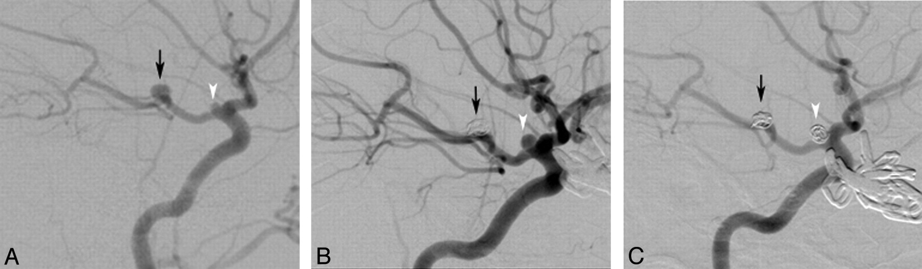
- Fig 1.
Multiple saccular intracranial aneurysms with enlargement. A 17-year-old female adolescent with MOPD II (patient 9 from On-line Tables 1 and 2) presented with SAH, was found to have 10 saccular intracranial aneurysms, and underwent surgical clipping of multiple left anterior circulation aneurysms (not shown). The lateral projection of a right ICA catheter angiogram (A) demonstrates filling of a basilar tip aneurysm (black arrow) across the PcomA as well as filling of a smaller aneurysm at the anterior origin of the PcomA (white arrowhead). The basilar tip aneurysm underwent selective endovascular coiling at the time of initial SAH and remained well occluded on 9-month asymptomatic surveillance angiography (black arrow, B). The previously identified right PcomA aneurysm, however, had increased in size from 1 mm in diameter to 2.5 mm in diameter (white arrowhead, A versus B) and was also treated with selective endovascular coiling (white arrowhead, C).
- Fig 2.
De novo aneurysm formation in a segment remote from the site of initial aneurysm treatment (remote segment vasculopathy). A 15-year-old female adolescent with history of remote head trauma years earlier (patient 8 from On-line Table 1) presented with pulsatile tinnitus and a vascular mass behind the right tympanic membrane. T2-weighted MR imaging (A) demonstrated a hyperintense mass (white arrow) in the right petrous bone; note that the basilar artery flow void (black arrowhead) in the prepontine cistern is normal in caliber. A right ICA angiogram in the lateral (B) projection confirms that the right petrous mass (black arrows) is a large fusiform aneurysm of the cervicopectoral ICA. The ICA was occluded with detachable balloons and the patient's tinnitus resolved. Six years later, the patient re-presented with diplopia and swallowing difficulties concerning for cranial neuropathies. T1-weighted axial (C) MR imaging performed at that time demonstrated a new mass (black arrowhead) centered in the prepontine cistern compressing the pons. The mass was confirmed on left vertebral artery angiography (not shown) to be a new giant fusiform aneurysm of the vertebrobasilar artery junction.
- Fig 3.
De novo aneurysm formation in a segment adjacent to a previously treated aneurysm (adjacent segment vasculopathy). A 3-year-old boy who fell from standing and hit his head (patient 1 from On-line Tables 1 and 2) was found on MR imaging to have a mass centered in the left ambient cistern (A). This mass was identified as a fusiform aneurysm of the left P2/3 PCA on conventional angiography (B). Endovascular coil occlusion of the aneurysm (C and D) was performed, resulting in parent artery occlusion of the PCA at and distal to the aneurysm. The patient developed no clinical deficit, and diffusion-weighted MR imaging (not shown) confirmed no infarct related to PCA occlusion in this young patient. Surveillance MR imaging (E) and MRA (F) 7 months later demonstrated no change in the ambient cistern coil mass and no patent aneurysm; a small amount of thrombus in the posterior portion of the aneurysm shines through on the time-of-flight MRA (white arrowheads, E and F). Surveillance MR imaging (G) and MRA (H) 4.5 years after initial aneurysm treatment, however, demonstrated a new mass compressing the left cerebral peduncle (G). This was confirmed on conventional angiography (I) to be a de novo aneurysm of the left P2 PCA just medial to the previously treated aneurysm. Selective endovascular coiling of the new aneurysm was performed (J), and a postcoiling unsubtracted x-ray image (K) confirms that the newly placed coils do not abut the previous coil mass, itself unchanged since initial therapy (D).


