Advertisement
  • Main menu
  • User menu
  • Search
  • English ▼
    • English
    • Afrikaans
    • Albanian
    • Amharic
    • Arabic
    • Armenian
    • Azerbaijani
    • Basque
    • Belarusian
    • Bengali
    • Bosnian
    • Bulgarian
    • Catalan
    • Cebuano
    • Chichewa
    • Chinese (Simplified)
    • Chinese (Traditional)
    • Corsican
    • Croatian
    • Czech
    • Danish
    • Dutch
    • Esperanto
    • Estonian
    • Filipino
    • Finnish
    • French
    • Frisian
    • Galician
    • Georgian
    • German
    • Greek
    • Gujarati
    • Haitian Creole
    • Hausa
    • Hawaiian
    • Hebrew
    • Hindi
    • Hmong
    • Hungarian
    • Icelandic
    • Igbo
    • Indonesian
    • Irish
    • Italian
    • Japanese
    • Javanese
    • Kannada
    • Kazakh
    • Khmer
    • Korean
    • Kurdish (Kurmanji)
    • Kyrgyz
    • Lao
    • Latin
    • Latvian
    • Lithuanian
    • Luxembourgish
    • Macedonian
    • Malagasy
    • Malay
    • Malayalam
    • Maltese
    • Maori
    • Marathi
    • Mongolian
    • Myanmar (Burmese)
    • Nepali
    • Norwegian
    • Pashto
    • Persian
    • Polish
    • Portuguese
    • Punjabi
    • Romanian
    • Russian
    • Samoan
    • Scottish Gaelic
    • Serbian
    • Sesotho
    • Shona
    • Sindhi
    • Sinhala
    • Slovak
    • Slovenian
    • Somali
    • Spanish
    • Sudanese
    • Swahili
    • Swedish
    • Tajik
    • Tamil
    • Telugu
    • Thai
    • Turkish
    • Ukrainian
    • Urdu
    • Uzbek
    • Vietnamese
    • Welsh
    • Xhosa
    • Yiddish
    • Yoruba
    • Zulu

Main menu

  • Home
  • Content
    • Current Issue
    • Accepted Manuscripts
    • Article Preview
    • Past Issue Archive
    • Video Articles
    • AJNR Case Collection
    • Case of the Week Archive
    • Case of the Month Archive
    • Classic Case Archive
  • Special Collections
    • AJNR Awards
    • Low-Field MRI
    • Alzheimer Disease
    • ASNR Foundation Special Collection
    • Photon-Counting CT
    • View All
  • Multimedia
    • AJNR Podcasts
    • AJNR SCANtastic
    • Trainee Corner
    • MRI Safety Corner
    • Imaging Protocols
  • For Authors
    • Submit a Manuscript
    • Submit a Video Article
    • Submit an eLetter to the Editor/Response
    • Manuscript Submission Guidelines
    • Statistical Tips
    • Fast Publishing of Accepted Manuscripts
    • Graphical Abstract Preparation
    • Imaging Protocol Submission
    • Author Policies
  • About Us
    • About AJNR
    • Editorial Board
    • Editorial Board Alumni
  • More
    • Become a Reviewer/Academy of Reviewers
    • Subscribers
    • Permissions
    • Alerts
    • Feedback
    • Advertisers
    • ASNR Home

User menu

  • Alerts
  • Log in

Search

  • Advanced search
American Journal of Neuroradiology

American Journal of Neuroradiology

ASHNR American Society of Functional Neuroradiology ASHNR American Society of Pediatric Neuroradiology ASSR
  • Alerts
  • Log in

Advanced Search

English ▼
  • English
  • Afrikaans
  • Albanian
  • Amharic
  • Arabic
  • Armenian
  • Azerbaijani
  • Basque
  • Belarusian
  • Bengali
  • Bosnian
  • Bulgarian
  • Catalan
  • Cebuano
  • Chichewa
  • Chinese (Simplified)
  • Chinese (Traditional)
  • Corsican
  • Croatian
  • Czech
  • Danish
  • Dutch
  • Esperanto
  • Estonian
  • Filipino
  • Finnish
  • French
  • Frisian
  • Galician
  • Georgian
  • German
  • Greek
  • Gujarati
  • Haitian Creole
  • Hausa
  • Hawaiian
  • Hebrew
  • Hindi
  • Hmong
  • Hungarian
  • Icelandic
  • Igbo
  • Indonesian
  • Irish
  • Italian
  • Japanese
  • Javanese
  • Kannada
  • Kazakh
  • Khmer
  • Korean
  • Kurdish (Kurmanji)
  • Kyrgyz
  • Lao
  • Latin
  • Latvian
  • Lithuanian
  • Luxembourgish
  • Macedonian
  • Malagasy
  • Malay
  • Malayalam
  • Maltese
  • Maori
  • Marathi
  • Mongolian
  • Myanmar (Burmese)
  • Nepali
  • Norwegian
  • Pashto
  • Persian
  • Polish
  • Portuguese
  • Punjabi
  • Romanian
  • Russian
  • Samoan
  • Scottish Gaelic
  • Serbian
  • Sesotho
  • Shona
  • Sindhi
  • Sinhala
  • Slovak
  • Slovenian
  • Somali
  • Spanish
  • Sudanese
  • Swahili
  • Swedish
  • Tajik
  • Tamil
  • Telugu
  • Thai
  • Turkish
  • Ukrainian
  • Urdu
  • Uzbek
  • Vietnamese
  • Welsh
  • Xhosa
  • Yiddish
  • Yoruba
  • Zulu
  • Home
  • Content
    • Current Issue
    • Accepted Manuscripts
    • Article Preview
    • Past Issue Archive
    • Video Articles
    • AJNR Case Collection
    • Case of the Week Archive
    • Case of the Month Archive
    • Classic Case Archive
  • Special Collections
    • AJNR Awards
    • Low-Field MRI
    • Alzheimer Disease
    • ASNR Foundation Special Collection
    • Photon-Counting CT
    • View All
  • Multimedia
    • AJNR Podcasts
    • AJNR SCANtastic
    • Trainee Corner
    • MRI Safety Corner
    • Imaging Protocols
  • For Authors
    • Submit a Manuscript
    • Submit a Video Article
    • Submit an eLetter to the Editor/Response
    • Manuscript Submission Guidelines
    • Statistical Tips
    • Fast Publishing of Accepted Manuscripts
    • Graphical Abstract Preparation
    • Imaging Protocol Submission
    • Author Policies
  • About Us
    • About AJNR
    • Editorial Board
    • Editorial Board Alumni
  • More
    • Become a Reviewer/Academy of Reviewers
    • Subscribers
    • Permissions
    • Alerts
    • Feedback
    • Advertisers
    • ASNR Home
  • Follow AJNR on Twitter
  • Visit AJNR on Facebook
  • Follow AJNR on Instagram
  • Join AJNR on LinkedIn
  • RSS Feeds

AJNR Awards, New Junior Editors, and more. Read the latest AJNR updates

Research ArticleSpine Imaging and Spine Image-Guided Interventions
Open Access

Quantitative Evaluation and Visualization of Lumbar Foraminal Nerve Root Entrapment by Using Diffusion Tensor Imaging: Preliminary Results

Y. Eguchi, S. Ohtori, S. Orita, H. Kamoda, G. Arai, T. Ishikawa, M. Miyagi, G. Inoue, M. Suzuki, Y. Masuda, H. Andou, M. Takaso, Y. Aoki, T. Toyone, A. Watanabe and K. Takahashi
American Journal of Neuroradiology November 2011, 32 (10) 1824-1829; DOI: https://doi.org/10.3174/ajnr.A2681
Y. Eguchi
  • Find this author on Google Scholar
  • Find this author on PubMed
  • Search for this author on this site
S. Ohtori
  • Find this author on Google Scholar
  • Find this author on PubMed
  • Search for this author on this site
S. Orita
  • Find this author on Google Scholar
  • Find this author on PubMed
  • Search for this author on this site
H. Kamoda
  • Find this author on Google Scholar
  • Find this author on PubMed
  • Search for this author on this site
G. Arai
  • Find this author on Google Scholar
  • Find this author on PubMed
  • Search for this author on this site
T. Ishikawa
  • Find this author on Google Scholar
  • Find this author on PubMed
  • Search for this author on this site
M. Miyagi
  • Find this author on Google Scholar
  • Find this author on PubMed
  • Search for this author on this site
G. Inoue
  • Find this author on Google Scholar
  • Find this author on PubMed
  • Search for this author on this site
M. Suzuki
  • Find this author on Google Scholar
  • Find this author on PubMed
  • Search for this author on this site
Y. Masuda
  • Find this author on Google Scholar
  • Find this author on PubMed
  • Search for this author on this site
H. Andou
  • Find this author on Google Scholar
  • Find this author on PubMed
  • Search for this author on this site
M. Takaso
  • Find this author on Google Scholar
  • Find this author on PubMed
  • Search for this author on this site
Y. Aoki
  • Find this author on Google Scholar
  • Find this author on PubMed
  • Search for this author on this site
T. Toyone
  • Find this author on Google Scholar
  • Find this author on PubMed
  • Search for this author on this site
A. Watanabe
  • Find this author on Google Scholar
  • Find this author on PubMed
  • Search for this author on this site
K. Takahashi
  • Find this author on Google Scholar
  • Find this author on PubMed
  • Search for this author on this site
  • Article
  • Figures & Data
  • Info & Metrics
  • Responses
  • References
  • PDF
Loading

Article Figures & Data

Figures

  • Tables
  • Fig 1.
    • Download figure
    • Open in new tab
    • Download powerpoint
    Fig 1.

    Echo-planar imaging image (A) and FA mapping (B) ROIs were placed on bilateral roots and FA values were calculated (B).

  • Fig 2.
    • Download figure
    • Open in new tab
    • Download powerpoint
    Fig 2.

    Coronal tractogram of lumbar nerve roots in a healthy volunteer. L3, L4, L5, and S1 indicate the third, fourth, and fifth lumbar root, and the first sacral root.

  • Fig 3.
    • Download figure
    • Open in new tab
    • Download powerpoint
    Fig 3.

    Tractograms of lumbar nerve roots in a 75-year-old man with right L5–S1 foraminal stenosis (referenced as patient 1 in Table 3) by ROI placement on bilateral L5 roots at the stenotic level. ROIs were placed both proximally and distally to the foraminal zone at the nonstenotic level of L3, L4, and S1 roots. On the entrapped side of the right L5 root, by placing the region of interest on the proximal side (A), nerve tracts were seen to be disrupted and no tracts were seen distal to the foramen (arrow). However, by placing the secondary region of interest on the distal side (B), though the nerve tracts were traced on the distal side, a deficit is seen in the foramen (arrow). In contrast, on the intact side of the left L5 root, there was no difference whether the ROI was proximal or distal.

  • Fig 4.
    • Download figure
    • Open in new tab
    • Download powerpoint
    Fig 4.

    Tractograms of 8 patients by placing the ROI on the proximal side of the foramen. In all patients, tracts show disruption of nerve fibers in the foramen (arrows).

  • Fig 5.
    • Download figure
    • Open in new tab
    • Download powerpoint
    Fig 5.

    Tractograms of 8 patients by placing secondary ROI on the distal side of the foramen. Nerve traces show abnormalities (white arrows) such as tract disruption (case 1), nerve narrowing (cases 2–6), and indentation (cases 7 and 8) in their course through the foramen.
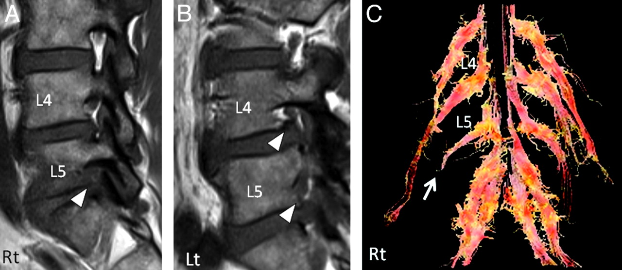
  • Fig 6.
    • Download figure
    • Open in new tab
    • Download powerpoint
    Fig 6.

    Sagittal T1-weighted MR images (A; right side, B; left side) and a diffusion tensor image (C) of a 62-year-old man with right L5–S1 foraminal stenosis (referenced as patient 4 in Table 3). Although asymptomatic foraminal stenosis on the left L4 and left L5 foramina (arrowheads in B) were found by MR imaging, abnormalities such as disruption of nerve fibers were only accurately detected at the symptomatic root by DTI (arrow in C).
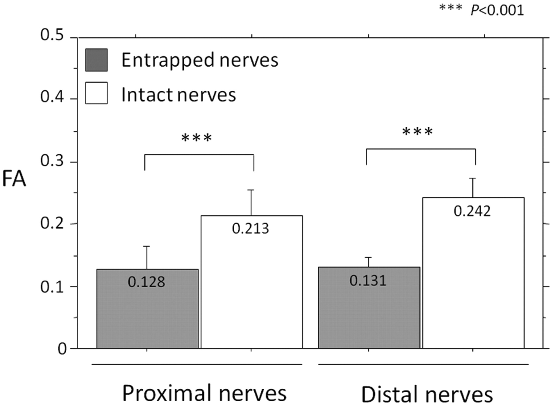
  • Fig 7.
    • Download figure
    • Open in new tab
    • Download powerpoint
    Fig 7.

    Mean FA values at the proximal nerve root and distal spinal nerve in patients with foraminal stenosis. The mean FA of proximal nerve roots on the side of entrapment was 0.155 ± 0.049 and is significantly lower than the 0.208 ± 0.036 on the intact side. The mean FA of distal spinal nerve roots on the side of entrapment was 0.131 ± 0.016 and significantly lower than the 0.240 ± 0.035 seen on the intact side (P < .001).

  • Fig 8.
    • Download figure
    • Open in new tab
    • Download powerpoint
    Fig 8.

    Bland-Altman plots of comparisons of FA values. Most observed differences are within mean ± 1.96 SD. Horizontal dashed lines indicate mean difference (middle line) and limits of agreement, defined as mean difference plus (top line) and minus (bottom line) 1.96 × SD of differences. A, Relationship between differences in the first analysis and second analysis (y-axis) and means of the first analysis and second analysis (x-axis). B, Relationship between differences in observer 1 and observer 2 (y-axis) and means of observer 1 and observer 2 (x-axis).

Tables

  • Figures
    • View popup
    Table 1:

    Mean FA values of healthy volunteers

    RootFA (Proximal)FA (Distal)
    RightLeftRightLeft
    L30.157 ± 0.0280.161 ± 0.0320.172 ± 0.0220.196 ± 0.047
    L40.183 ± 0.0170.190 ± 0.0270.188 ± 0.0310.185 ± 0.029
    L50.196 ± 0.0200.192 ± 0.0200.220 ± 0.0300.214 ± 0.037
    S10.195 ± 0.0300.192 ± 0.0400.212 ± 0.0320.205 ± 0.040
    • View popup
    Table 2:

    Incidence of detected foraminal narrowing in patients on MR imaging and DTI

    L4L5Total
    MR imaging
        Asymptomatic foramina (n = 24)5/166/811/24
        Symptomatic foramina (n = 8)0/08/88/8
        False-positive rate (%)45.80
    DTIL4L5Total
        Asymptomatic foramina (n = 24)0/160/80/24
        Symptomatic foramina (n = 8)0/08/88/8
        False-positive rate (%)0.00
    • View popup
    Table 3:

    Patient summary

    No.Age (yr)SexSymptomatic RootDisease Duration (mo)VAS (Leg Pain)DTI FindingsFA
    ProximalDistal
    EntrappedIntactEntrappedIntact
    175ML5 (Right)1560Tract disruption0.06980.1620.1170.195
    264FL5 (Left)1890Tract disruption0.0860.1850.1480.278
    366ML5 (Right)870Tract disruption0.1120.2710.1300.252
    462ML5 (Right)1490Tract disruption0.1650.2380.1240.223
    547ML5 (Right)2480Tract disruption0.1350.1740.1060.238
    644ML5 (Left)760Tract disruption0.1280.2000.1380.242
    764FL5 (Right)24100Tract disruption0.160.2710.1410.292
    868FL5 (Right)1260Tract disruption0.1660.2030.1430.214
        Mean6115.276.30.1280.2130.1310.242
PreviousNext
Back to top

In this issue

American Journal of Neuroradiology: 32 (10)
American Journal of Neuroradiology
Vol. 32, Issue 10
1 Nov 2011
  • Table of Contents
  • Index by author
Advertisement
Print
Download PDF
Email Article
Cite this article
0 Responses
Respond to this article
Share
Bookmark this article
Quantitative Evaluation and Visualization of Lumbar Foraminal Nerve Root Entrapment by Using Diffusion Tensor Imaging: Preliminary Results
Y. Eguchi, S. Ohtori, S. Orita, H. Kamoda, G. Arai, T. Ishikawa, M. Miyagi, G. Inoue, M. Suzuki, Y. Masuda, H. Andou, M. Takaso, Y. Aoki, T. Toyone, A. Watanabe, K. Takahashi
American Journal of Neuroradiology Nov 2011, 32 (10) 1824-1829; DOI: 10.3174/ajnr.A2681
del.icio.us logo Twitter logo Facebook logo Mendeley logo
  • Tweet Widget
Purchase

Jump to section

  • Article
    • Abstract
    • Abbreviations
    • Materials and Methods
    • Results
    • Discussion
    • Conclusions
    • Footnotes
    • References
  • Figures & Data
  • Info & Metrics
  • Responses
  • References
  • PDF

Related Articles

  • No related articles found.
  • PubMed
  • Google Scholar

Cited By...

  • Diffusion tensor imaging of radiculopathy in patients with lumbar disc herniation: preliminary results
  • Crossref
  • Google Scholar

This article has not yet been cited by articles in journals that are participating in Crossref Cited-by Linking.

More in this TOC Section

  • Dynamic CT Myelography: Patient Positioning
  • Evaluation of SIH MR Scoring Systems in Normals
  • Characteristics of SIH Type I Culprit Lesions
Show more SPINE IMAGING AND SPINE IMAGE-GUIDED INTERVENTIONS

Similar Articles

Advertisement

Indexed Content

  • Current Issue
  • Accepted Manuscripts
  • Article Preview
  • Past Issues
  • Editorials
  • Editor's Choice
  • Fellows' Journal Club
  • Letters to the Editor
  • Video Articles

Cases

  • Case Collection
  • Archive - Case of the Week
  • Archive - Case of the Month
  • Archive - Classic Case

More from AJNR

  • Trainee Corner
  • Imaging Protocols
  • MRI Safety Corner
  • Book Reviews

Multimedia

  • AJNR Podcasts
  • AJNR Scantastics

Resources

  • Turnaround Time
  • Submit a Manuscript
  • Submit a Video Article
  • Submit an eLetter to the Editor/Response
  • Manuscript Submission Guidelines
  • Statistical Tips
  • Fast Publishing of Accepted Manuscripts
  • Graphical Abstract Preparation
  • Imaging Protocol Submission
  • Evidence-Based Medicine Level Guide
  • Publishing Checklists
  • Author Policies
  • Become a Reviewer/Academy of Reviewers
  • News and Updates

About Us

  • About AJNR
  • Editorial Board
  • Editorial Board Alumni
  • Alerts
  • Permissions
  • Not an AJNR Subscriber? Join Now
  • Advertise with Us
  • Librarian Resources
  • Feedback
  • Terms and Conditions
  • AJNR Editorial Board Alumni

American Society of Neuroradiology

  • Not an ASNR Member? Join Now

© 2025 by the American Society of Neuroradiology All rights, including for text and data mining, AI training, and similar technologies, are reserved.
Print ISSN: 0195-6108 Online ISSN: 1936-959X

Powered by HighWire
Email this Article

Thank you for your interest in spreading the word on American Journal of Neuroradiology.

NOTE: We only request your email address so that the person you are recommending the page to knows that you wanted them to see it, and that it is not junk mail. We do not capture any email address.

Enter multiple addresses on separate lines or separate them with commas.
Quantitative Evaluation and Visualization of Lumbar Foraminal Nerve Root Entrapment by Using Diffusion Tensor Imaging: Preliminary Results
(Your Name) has sent you a message from American Journal of Neuroradiology
(Your Name) thought you would like to see the American Journal of Neuroradiology web site.
CAPTCHA
This question is for testing whether or not you are a human visitor and to prevent automated spam submissions.
Cite this article
Y. Eguchi, S. Ohtori, S. Orita, H. Kamoda, G. Arai, T. Ishikawa, M. Miyagi, G. Inoue, M. Suzuki, Y. Masuda, H. Andou, M. Takaso, Y. Aoki, T. Toyone, A. Watanabe, K. Takahashi
Quantitative Evaluation and Visualization of Lumbar Foraminal Nerve Root Entrapment by Using Diffusion Tensor Imaging: Preliminary Results
American Journal of Neuroradiology Nov 2011, 32 (10) 1824-1829; DOI: 10.3174/ajnr.A2681

Citation Manager Formats

  • BibTeX
  • Bookends
  • EasyBib
  • EndNote (tagged)
  • EndNote 8 (xml)
  • Medlars
  • Mendeley
  • Papers
  • RefWorks Tagged
  • Ref Manager
  • RIS
  • Zotero

We use cookies on this site to enhance your user experience. By clicking any link on this page you are giving your consent for us to set cookies.