
Article Figures & Data
Figures
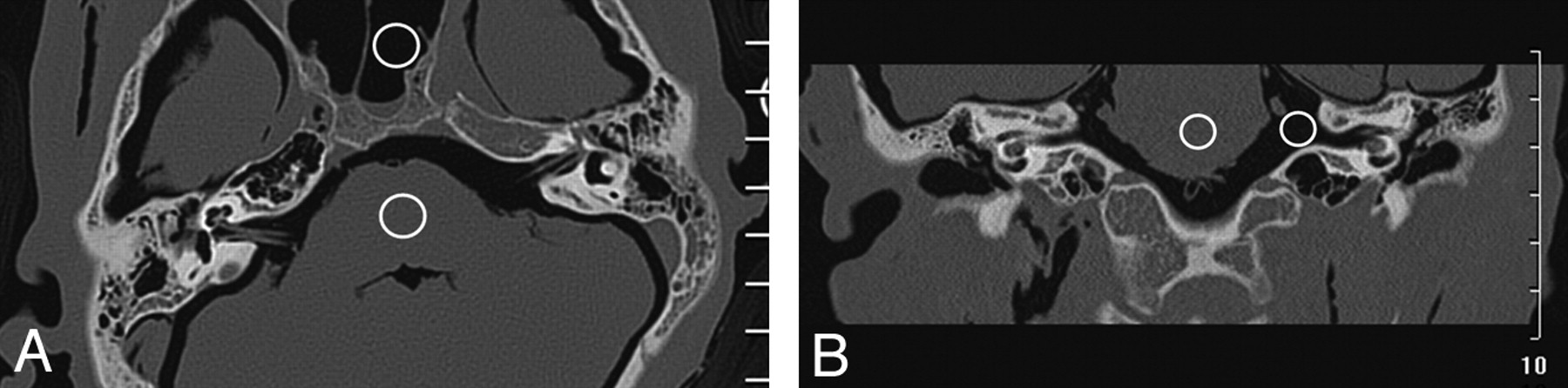
- Fig 1.
ROIs in the brain stem and air were used to calculate the CNR for each image. Example ROIs are shown in an axial (A) and the coronal (B) reformatted images here.
- Fig 2.
Axial (A–C) and coronal (D–F) images reformatted from helical temporal bone scans. Each scan was performed with a similar CTDIvol but a different tube voltage. The images shown were acquired at 80 (A, D), 120 (B, E), and 140 kVp (C, F), respectively.


Tables
- Table 1:
Radiation dose indices, noise, and CNR of temporal bone axial scans with different detector collimations
Detector Collimation (mm) CTDIvol(mGy) DLP (mGy · cm) Noise (HU) CNR Brain Stem Sphenoid 2 × 0.625 47.2 1898.6 77.9 41.4 16.1 12 × 0.625 20.8 935.6 18.7 15.0 60.5 16 × 0.625 22.6 902.8 82.9 43.4 15.1 40 × 0.625 24.2 1211.5 75.3 34.6 17.2 64 × 0.625 19.4 775.6 80.4 49.6 14.8 Note:—When the 12 × 0.625-mm collimation was selected, the system could not choose 180 mAs/section. The closest tube mAs, 165 mAs/section, was selected. In addition, the 12 × 0.625-mm collimation used the system's “detailed resolution” setting and got substantially different noise and CNR, with no comparability with other collimations. The 12 × 0.625-mm detector collimation was not available in helical mode and was not included in the comparison.
- Table 2:
Radiation dose indices, noise, and CNR of temporal bone helical scans with different detector collimations
Detector Collimation (mm) CTDIvol (mGy) DLP (mGy · cm) Noise (HU) CNR Axial Coronal Brain Stem Sphenoid Brain Stem Air Axial Coronal 2 × 0.5 32.2 1296.7 186.2 60.4 76.5 27.5 7.3 18.1 16 × 0.625 25.5 1026.9 97.8 41.8 64.5 55.8 13.0 15.8 20 × 0.625 16.3 656.4 156.6 65.4 82.8 60.7 8.1 13.2 40 × 0.625 23.3 1169.0 92.3 40.7 73.9 56.8 13.6 14.3 64 × 0.625 22.9 1836.6 99.9 45.9 62.7 60.5 12.3 15.6 -
Note:—The 2 × 0.5- and 20 × 0.625-mm detector collimations used the system's “ultrahigh resolution” setting and pitches of 0.7 and 0.656, respectively. So, the noise and CNR of axial images were significantly different from the other detector collimations utilizing the system's “high resolution” setting.
-
- Table 3:
Radiation dose indices, noise, and CNR of temporal bone helical scans with different kVp
kVp Effective mAs CTDIvol (mGy) Noise (HU) CNR Axial Coronal Brain Stem Sphenoid Brain Stem Air Axial Coronal 80 540 14.5 166.3 55.7 96.8 66.8 7.6 11.1 120 240 25.4 103.4 45.2 73.8 51.6 12.3 15.1 140 180 25.5 97.8 41.8 64.5 55.8 12.9 15.8 - Table 4:
Radiation dose indices, noise, and CNR of temporal bone helical scans with different effective tube mAs
Effective mAs CTDIvol (mGy) Noise (HU) CNR Axial Coronal Brain Stem Sphenoid Brain Stem Air Axial Coronal 40 4.2 268.1 110.0 178.6 97.5 4.4 6.0 80 8.7 191.3 66.4 113.8 70.0 6.6 9.7 120 13.3 155.0 62.9 102.3 67.8 8.1 10.7 160 17.1 130.3 62.8 88.8 59.1 9.3 12.7 200 20.7 105.6 50.2 82.5 59.8 11.8 13.1 240 25.4 103.4 45.2 73.8 51.6 12.3 15.1 280 29.1 99.2 43.5 65.5 50.8 12.8 16.4 320 35.2 88.1 42.5 60.5 49.9 14.1 17.2 360 38.2 88.9 43.3 57.2 44.9 14.0 18.9 Effective mAs Axial Image Grade Coronal Image Grade 40 III III III III III III 80 III II II II III II 120 II II II I II I 160 I I I I I I 200 I I I I I I 240 I I I I I I 280 I I I I I I 320 I I I I I I 360 I I I I I I - Table 6:
Radiation dose indices, noise, and CNR of temporal bone helical scans with fixed effective mAs and different pitches
Pitch CTDIvol (mGy) Noise CNR Axial Coronal Brain Stem Sphenoid Brain Stem Air Axial Coronal 0.19 20.2 152.4 46.4 72.3 47.5 8.7 15.5 0.31 20.4 134.0 47.2 68.8 41.6 9.7 16.8 0.44 20.3 106.7 49.5 72.1 48.7 11.7 15.5 0.685 20.1 109.3 45.3 72.7 51.3 11.5 17.2 0.935 20.1 123.3 48.7 76.3 60.5 10.3 13.6 1.19 21.4 131.2 48.5 70.5 50.7 9.8 15.5 1.315 21.8 117.5 51.7 73.7 65.3 10.6 13.5
In this issue


Jump to section
Related Articles
Cited By...
- No citing articles found.