Advertisement

Main menu

  • Home
  • Content
    • Current Issue
    • Accepted Manuscripts
    • Article Preview
    • Past Issue Archive
    • Video Articles
    • AJNR Case Collection
    • Case of the Week Archive
    • Case of the Month Archive
    • Classic Case Archive
  • Special Collections
    • AJNR Awards
    • Low-Field MRI
    • Alzheimer Disease
    • ASNR Foundation Special Collection
    • Photon-Counting CT
    • View All
  • Multimedia
    • AJNR Podcasts
    • AJNR SCANtastic
    • Trainee Corner
    • MRI Safety Corner
    • Imaging Protocols
  • For Authors
    • Submit a Manuscript
    • Submit a Video Article
    • Submit an eLetter to the Editor/Response
    • Manuscript Submission Guidelines
    • Statistical Tips
    • Fast Publishing of Accepted Manuscripts
    • Graphical Abstract Preparation
    • Imaging Protocol Submission
    • Author Policies
  • About Us
    • About AJNR
    • Editorial Board
    • Editorial Board Alumni
  • More
    • Become a Reviewer/Academy of Reviewers
    • Subscribers
    • Permissions
    • Alerts
    • Feedback
    • Advertisers
    • ASNR Home

User menu

  • Alerts
  • Log in

Search

  • Advanced search
American Journal of Neuroradiology

American Journal of Neuroradiology

ASHNR American Society of Functional Neuroradiology ASHNR American Society of Pediatric Neuroradiology ASSR
  • Alerts
  • Log in

Advanced Search

  • Home
  • Content
    • Current Issue
    • Accepted Manuscripts
    • Article Preview
    • Past Issue Archive
    • Video Articles
    • AJNR Case Collection
    • Case of the Week Archive
    • Case of the Month Archive
    • Classic Case Archive
  • Special Collections
    • AJNR Awards
    • Low-Field MRI
    • Alzheimer Disease
    • ASNR Foundation Special Collection
    • Photon-Counting CT
    • View All
  • Multimedia
    • AJNR Podcasts
    • AJNR SCANtastic
    • Trainee Corner
    • MRI Safety Corner
    • Imaging Protocols
  • For Authors
    • Submit a Manuscript
    • Submit a Video Article
    • Submit an eLetter to the Editor/Response
    • Manuscript Submission Guidelines
    • Statistical Tips
    • Fast Publishing of Accepted Manuscripts
    • Graphical Abstract Preparation
    • Imaging Protocol Submission
    • Author Policies
  • About Us
    • About AJNR
    • Editorial Board
    • Editorial Board Alumni
  • More
    • Become a Reviewer/Academy of Reviewers
    • Subscribers
    • Permissions
    • Alerts
    • Feedback
    • Advertisers
    • ASNR Home
  • Follow AJNR on Twitter
  • Visit AJNR on Facebook
  • Follow AJNR on Instagram
  • Join AJNR on LinkedIn
  • RSS Feeds

AJNR Awards, New Junior Editors, and more. Read the latest AJNR updates

Research ArticleBrain
Open Access

Diffusion Tensor Group Tractography of the Corpus Callosum in Clinically Isolated Syndrome

F. Lin, C. Yu, Y. Liu, K. Li and H. Lei
American Journal of Neuroradiology January 2011, 32 (1) 92-98; DOI: https://doi.org/10.3174/ajnr.A2273
F. Lin
  • Find this author on Google Scholar
  • Find this author on PubMed
  • Search for this author on this site
C. Yu
  • Find this author on Google Scholar
  • Find this author on PubMed
  • Search for this author on this site
Y. Liu
  • Find this author on Google Scholar
  • Find this author on PubMed
  • Search for this author on this site
K. Li
  • Find this author on Google Scholar
  • Find this author on PubMed
  • Search for this author on this site
H. Lei
  • Find this author on Google Scholar
  • Find this author on PubMed
  • Search for this author on this site
  • Article
  • Figures & Data
  • Info & Metrics
  • Responses
  • References
  • PDF
Loading

Article Figures & Data

Figures

  • Tables
  • Fig 1.
    • Download figure
    • Open in new tab
    • Download powerpoint
    Fig 1.

    The size of the midsagittal CC for NC and CIS suggestive of MS groups. A, One example of midsagittal CC and its subregions (yellow indicates the genu; red, the body; green, the splenium). B, The midsagittal CC area for the 2 groups. C, The normalized midsagittal CC area by midsagittal brain for the 2 groups.

  • Fig 2.
    • Download figure
    • Open in new tab
    • Download powerpoint
    Fig 2.

    The probability maps for the entire CC and its subregions. A, The entire CC probability map. B, The genu probability map. C, The body probability map. D, The splenium probability map. The gray-scale overlay indicates the probability of a voxel being part of the CC or its subregions.
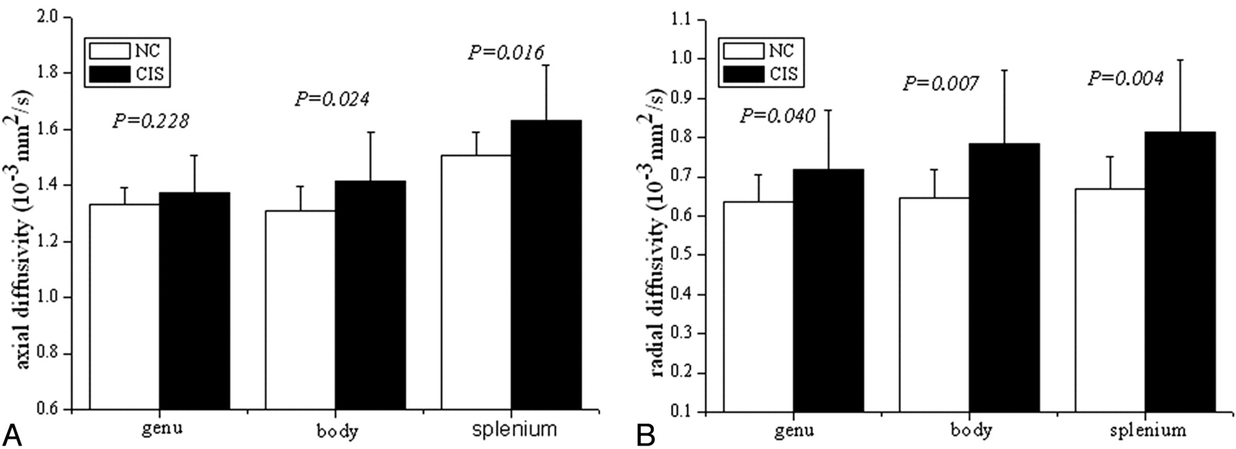
  • Fig 3.
    • Download figure
    • Open in new tab
    • Download powerpoint
    Fig 3.

    The λ1 and λ23 distributions for the 3 subregions of the CC in CIS suggestive of MS and NCs. A, λ1 (×10−3 mm2/s) distribution for the 3 subregions of the CC for these 2 groups. B, λ23 (×10−3 mm2/s) distribution for the 3 subregions of the CC for these 2 groups.

Tables

  • Figures
  • Comparison of diffusion indices measured in subregions of CC of NC and CIS

    NC (n = 19)CIS (n = 19)Pa
    Mean ± SDMean ± SD
    FA
        Whole CC0.46 ± 0.030.42 ± 0.04.001
        Genu0.458 ± 0.0380.419 ± 0.053.011
        Body0.442 ± 0.0320.399 ± 0.046.002
        Splenium0.494 ± 0.0340.453 ± 0.062.020
    MDb
        Whole CC0.90 ± 0.061.02 ± 0.16.004
        Genu0.868 ± 0.0620.937 ± 0.142.064
        Body0.868 ± 0.0700.995 ± 0.182.009
        Splenium0.948 ± 0.0781.087 ± 0.181.005
    λ1c
        Whole CC1.38 ± 0.061.49 ± 0.15.010
        Genu1.333 ± 0.0591.417 ± 0.175.228
        Body1.310 ± 0.0871.409 ± 0.177.024
        Splenium1.505 ± 0.0861.633 ± 0.197.016
    λ23d
        Whole CC0.66 ± 0.070.79 ± 0.16.003
        Genu0.635 ± 0.0710.718 ± 0.151.040
        Body0.648 ± 0.0700.785 ± 0.188.007
        Splenium0.670 ± 0.0820.814 ± 0.183.004
    • a A P value < .01 was considered statistically significant, while a value between .01 and .05 was considered a trend.

    • b MD = ×10−3 mm2/s.

    • c λ1= ×10−3 mm2/s.

    • d λ23 = ×10−3 mm2/s.

PreviousNext
Back to top

In this issue

American Journal of Neuroradiology: 32 (1)
American Journal of Neuroradiology
Vol. 32, Issue 1
1 Jan 2011
  • Table of Contents
  • Index by author
Advertisement
Print
Download PDF
Email Article

Thank you for your interest in spreading the word on American Journal of Neuroradiology.

NOTE: We only request your email address so that the person you are recommending the page to knows that you wanted them to see it, and that it is not junk mail. We do not capture any email address.

Enter multiple addresses on separate lines or separate them with commas.
Diffusion Tensor Group Tractography of the Corpus Callosum in Clinically Isolated Syndrome
(Your Name) has sent you a message from American Journal of Neuroradiology
(Your Name) thought you would like to see the American Journal of Neuroradiology web site.
CAPTCHA
This question is for testing whether or not you are a human visitor and to prevent automated spam submissions.
Cite this article
F. Lin, C. Yu, Y. Liu, K. Li, H. Lei
Diffusion Tensor Group Tractography of the Corpus Callosum in Clinically Isolated Syndrome
American Journal of Neuroradiology Jan 2011, 32 (1) 92-98; DOI: 10.3174/ajnr.A2273

Citation Manager Formats

  • BibTeX
  • Bookends
  • EasyBib
  • EndNote (tagged)
  • EndNote 8 (xml)
  • Medlars
  • Mendeley
  • Papers
  • RefWorks Tagged
  • Ref Manager
  • RIS
  • Zotero
0 Responses
Respond to this article
Share
Bookmark this article
Diffusion Tensor Group Tractography of the Corpus Callosum in Clinically Isolated Syndrome
F. Lin, C. Yu, Y. Liu, K. Li, H. Lei
American Journal of Neuroradiology Jan 2011, 32 (1) 92-98; DOI: 10.3174/ajnr.A2273
del.icio.us logo Twitter logo Facebook logo Mendeley logo
  • Tweet Widget
  • Facebook Like
  • Google Plus One
Purchase

Jump to section

  • Article
    • Abstract
    • Abbreviations
    • Materials and Methods
    • Results
    • Discussion
    • Conclusions
    • Footnotes
    • References
  • Figures & Data
  • Info & Metrics
  • Responses
  • References
  • PDF

Related Articles

  • No related articles found.
  • PubMed
  • Google Scholar

Cited By...

  • No citing articles found.
  • Crossref (4)
  • Google Scholar

This article has been cited by the following articles in journals that are participating in Crossref Cited-by Linking.

  • Position Sense Deficits at the Lower Limbs in Early Multiple Sclerosis: Clinical and Neural Correlates
    Riccardo Iandolo, Giulia Bommarito, Laura Falcitano, Simona Schiavi, Niccolò Piaggio, Giovanni Luigi Mancardi, Maura Casadio, Matilde Inglese
    Neurorehabilitation and Neural Repair 2020 34 3
  • Gadolinium‐Enhancing Lesions Lead to Decreases in White Matter Tract Fractional Anisotropy in Multiple Sclerosis
    Gloria C. Chiang, Soniya Pinto, Joseph P. Comunale, Susan A. Gauthier
    Journal of Neuroimaging 2016 26 3
  • Exploring mania-associated white matter injury by comparison with multiple sclerosis: a diffusion tensor imaging study
    Niccolò Piaggio, Simona Schiavi, Matteo Martino, Giulia Bommarito, Matilde Inglese, Paola Magioncalda
    Psychiatry Research: Neuroimaging 2018 281
  • Composite MRI measures and short-term disability in patients with clinically isolated syndrome suggestive of MS
    Giulia Bommarito, Alessandro Bellini, Matteo Pardini, Claudio Solaro, Luca Roccatagliata, Alice Laroni, Elisabetta Capello, Giovanni Luigi Mancardi, Antonio Uccelli, Matilde Inglese
    Multiple Sclerosis Journal 2018 24 5

More in this TOC Section

  • Evaluating the Effects of White Matter Multiple Sclerosis Lesions on the Volume Estimation of 6 Brain Tissue Segmentation Methods
  • Quiet PROPELLER MRI Techniques Match the Quality of Conventional PROPELLER Brain Imaging Techniques
  • Predictors of Reperfusion in Patients with Acute Ischemic Stroke
Show more BRAIN

Similar Articles

Advertisement

Indexed Content

  • Current Issue
  • Accepted Manuscripts
  • Article Preview
  • Past Issues
  • Editorials
  • Editor's Choice
  • Fellows' Journal Club
  • Letters to the Editor
  • Video Articles

Cases

  • Case Collection
  • Archive - Case of the Week
  • Archive - Case of the Month
  • Archive - Classic Case

More from AJNR

  • Trainee Corner
  • Imaging Protocols
  • MRI Safety Corner
  • Book Reviews

Multimedia

  • AJNR Podcasts
  • AJNR Scantastics

Resources

  • Turnaround Time
  • Submit a Manuscript
  • Submit a Video Article
  • Submit an eLetter to the Editor/Response
  • Manuscript Submission Guidelines
  • Statistical Tips
  • Fast Publishing of Accepted Manuscripts
  • Graphical Abstract Preparation
  • Imaging Protocol Submission
  • Evidence-Based Medicine Level Guide
  • Publishing Checklists
  • Author Policies
  • Become a Reviewer/Academy of Reviewers
  • News and Updates

About Us

  • About AJNR
  • Editorial Board
  • Editorial Board Alumni
  • Alerts
  • Permissions
  • Not an AJNR Subscriber? Join Now
  • Advertise with Us
  • Librarian Resources
  • Feedback
  • Terms and Conditions
  • AJNR Editorial Board Alumni

American Society of Neuroradiology

  • Not an ASNR Member? Join Now

© 2025 by the American Society of Neuroradiology All rights, including for text and data mining, AI training, and similar technologies, are reserved.
Print ISSN: 0195-6108 Online ISSN: 1936-959X

Powered by HighWire