
Article Figures & Data
Figures
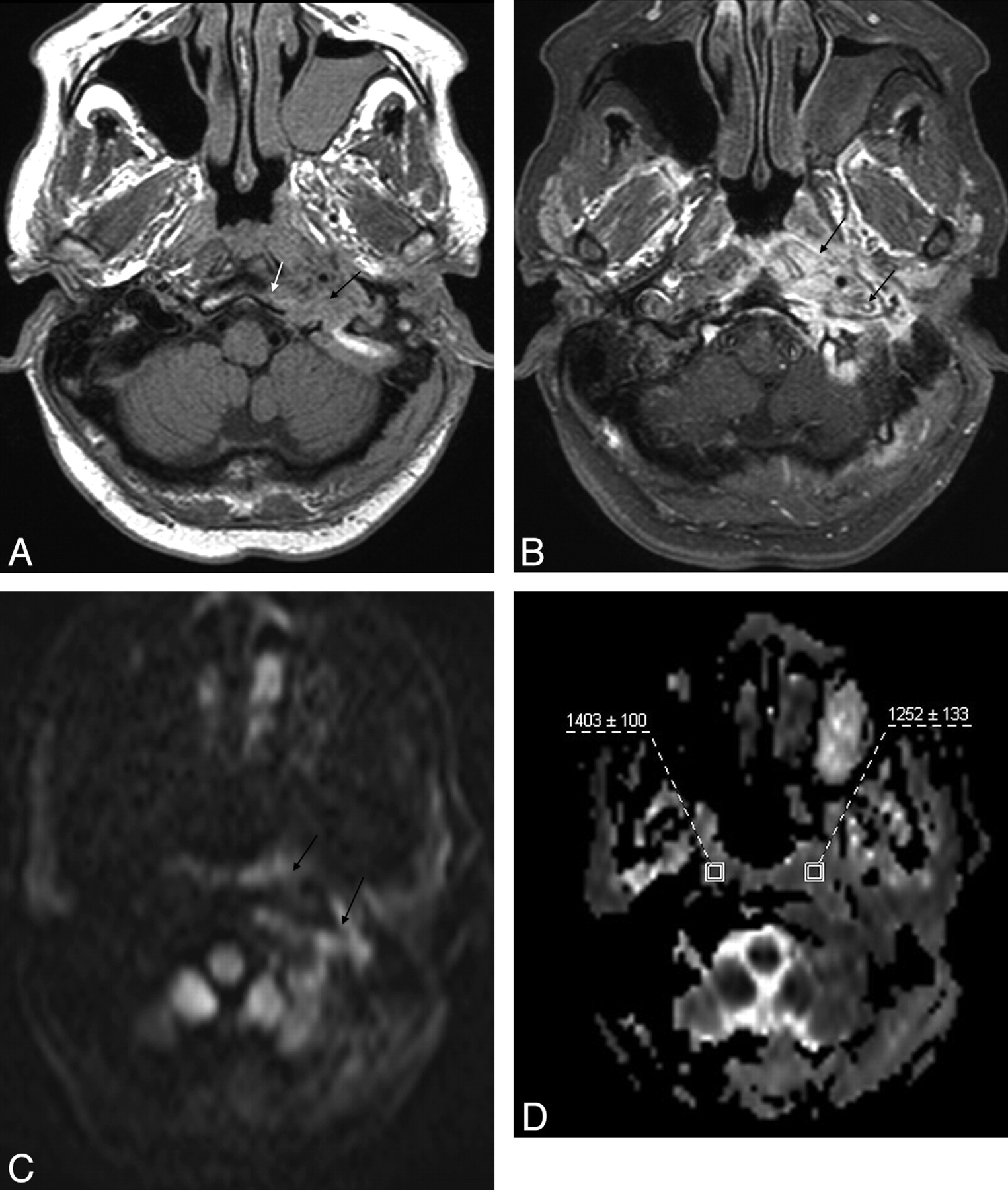
- Fig 1.
MR images in a case of SBO (patient 8). A, Axial T1-weighted image demonstrates infiltration with decreased T1 signal intensity of the left jugular foramen (black arrow) extending to the poststyloid parapharyngeal space with loss of the cortical margin of the clivus on the left (white arrow). B, Axial postcontrast and fat-suppressed T1-weighted image reveals extensive enhancement of the affected region (arrows). C, Diffusion TRACE image of the same region shows increased signal intensity (arrows). D, Corresponding ADC map with the regions of interest placed to measure the ADC values from the abnormal soft tissues (on the left) and normal-appearing soft tissues (on the right).
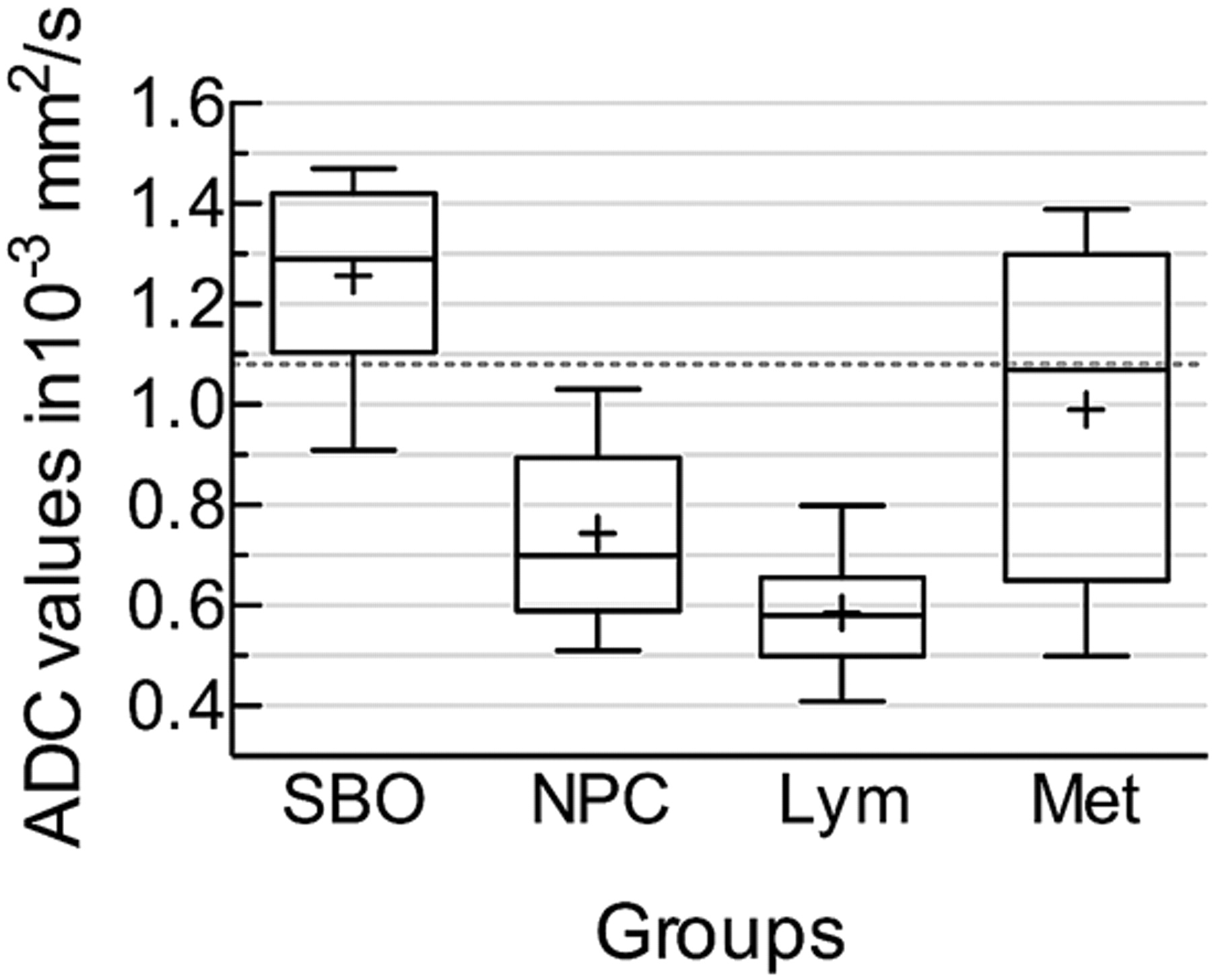
- Fig 2.
Box-and-whisker plot comparing the mean ADC values of the affected soft tissues in SBO, NPC, lymphoma, and metastatic lesions. The horizontal line is the median (50th percentile) of the measured values; the top and bottom of the box represent the 25th and 75th percentiles, respectively; and whiskers indicate the range from the largest to the smallest observed data points. The plus sign within the whiskers indicates the mean value for each group. Note that despite the overlap between the ADC values of different groups, the ADCs of SBO are significantly higher than those of lymphoma and NPC. The dotted line represents the cutoff value (1.08 × 10⁻³ mm2/s) distinguishing SBO from NPC and lymphoma.
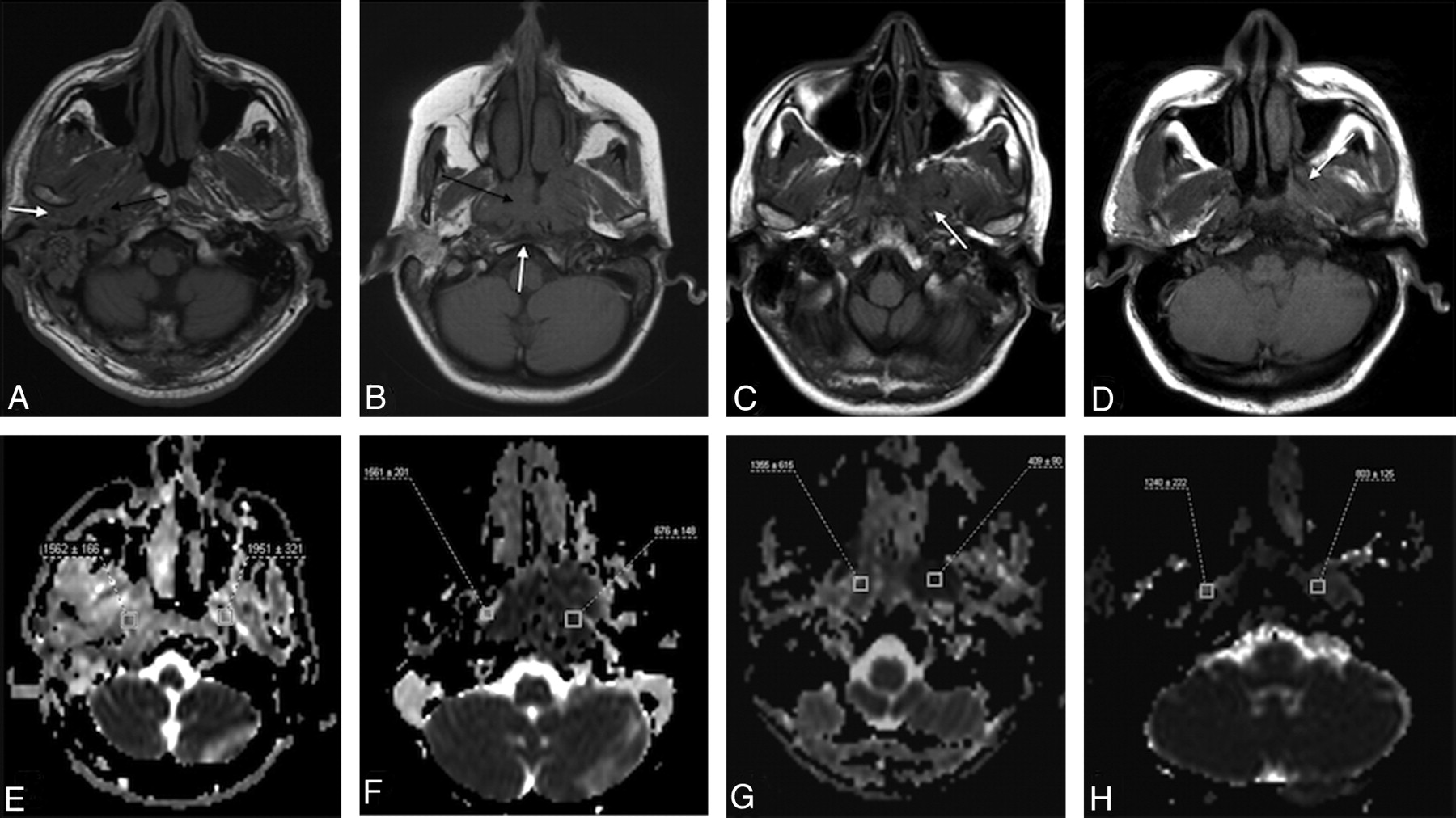
- Fig 3.
MR images (anatomic images and ADC maps) of patients from each group. A, Axial T1-weighted image of a patient with SBO demonstrates infiltration of the soft tissues around the EAC, in the retrocondylar fat space (white arrow), extending to the poststyloid parapharyngeal space and jugular foramen (black arrow). B, Axial T1-weighted image of a patient with NPC reveals a large infiltrative mass in the nasopharynx (black arrow) with a small area of clival infiltration (white arrow). C, Axial T1-weighted image of a patient with lymphoma with a low T1-signal-intensity mass around the left pterygoid process (arrow), extending to the masticator space. D, Axial T1-weighted image of a patient with undifferentiated carcinoma metastatic to the skull base with the low T1-signal-intensity metastases on the left (arrow). E−H, Corresponding ADC maps of the patients with SBO, NPC, lymphoma, and metastases, respectively, with the regions of interest placed to measure the ADC values.


Tables
Group/Patient No. Age (yr) Sex Histopathologic diagnosis SBO 1 66 M Chronic inflammation and fibrosis 2 41 F Active chronic inflammation and fibrosis 3 73 M Active chronic inflammation and fibrosis 4 63 M Active chronic inflammation and fibrosis 5 56 M Chronic inflammation and fibrosis 6 56 M Chronic inflammation and fibrosis 7 81 M Chronic inflammation and fibrosis 8 57 F Chronic inflammation and fibrosis 9 61 M Chronic inflammation, giant cell reaction, fibrosis NPC 1 47 M SCC, undifferentiated type 2 47 F SCC, undifferentiated type 3 44 M SCC, undifferentiated type 4 61 M SCC, undifferentiated type 5 60 M SCC, undifferentiated type 6 37 M SCC, moderately differentiated type 7 24 F SCC, undifferentiated type 8 62 M SCC, undifferentiated type 9 12 M SCC, undifferentiated type Lymphoma 1 14 M B-cell lymphoma 2 14 M B-cell lymphoma 3 14 M T-cell lymphoblastic-type lymphoma 4 43 F Diffuse large B-cell, immunoblastic type lymphoma 5 83 F Diffuse B-cell lymphoma 6 37 F Diffuse B-cell lymphoma 7 26 M T-cell lymphoblastic-type lymphoma 8 20 M B-cell lymphoma 9 49 M Diffuse large B-cell lymphoma Metastasis 1 60 M Undifferentiated carcinoma 2 53 M Undifferentiated carcinoma 3 58 M Prostate carcinoma 4 57 M Undifferentiated carcinoma 5 50 M Moderately differentiated carcinoma 6 54 F Multiple myeloma 7 46 M Multiple myeloma 8 48 M Multiple myeloma 9 71 M Prostate carcinoma Dx ADCST ADCNST ADCST/ADCNST ADCPONS SBO 1.26 ± 0.19 (0.91–1.45) 1.47 ± 0.18 (1.23–1.80) 0.86 ± 0.10 (0.70–1.02) 0.77 ± 0.09 (0.64–0.89) NPC 0.74 ± 0.18 (0.51–1.03) 1.53 ± 0.28 (1.23–1.9) 0.46 ± 0.09 (0.27–0.57) 0.75 ± 0.13 (0.61–1.05) Lymphoma 0.59 ± 0.11 (0.41–0.80) 1.53 ± 0.23 (1.20–1.87) 0.37 ± 0.06 (0.24–57) 0.79 ± 0.21 (0.67–0.86) Metastasis 0.99 ± 0.34 (0.5–1.39) 1.53 ± 0.35 (1.01–2.12) 0.64 ± 0.32 (0.41–1.38) 0.79 ± 0.04 (0.71–0.85) -
The ADC values are expressed as mean ± SD (minimum ADC value − maximum ADC value) × 10−3 mm2/s; the ADC ratios, as mean ± SD (minimum − maximum).
-
In this issue


Jump to section
Related Articles
Cited By...
- Dynamic Contrast-Enhanced MRI Parameters and Normalized ADC Values Could Aid Differentiation of Skull Base Osteomyelitis from Nasopharyngeal Cancer
- Skull Base Osteomyelitis: A Comprehensive Imaging Review
- Role of diffusion-weighted MRI in differentiation of masticator space malignancy from infection
- A diagnostic dilemma of central skull base osteomyelitis mimicking neoplasia in a diabetic patient