
Article Figures & Data
Figures
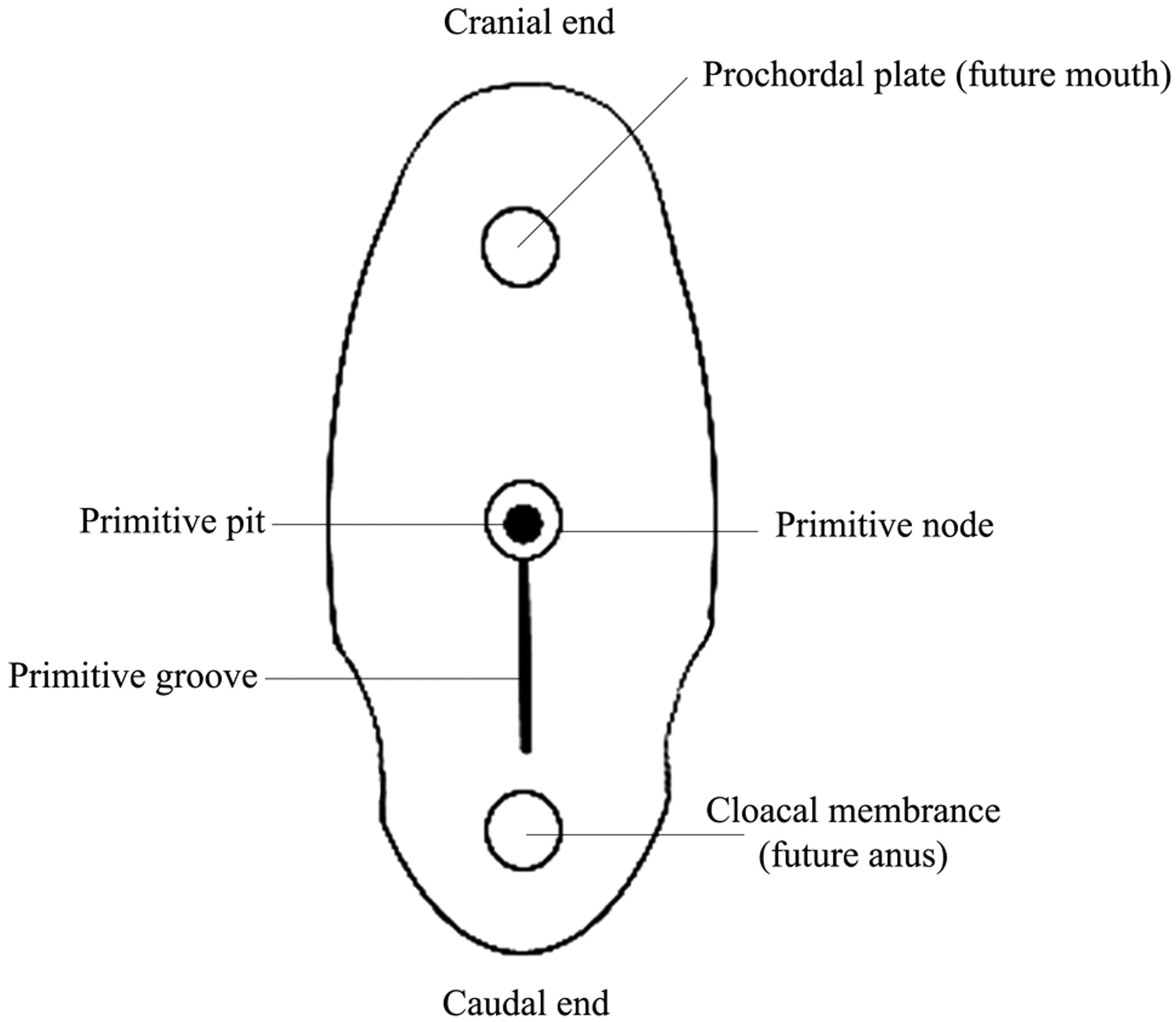
- Fig 1.
Dorsal aspect of the germ disk from an approximately 15-day embryo.
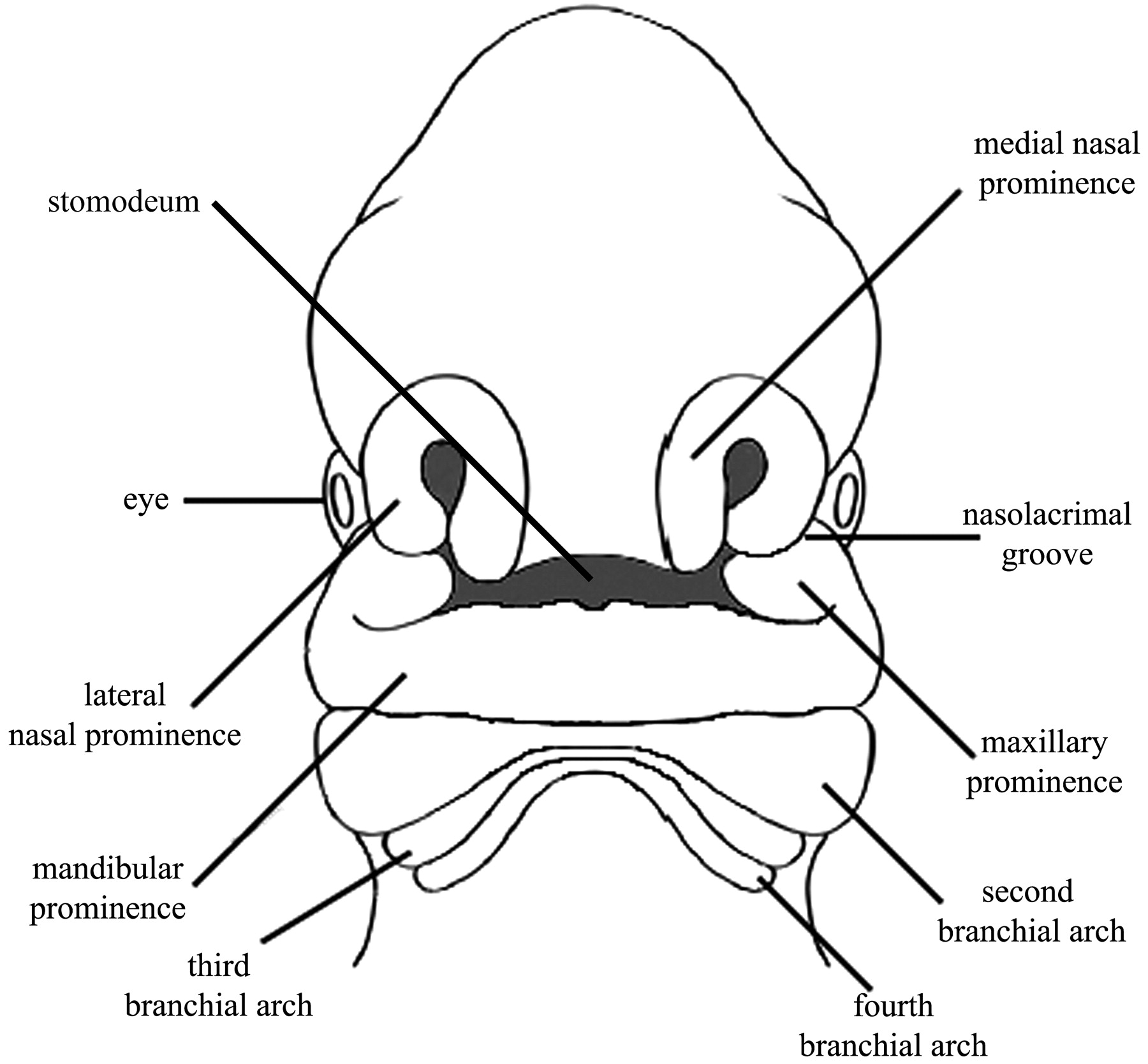
- Fig 2.
Frontal view of an approximately 30-day embryo showing the positions of the stomodeum relative to the medial and lateral nasal prominence and the maxillary and mandibular prominences.
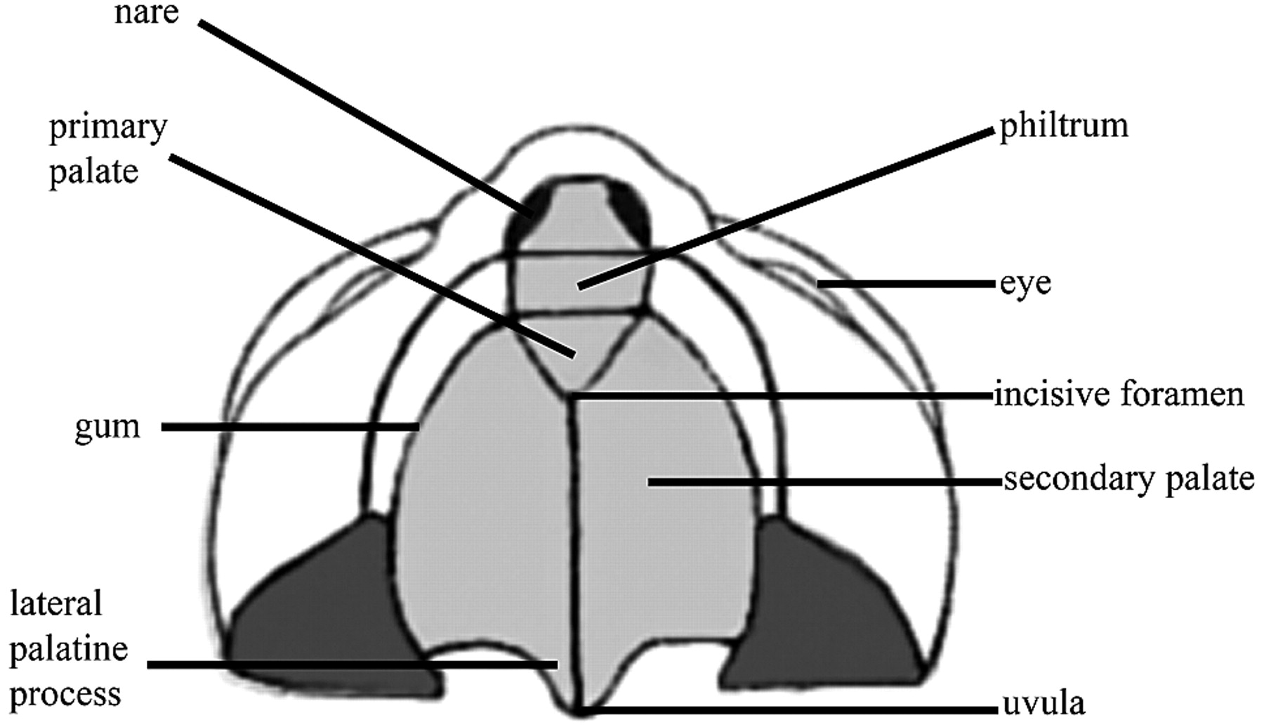
- Fig 3.
Ventral illustration of the palate, incisive foramen, gum, lip, and nose.
- Fig 4.
A, A 44-year-old woman with CP. 3D bony reconstruction shows a bony cleft (arrow) extending from the left aspect of an asymmetrically enlarged pyriform aperture to the alveolar surface. B, Axial CT image shows a bony cleft (arrow) between the left central and lateral maxillary incisors. C, Coronal CT image shows the extension of the bony clefting (arrow) to involve the primary palate.
- Fig 5.
Auricular atresia in various degrees of severity. A, Axial CT image in a 64-year-old woman with nonsyndromic EAC atresia shows marked narrowing of the bony EAC (arrow). B, Axial CT image in a 9-year-old girl shows severe atresia with a lateral bony plate (arrow). The middle ear cavity is small and dysplastic (arrowhead). There is also ipsilateral microtia. C, Axial CT image in a 3-year-old boy with Goldenhar syndrome shows complete bony atresia of the right EAC. D, Axial CT image in a 6-year-old boy with unilateral auricular atresia with associated ossicular chain fusion (arrow) and microtia (not shown).
- Fig 6.
A 6-year-old boy with syndromic micrognathia. A−C, 3D bony reconstructions show mandibular hypoplasia and abnormal temporomandibular joints, condyles, and coronoid processes. D, Axial CT image shows severe micrognathia and malocclusion.


Tables
Location Cleft Arch Nerve Pouch First External ear canal Mandible, muscles of mastication, 5th cranial nerve, malleus, and incus Trigeminal nerve (V2 and V3) Eustachian tube, tympanic cavity, mastoid air cells Second Cervical sinus of His Muscles of facial expression, body and lesser horns of hyoid, 7th and 8th cranial nerves, stapes Facial nerve (VII) Palatine tonsil Third Cervical sinus of His Superior constrictor muscles, internal carotid artery, 9th cranial nerve, greater horn, and body of hyoid Glossopharyngeal nerve (IX) Inferior parathyroid, thymus, pyriform fossa Fourth Cervical sinus of His Thyroid and cuneiform cartilages, 10th cranial nerve, aortic arch and right subclavian artery, part of laryngeal muscles Vagus nerve (X), superior laryngeal nerve Superior parathyroid, apex of pyriform sinus Fifth and sixth None Portions of the laryngeal muscles and skeleton, inferior pharyngeal constrictor muscles, 11th cranial nerve Vagus nerve (X), recurrent laryngeal nerve Parafollicular ″C″ cells of thyroid gland - Table 2:
System of Jahrsdoerfer et al for preoperative evaluation of aural atresia and stenosis as assessed using high-resolution CT of the temporal bonea
Parameter Points Score Candidate Stapes present 2 10 Excellent Oval window 1 9 Very good Round window 1 8 Good Middle ear space 1 7 Regular Mastoid pneumatization 1 6 Borderline Facial nerve course 1 ≤5 Poor Malleus-incus complex 1 Incus-stapes articulation 1 Auricle appearance 1 -
a The percentage of successful surgeries corresponds roughly with the rating scale (ie, score of 8 equals approximately 80% chance of postoperative hearing at normal or near-normal levels). Adapted from Jahrsdoerfer et al, 1992.34
-


