
Article Figures & Data
Figures
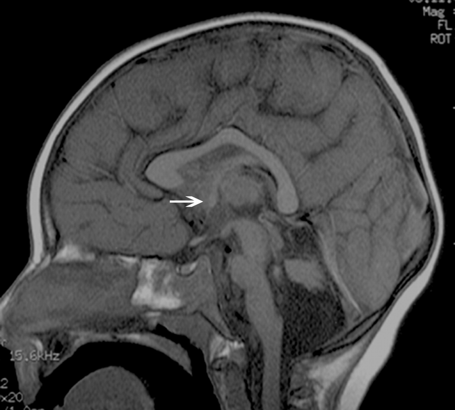
- Fig 1.
T1-weighted midsagittal image shows a hypogenetic CC in a patient with (p)LIS1. The rostrum is absent, the inferior part of the genu is small, and the splenium is thin. Note that splenium is also vertical; this was a common finding in classic lissencephalies. A normal AC is seen (arrow).
- Fig 2.
Midline sagittal T1-weighted image of a patient with a VLDLR; this is a dysmorphic CC with an abnormal shape and a convex upward callosal body configuration. The brain stem and vermis are hypoplastic, and the tentorium is displaced inferiorly. The AC is small (arrow).
- Fig 3.
Midsagittal T1-weighted image of a patient with the muscle-eye-brain disease phenotype of dystroglycanopathy. The splenium is thin, and the body is arched. This type of CC was classified as having a convex upward callosal body shape. Note the small pons, large tectum, and dysmorphic cerebellar vermis. The AC is normal (arrow); the patient has hydrocephalus.
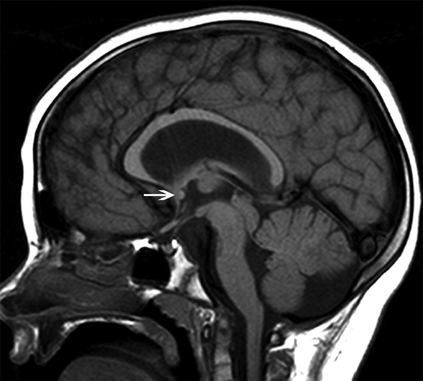
- Fig 4.
The image of the (p)LIS case shows no rostrum, a small inferior genu, a flat corpus, and a vertically angled splenium. The case was classified as having a thin flat body and a vertical splenium shape. The CC has a sharp angle (approximately 90°) between the flat callosal body and the splenium. A small AC is seen (arrow).


Tables
Significant differences among the major types of lissencephalies for each structure and specific features
Structure Groups Compared and Associated Features P Value Splenium cLIS, vertical or thin <.001 vLIS, absent or thin, and small Brain involvement cLIS, pachygyria (P > A, central, A > P) <.001 vLIS, pachygyria entire brain Myelination cLIS, normal <.001 vLIS, delayed CC genu cLIS, small inferior or N .002 vLIS, absent or small inferior CBSC, normal genu HC cLIS, normal .003 AC cLIS, normal or small .047 vLIS, absent CC body cLIS, normal or flat thin .011 vLIS, convex upward


