Advertisement

Main menu

  • Home
  • Content
    • Current Issue
    • Accepted Manuscripts
    • Article Preview
    • Past Issue Archive
    • Video Articles
    • AJNR Case Collection
    • Case of the Week Archive
    • Case of the Month Archive
    • Classic Case Archive
  • Special Collections
    • AJNR Awards
    • Low-Field MRI
    • Alzheimer Disease
    • ASNR Foundation Special Collection
    • Photon-Counting CT
    • View All
  • Multimedia
    • AJNR Podcasts
    • AJNR SCANtastic
    • Trainee Corner
    • MRI Safety Corner
    • Imaging Protocols
  • For Authors
    • Submit a Manuscript
    • Submit a Video Article
    • Submit an eLetter to the Editor/Response
    • Manuscript Submission Guidelines
    • Statistical Tips
    • Fast Publishing of Accepted Manuscripts
    • Graphical Abstract Preparation
    • Imaging Protocol Submission
    • Author Policies
  • About Us
    • About AJNR
    • Editorial Board
    • Editorial Board Alumni
  • More
    • Become a Reviewer/Academy of Reviewers
    • Subscribers
    • Permissions
    • Alerts
    • Feedback
    • Advertisers
    • ASNR Home

User menu

  • Alerts
  • Log in

Search

  • Advanced search
American Journal of Neuroradiology

American Journal of Neuroradiology

ASHNR American Society of Functional Neuroradiology ASHNR American Society of Pediatric Neuroradiology ASSR
  • Alerts
  • Log in

Advanced Search

  • Home
  • Content
    • Current Issue
    • Accepted Manuscripts
    • Article Preview
    • Past Issue Archive
    • Video Articles
    • AJNR Case Collection
    • Case of the Week Archive
    • Case of the Month Archive
    • Classic Case Archive
  • Special Collections
    • AJNR Awards
    • Low-Field MRI
    • Alzheimer Disease
    • ASNR Foundation Special Collection
    • Photon-Counting CT
    • View All
  • Multimedia
    • AJNR Podcasts
    • AJNR SCANtastic
    • Trainee Corner
    • MRI Safety Corner
    • Imaging Protocols
  • For Authors
    • Submit a Manuscript
    • Submit a Video Article
    • Submit an eLetter to the Editor/Response
    • Manuscript Submission Guidelines
    • Statistical Tips
    • Fast Publishing of Accepted Manuscripts
    • Graphical Abstract Preparation
    • Imaging Protocol Submission
    • Author Policies
  • About Us
    • About AJNR
    • Editorial Board
    • Editorial Board Alumni
  • More
    • Become a Reviewer/Academy of Reviewers
    • Subscribers
    • Permissions
    • Alerts
    • Feedback
    • Advertisers
    • ASNR Home
  • Follow AJNR on Twitter
  • Visit AJNR on Facebook
  • Follow AJNR on Instagram
  • Join AJNR on LinkedIn
  • RSS Feeds

AJNR Awards, New Junior Editors, and more. Read the latest AJNR updates

LetterLetter

Immune Reconstitution Inflammatory Syndrome and Cerebral Toxoplasmosis

R.F. Cabral, P.R. Valle Bahia, E.L. Gasparetto and L. Chimelli
American Journal of Neuroradiology August 2010, 31 (7) E65-E69; DOI: https://doi.org/10.3174/ajnr.A2158
R.F. Cabral
  • Find this author on Google Scholar
  • Find this author on PubMed
  • Search for this author on this site
P.R. Valle Bahia
  • Find this author on Google Scholar
  • Find this author on PubMed
  • Search for this author on this site
E.L. Gasparetto
  • Find this author on Google Scholar
  • Find this author on PubMed
  • Search for this author on this site
L. Chimelli
  • Find this author on Google Scholar
  • Find this author on PubMed
  • Search for this author on this site
  • Article
  • Figures & Data
  • Info & Metrics
  • Responses
  • References
  • PDF
Loading

Immune reconstitution inflammatory syndrome (IRIS) is a clinical deterioration that occurs despite increasing CD4+ T-lymphocyte counts and decreasing plasma human immunodeficiency virus-1 (HIV-1) viral loads. It results from an inflammatory response or “dysregulation” of the immune system to both intact subclinical pathogens and residual antigens. Resulting clinical manifestations of this syndrome are diverse and depend on the infectious or noninfectious agent involved.1 Neurologic symptoms and signs of IRIS have been reported in association with clinical and subclinical infections, such as cryptococcosis, tuberculosis, mycobacteriosis, and progressive multifocal leukoencephalopathy,2 as well as noninfectious phenomena. There are very rare reports of neurologic IRIS associated with cerebral toxoplasmosis.3,4

A 26-year-old woman with HIV-1 infection for 8 years presented with a 1-month history of ataxia, left-sided weakness, and hyperreflexia. She was not taking highly active antiretroviral therapy (HAART) regularly and was referred to treatment for cerebral toxoplasmosis 4 years before with clinical and radiologic improvement. Currently, a CT scan showed scattered calcified lesions in the brain parenchyma with no perilesional edema or contrast enhancement. On clinical examination, her mental status was normal, and no papilledema was present. Relevant laboratory tests were a positive HIV-1 test result, a CD4 cell count of 276 × 109 cells/L, and a plasma HIV-1 ribonucleic acid (RNA) load of 105.452 copies/mL. CSF evaluation revealed the following values: 3 cells/mL; protein, 32 mg/dL; glucose, 44 mg/dL; sterile cultures; and negative polymerase chain reaction for JC virus (JCV), cytomegalovirus (CMV), herpes simplex virus (HSV), Toxoplasma gondii, and Epstein-Barr virus. Brain MR imaging showed multiple areas of high signal intensity on fluid-attenuated inversion recovery (FLAIR) images, some presenting nodular or ring enhancement, located mainly in the subcortical white matter and deep gray matter (Fig 1A, -B). The larger lesion, seen in the left frontal lobe, had marked low signal intensity on the T2*-weighted images, suggesting calcification, corroborating the CT findings. Treatment was initiated with sulfadiazine, pyrimethamine, and folinic acid. HAART was restored with tenofovir, lamivudine, lopinavir, and ritonavir. She was treated for 3 weeks without clinical or radiologic improvement.

Fig 1.
  • Download figure
  • Open in new tab
  • Download powerpoint
Fig 1.

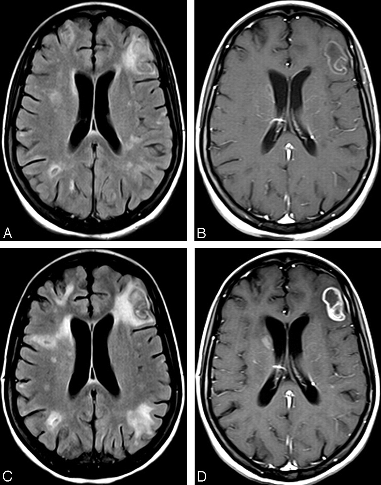
A, FLAIR image at admission shows multiple areas of high signal intensity, some with a central area of low signal intensity, located in the subcortical and periventricular white matter. B, T1-weighted image after gadolinium administration demonstrates mild ring enhancement in the left frontal lesion, as well as in the lesion in the head of the right caudate nucleus. C, FLAIR image 1 month later shows significant increase in the size of the areas of high signal intensity in the white matter. D, T1-weighted image after gadolinium administration demonstrates stronger enhancement in the lesions at the left frontal lobe and right caudate nucleus, as well as new nodular areas of enhancement bilaterally.

One month after the beginning of treatment for cerebral toxoplasmosis, she presented with worsening of clinical signs and symptoms. A new brain MR imaging revealed enlargement of most of the lesions, mainly with perilesional high signal intensity on FLAIR images, as well as stronger contrast enhancement (Fig 1 C, -D). At this time, the CD4 cell count increased to 495 cells/mL, and the HIV-1 RNA load decreased to 768 copies/mL. A stereotactic brain biopsy of the left frontal lobe lesion revealed reactive gliosis, collections of histiocytic giant multinucleated cells, and marked perivascular lymphocytic infiltrates with a predominance of CD8 lymphocytes. No fungal elements were identified on periodic-acid-Schiff or Grocott stains; no toxoplasma pseudocysts, tachyzoites, or cytopathic changes were seen in neuronal or glial cells suggestive of infection by CMV, HSV, or JCV. A culture for mycobacterial organisms or fungi was also negative. About a month after the biopsy, even without using steroids, the patient showed significant improvement in clinical status, fulfilling the diagnostic criteria for cerebral toxoplasmosis associated with IRIS.

IRIS is a paradoxic worsening or onset of systemic inflammatory clinical signs and symptoms among patients with HIV/AIDS. It occurs after the initiation of HAART, particularly in those patients with profound immune suppression, and can occur with or without a concurrent opportunistic infection. Whether elicited by an infectious or noninfectious agent, the presence of an antigenic stimulus for development of the syndrome appears necessary. This antigenic stimulus can be intact “clinically silent” organisms or dead organisms and their residual antigens.1–4 Conventional and advanced MR imaging techniques are useful in evaluating these phenomena. Typical MR imaging patterns of some infections can be modified during IRIS. The most frequent findings are a paradoxic increase in the size and number of lesions, perilesional edema, and greater enhancement in T1-weighted images post–gadolinium administration. A combination of clinical, laboratory, and MR imaging findings with follow-up studies is recommended in these patients. The use of MR imaging in association with clinical and laboratory data can reduce the number of unnecessary cerebral biopsies in these clinical scenarios.

References

  1. 1.↵
    1. Murdoch DM,
    2. Venter WD,
    3. Van Rie A,
    4. et al
    . Immune reconstitution inflammatory syndrome (IRIS): review of common infectious manifestations and treatment options. AIDS Res Ther 2007;4:9
  2. 2.↵
    1. Thurnher MM,
    2. Post MJ,
    3. Rieger A,
    4. et al
    . Initial and follow-up MR imaging findings in AIDS-related progressive multifocal leukoencephalopathy treated with highly active antiretroviral therapy. AJNR Am J Neuroradiol 2001;22:977–84
    Abstract/FREE Full Text
  3. 3.↵
    1. Pfeffer G,
    2. Prout A,
    3. Hooge J,
    4. et al
    . Biopsy-proven immune reconstitution syndrome in a patient with AIDS and cerebral toxoplasmosis. Neurology 2009;73:321–22
    FREE Full Text
  4. 4.↵
    1. Tremont-Lukats IW,
    2. Garciarena P,
    3. Juarbe R,
    4. et al
    . The immune inflammatory reconstitution syndrome and central nervous system toxoplasmosis. Ann Intern Med 2009;150:656–57
    CrossRefPubMed
  • Copyright © American Society of Neuroradiology
PreviousNext
Back to top

In this issue

American Journal of Neuroradiology: 31 (7)
American Journal of Neuroradiology
Vol. 31, Issue 7
1 Aug 2010
  • Table of Contents
  • Index by author
Advertisement
Print
Download PDF
Email Article

Thank you for your interest in spreading the word on American Journal of Neuroradiology.

NOTE: We only request your email address so that the person you are recommending the page to knows that you wanted them to see it, and that it is not junk mail. We do not capture any email address.

Enter multiple addresses on separate lines or separate them with commas.
Immune Reconstitution Inflammatory Syndrome and Cerebral Toxoplasmosis
(Your Name) has sent you a message from American Journal of Neuroradiology
(Your Name) thought you would like to see the American Journal of Neuroradiology web site.
CAPTCHA
This question is for testing whether or not you are a human visitor and to prevent automated spam submissions.
Cite this article
R.F. Cabral, P.R. Valle Bahia, E.L. Gasparetto, L. Chimelli
Immune Reconstitution Inflammatory Syndrome and Cerebral Toxoplasmosis
American Journal of Neuroradiology Aug 2010, 31 (7) E65-E69; DOI: 10.3174/ajnr.A2158

Citation Manager Formats

  • BibTeX
  • Bookends
  • EasyBib
  • EndNote (tagged)
  • EndNote 8 (xml)
  • Medlars
  • Mendeley
  • Papers
  • RefWorks Tagged
  • Ref Manager
  • RIS
  • Zotero
0 Responses
Respond to this article
Share
Bookmark this article
Immune Reconstitution Inflammatory Syndrome and Cerebral Toxoplasmosis
R.F. Cabral, P.R. Valle Bahia, E.L. Gasparetto, L. Chimelli
American Journal of Neuroradiology Aug 2010, 31 (7) E65-E69; DOI: 10.3174/ajnr.A2158
del.icio.us logo Twitter logo Facebook logo Mendeley logo
  • Tweet Widget
  • Facebook Like
  • Google Plus One
Purchase

Jump to section

  • Article
    • References
  • Figures & Data
  • Info & Metrics
  • Responses
  • References
  • PDF

Related Articles

  • No related articles found.
  • PubMed
  • Google Scholar

Cited By...

  • Coinfection of cerebral toxoplasmosis and neurosyphilis as the first manifestation of AIDS
  • Crossref
  • Google Scholar

This article has not yet been cited by articles in journals that are participating in Crossref Cited-by Linking.

More in this TOC Section

  • Letter to the Editor regarding “Automated Volumetric Software in Dementia: Help or Hindrance to the Neuroradiologist?”
  • Reply:
  • Brain AVM’s Nidus: What if We Hadn’t Understood Anything?
Show more LETTERS

Similar Articles

Advertisement

Indexed Content

  • Current Issue
  • Accepted Manuscripts
  • Article Preview
  • Past Issues
  • Editorials
  • Editor's Choice
  • Fellows' Journal Club
  • Letters to the Editor
  • Video Articles

Cases

  • Case Collection
  • Archive - Case of the Week
  • Archive - Case of the Month
  • Archive - Classic Case

More from AJNR

  • Trainee Corner
  • Imaging Protocols
  • MRI Safety Corner
  • Book Reviews

Multimedia

  • AJNR Podcasts
  • AJNR Scantastics

Resources

  • Turnaround Time
  • Submit a Manuscript
  • Submit a Video Article
  • Submit an eLetter to the Editor/Response
  • Manuscript Submission Guidelines
  • Statistical Tips
  • Fast Publishing of Accepted Manuscripts
  • Graphical Abstract Preparation
  • Imaging Protocol Submission
  • Evidence-Based Medicine Level Guide
  • Publishing Checklists
  • Author Policies
  • Become a Reviewer/Academy of Reviewers
  • News and Updates

About Us

  • About AJNR
  • Editorial Board
  • Editorial Board Alumni
  • Alerts
  • Permissions
  • Not an AJNR Subscriber? Join Now
  • Advertise with Us
  • Librarian Resources
  • Feedback
  • Terms and Conditions
  • AJNR Editorial Board Alumni

American Society of Neuroradiology

  • Not an ASNR Member? Join Now

© 2025 by the American Society of Neuroradiology All rights, including for text and data mining, AI training, and similar technologies, are reserved.
Print ISSN: 0195-6108 Online ISSN: 1936-959X

Powered by HighWire