
Article Figures & Data
Figures
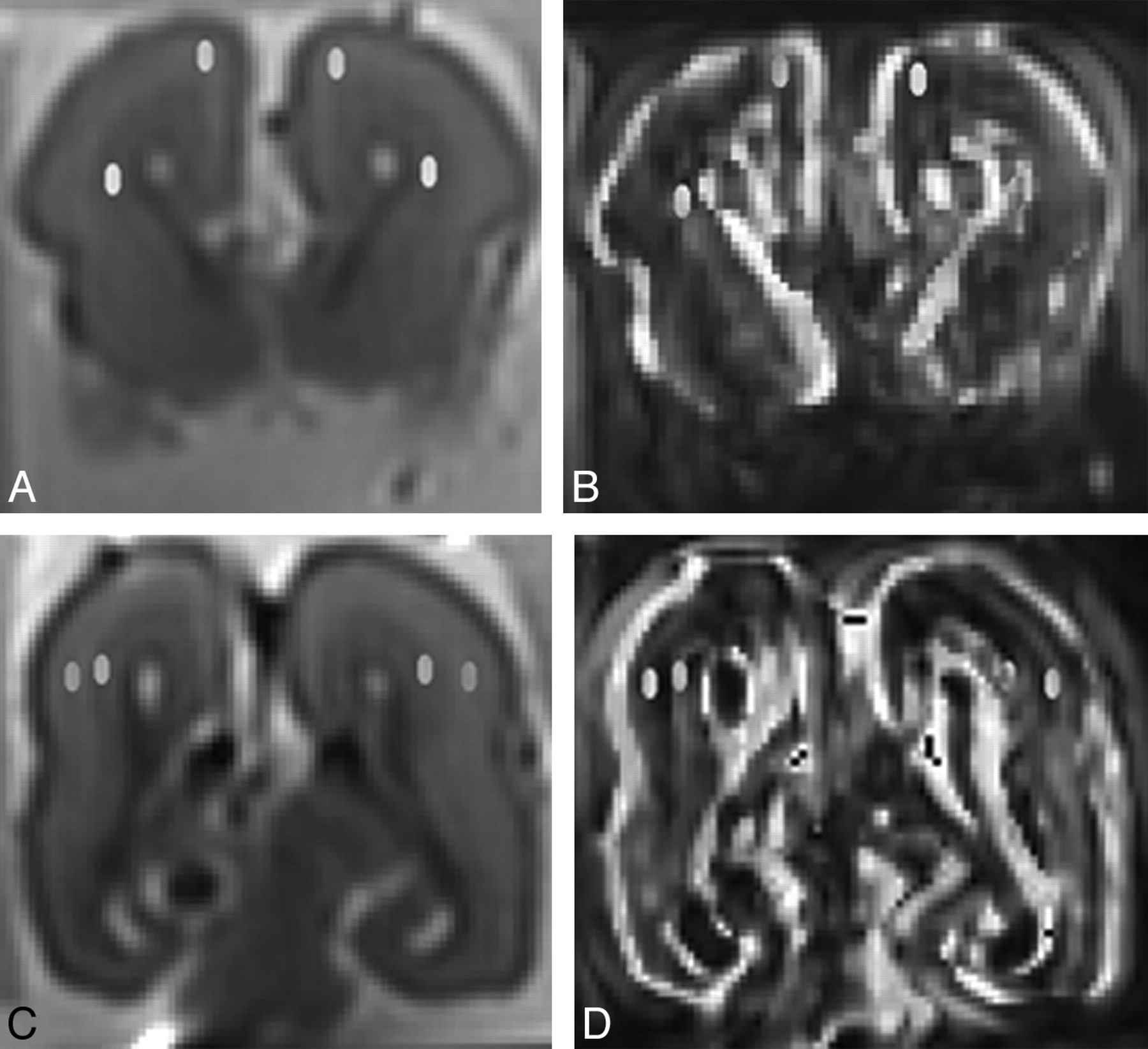
- Fig 1.
Postmortem coronal (A and C) b = 0 images and fractional anisotropy maps (B and D) demonstrating placement of regions of interest in the subplate layer and intermediate zone anteriorly (A and B) and posteriorly (C and D). The corpus callosum has been injured during autopsy.
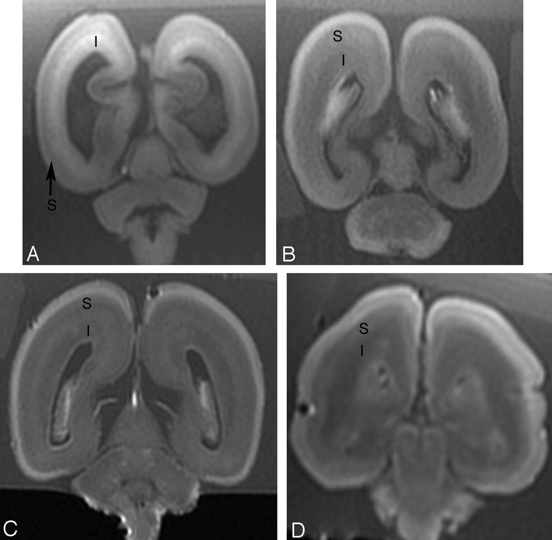
- Fig 2.
Postmortem coronal T1-weighted images at (A) 18 weeks, (B) 22 weeks, (C) 23 weeks, and (D) 25 weeks gestational age. At 18 gestational weeks, the intermediate zone (I) is of higher T1 signal intensity and the subplate layer (S) is of lower T1 signal intensity. With increasing gestational age, there is a reduction in the high T1 signal intensity of the intermediate zone and an increase in the signal intensity of the subplate layer. At approximately 22 weeks, the distinction between the intermediate zone and subplate layer is decreased on T1.
- Fig 3.
Postmortem coronal T2-weighted images at (A) 18 weeks, (B) 22 weeks, (C) 23 weeks, and (D) 25 weeks gestational age. At 18 gestational weeks, the intermediate zone (I) is of lower T2 signal intensity and the subplate layer (S) is of higher T2 signal intensity. With increasing gestational age, there is an increase in the T2 signal intensity of the intermediate zone, but the subplate layer remains of higher T2 signal intensity with no appreciable signal intensity alteration on visual assessment.
- Fig 4.
Scatterplots demonstrating fractional anisotropy of (A) subplate layer and (B) intermediate zone anteriorly and posteriorly at different gestational age.
- Fig 5.
Photomicrographs of fetal cerebrum at (A) 18, (B) 22, (C) 23, and (D) 25 gestational weeks. Images are composite photographs of a transect of the cerebral mantle, and the images have been rescaled for illustration purposes. All original photography is at ×12.5 original magnification, and the superficial aspect of the brain is on the left with the ventricle on the right. At 18 weeks, the intermediate zone has a very cellular superficial region, which is separated from the deeper aspects of the intermediate zone by the developing fiber systems. In contrast, the subplate layer has an abundant extracellular matrix at 18 weeks gestational age, which decreases with increasing gestation. As development progresses, the intermediate zone becomes less cellular, and the deep projecting fiber system becomes thicker such that the cellular superficial region of the intermediate zone becomes harder to distinguish from the subplate layer.
- Fig 6.
Whole mounts of fetal cerebrum at (A) 18 and (B) 25 gestational weeks stained with neurofilament to demonstrate axons and vimentin to demonstrate cell processes and radial glia. By 25 gestational weeks, the deep subplate merged with the intermediate zone with an increase in cell processes in both the subplate layer and intermediate zone. Neurofilament stain shows increased fibers (reddish brown) within the intermediate zone.
- Fig 7.
Whole mounts of fetal cerebrum at (A) 18 and (B) 25 gestational weeks stained with Alcian blue to demonstrate acid mucopolysaccharide, which forms most extracellular matrix in the developing brain. At 18 gestational weeks, acid mucopolysaccharide (blue) is abundant in the subplate layer but absent in the intermediate zone. By 25 gestational weeks, acid mucopolysaccharide is present in the intermediate zone.
- Fig 8.
Antenatal coronal T2-weighted images at (A) 20 weeks, (B) 22 weeks, (C) 23 weeks, and (D) 25 weeks gestational age. At 20 gestational weeks, the intermediate zone (I) is of lower T2 signal intensity and the subplate layer (S) is of higher T2 signal intensity. With increasing gestational age, there is an increase in the T2 signal intensity of the intermediate zone, but the subplate layer remains of higher T2 signal intensity with no appreciable signal intensity alteration on visual assessment.


Tables
Spearman correlation between signal intensity and fractional anisotropy of subplate layer and intermediate zone on postmortem MR imaging with gestational age, and also between signal intensity of the subplate layer and intermediate zone on antenatal MR imaging with gestational age
Spearman Correlation with Gestational Age Postmortem MR imaging Signal intensity on T1 Subplate layer r = 0.66, P = .012 Intermediate zone r = −0.78, P = .001 Signal intensity on T2 Subplate layer No correlation,a P > .05 Intermediate zone r = 0.48, P = .086 Fractional anisotropy Subplate layer r = 0.55, P < .001 Intermediate zone r = −0.64, P < .0001 Antenatal MR imaging Signal intensity on T2 Subplate layer No correlation,a P > .05 Intermediate zone r = 0.67, P = .003 -
a No measurable correlation, as signal intensity of the subplate layer remains constant.
-
In this issue


Jump to section
Related Articles
Cited By...
- Neurodevelopmental Patterns of Early Postnatal White Matter Maturation Represent Distinct Underlying Microstructure and Histology
- Does 3T Fetal MRI Improve Image Resolution of Normal Brain Structures between 20 and 24 Weeks' Gestational Age?
- Local Tissue Growth Patterns Underlying Normal Fetal Human Brain Gyrification Quantified In Utero
- Corroboration of Normal and Abnormal Fetal Cerebral Lamination on Postmortem MR Imaging with Postmortem Examination