Advertisement

Main menu

  • Home
  • Content
    • Current Issue
    • Accepted Manuscripts
    • Article Preview
    • Past Issue Archive
    • Video Articles
    • AJNR Case Collection
    • Case of the Week Archive
    • Case of the Month Archive
    • Classic Case Archive
  • Special Collections
    • Advancing NeuroMRI with High-Relaxivity Contrast Agents
    • AJNR Awards
    • Low-Field MRI
    • Alzheimer Disease
    • ASNR Foundation Special Collection
    • Photon-Counting CT
    • View All
  • Multimedia
    • AJNR Podcasts
    • AJNR SCANtastic
    • Trainee Corner
    • MRI Safety Corner
    • Imaging Protocols
  • For Authors
    • Submit a Manuscript
    • Submit a Video Article
    • Submit an eLetter to the Editor/Response
    • Manuscript Submission Guidelines
    • Statistical Tips
    • Fast Publishing of Accepted Manuscripts
    • Graphical Abstract Preparation
    • Imaging Protocol Submission
    • Author Policies
  • About Us
    • About AJNR
    • Editorial Board
    • Editorial Board Alumni
  • More
    • Become a Reviewer/Academy of Reviewers
    • Subscribers
    • Permissions
    • Alerts
    • Feedback
    • Advertisers
    • ASNR Home

User menu

  • Alerts
  • Log in

Search

  • Advanced search
American Journal of Neuroradiology

American Journal of Neuroradiology

ASHNR American Society of Functional Neuroradiology ASHNR American Society of Pediatric Neuroradiology ASSR
  • Alerts
  • Log in

Advanced Search

  • Home
  • Content
    • Current Issue
    • Accepted Manuscripts
    • Article Preview
    • Past Issue Archive
    • Video Articles
    • AJNR Case Collection
    • Case of the Week Archive
    • Case of the Month Archive
    • Classic Case Archive
  • Special Collections
    • Advancing NeuroMRI with High-Relaxivity Contrast Agents
    • AJNR Awards
    • Low-Field MRI
    • Alzheimer Disease
    • ASNR Foundation Special Collection
    • Photon-Counting CT
    • View All
  • Multimedia
    • AJNR Podcasts
    • AJNR SCANtastic
    • Trainee Corner
    • MRI Safety Corner
    • Imaging Protocols
  • For Authors
    • Submit a Manuscript
    • Submit a Video Article
    • Submit an eLetter to the Editor/Response
    • Manuscript Submission Guidelines
    • Statistical Tips
    • Fast Publishing of Accepted Manuscripts
    • Graphical Abstract Preparation
    • Imaging Protocol Submission
    • Author Policies
  • About Us
    • About AJNR
    • Editorial Board
    • Editorial Board Alumni
  • More
    • Become a Reviewer/Academy of Reviewers
    • Subscribers
    • Permissions
    • Alerts
    • Feedback
    • Advertisers
    • ASNR Home
  • Follow AJNR on Twitter
  • Visit AJNR on Facebook
  • Follow AJNR on Instagram
  • Join AJNR on LinkedIn
  • RSS Feeds

AJNR Awards, New Junior Editors, and more. Read the latest AJNR updates


Improved Turnaround Times | Median time to first decision: 12 days

Research ArticlePatient SafetyF

Dose Exposure of Patients Undergoing Comprehensive Stroke Imaging by Multidetector-Row CT: Comparison of 320-Detector Row and 64-Detector Row CT Scanners

S. Diekmann, E. Siebert, R. Juran, M. Roll, W. Deeg, H.-C. Bauknecht, F. Diekmann, R. Klingebiel and G. Bohner
American Journal of Neuroradiology June 2010, 31 (6) 1003-1009; DOI: https://doi.org/10.3174/ajnr.A1971
S. Diekmann
  • Find this author on Google Scholar
  • Find this author on PubMed
  • Search for this author on this site
E. Siebert
  • Find this author on Google Scholar
  • Find this author on PubMed
  • Search for this author on this site
R. Juran
  • Find this author on Google Scholar
  • Find this author on PubMed
  • Search for this author on this site
M. Roll
  • Find this author on Google Scholar
  • Find this author on PubMed
  • Search for this author on this site
W. Deeg
  • Find this author on Google Scholar
  • Find this author on PubMed
  • Search for this author on this site
H.-C. Bauknecht
  • Find this author on Google Scholar
  • Find this author on PubMed
  • Search for this author on this site
F. Diekmann
  • Find this author on Google Scholar
  • Find this author on PubMed
  • Search for this author on this site
R. Klingebiel
  • Find this author on Google Scholar
  • Find this author on PubMed
  • Search for this author on this site
G. Bohner
  • Find this author on Google Scholar
  • Find this author on PubMed
  • Search for this author on this site
  • Article
  • Figures & Data
  • Info & Metrics
  • Responses
  • References
  • PDF
Loading

Article Figures & Data

Figures

  • Tables
  • Fig 1.
    • Download figure
    • Open in new tab
    • Download powerpoint
    Fig 1.

    Alderson-Rando phantom. Numbers indicate the sections in which TLDs were placed (Table 1).
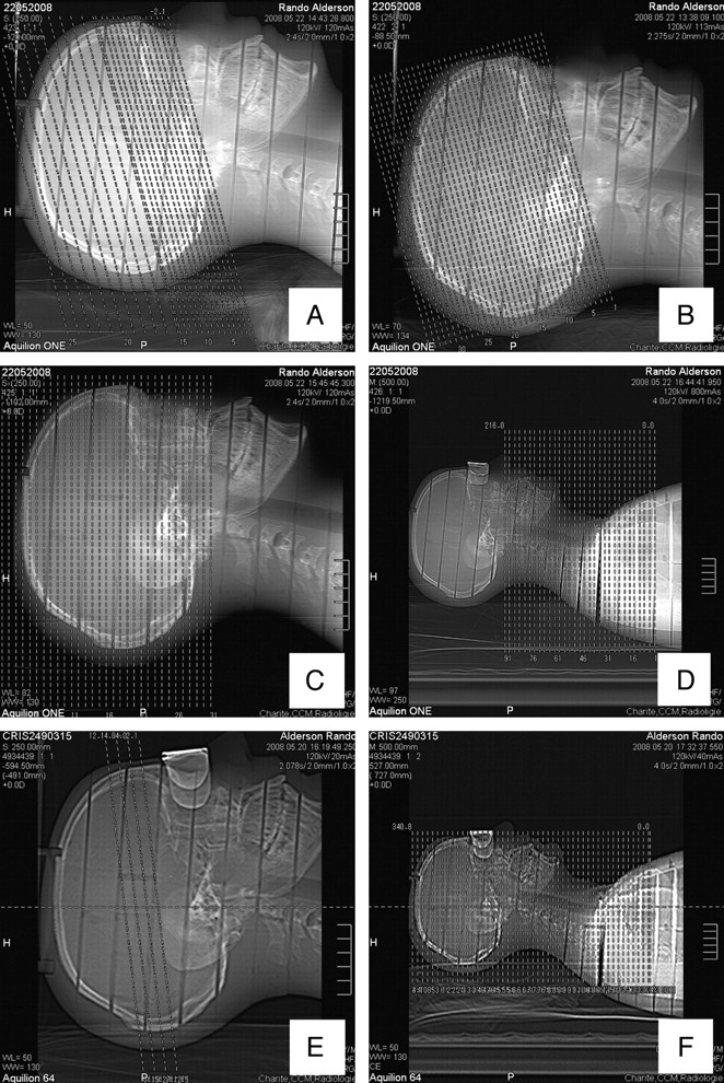
  • Fig 2.
    • Download figure
    • Open in new tab
    • Download powerpoint
    Fig 2.

    Scanograms of the 320-detector row CT scanner: A, incremental CCT; B, volume CCT; C, combo protocol; D, neck CT. Scanograms of the 64-detector row CT scanner: E, CTP; F, CTA (head and neck) (incremental CCT is analogous to A).

Tables

  • Figures
    • View popup
    Table 1:

    Doses measured for all protocols on 64-row scanner (Toshiba Aquilion 64)

    OrganW (T)Noncontrast Head CTPerfusionCTA
    Dose (mGy)Weighted Dose (mSv)Dose (mGy)Weighted Dose (mSv)Dose (mGy)Weighted Dose (mSv)
    Hemispheres0.0133.310.3366.220.6624.340.24
    Skin (within scan area)33.1775.4126.27
    Skin (total)0.0117.430.1737.90.3813.870.14
    Eye lenses41.696.7414.86
    Pituitary gland33.714.2620.07
    Bone marrow (scan area)8.44.122.05
    Bone marrow (total)0.124.60.562.360.2811.881.43
    Thyroid gland0.041.490.061.040.0439.171.57
    Lung0.120.80.10.480.062.990.36
    Esophagus0.040.360.010.550.022.780.11
    Breast0.121.690.20.390.051.470.18
    Liver0.050.370.020.280.010.940.05
    Stomach0.120.530.060.440.050.920.11
    Ovaries0.080.280.020.330.030.350.03
    Lower colon0.120.740.090.280.030.240.03
    Bladder0.040.730.030.340.010.570.02
    Testes0.080.310.020.270.020.280.02
    Effective dose (women)1.651.624.27
    Effective dose (men)1.651.614.26
    • View popup
    Table 2:

    Doses measured for all protocols on a 320-row scanner (Toshiba Aquilion ONE)

    OrganW (T)Noncontrast Head CT VolumeNoncontrast Head CT IncrementalCTP + CTA (Intractranial)CTA (Neck)
    Dose (mGy)Weighted Dose (mSv)Dose (mGy)Weighted Dose (mSv)Dose (mGy)Weighted Dose (mSv)Dose (mGy)Weighted Dose (mSv)
    Hemispheres0.0131.420.3141.910.4269.910.73.340.03
    Skin (scan area)36.8642.34923.84
    Skin (total)0.0119.190.1921.470.2146.490.4630.03
    Eye lenses40.2344.3144.012.39
    Pituitary gland25.2438.9352.695.3
    Bone marrow (scan area)7.6712.7730.1714.8
    Bone marrow (total)0.124.20.56.970.8415.811.98.571.03
    Thyroid gland0.042.540.13.390.145.370.2158.62.3
    Lung0.120.780.090.40.050.610.074.360.5
    Esophagus0.040.50.020.630.030.460.023.510.14
    Breast0.121.520.180.590.070.980.122.170.26
    Liver0.050.770.040.420.020.290.011.160.06
    Stomach0.120.630.080.30.040.290.031.180.14
    Ovaries0.080.480.041.010.080.420.030.360.03
    Lower colon0.120.460.060.430.510.230.030.290.03
    Bladder0.040.680.030.590.020.740.030.330.01
    Testes0.080.290.020.530.040.430.030.340.03
    Effective dose (women)1.642.433.614.56
    Effective dose (men)1.622.393.614.56
    • View popup
    Table 3:

    Parameters of the protocols used on the 64-row CT scanner

    ParameterNoncontrast Head CT ScanCTPCTA
    InfratentorialSupratentorial
    Lens protectionNoYesYes
    Collimation (mm)2 × 44 × 80.5 × 64
    Table increment (mm)8 mm8 mmNone20.5
    Pitch1No0,614
    Tube current (mAseff)37525050150
    Tube current (mA)25050300
    Voltage (kV)120120a120
    Rotation time1.5 s1.0 s1 s0.5 s
    Scan length (mm)14032340.8
    Total scan time24.8 s45.7 s23 s
    Gantry angulationYesYesNo
    • a The Aquilion 64 scanner at the Department of Radiology, Charité Campus Mitte (Berlin, Germany) was one of the first scanners of this type installed. Due to a technical limitation, this Aquilion 64 scanner does not support CTP with 80 kV. Newer versions of the Aquilion 64 scanner allow performing CTP with 80 kV in the same way as the Aquilion ONE does.

    • View popup
    Table 4:

    Parameters of the protocols used on the 320-row CT scanner

    ParameterNoncontrast Head CT Scan (Volume)Noncontrast Head CT (Incremental)Combo (Cerebral CTA + CTP)Neck CTA
    InfratentorialSupratentorial
    Lens protectionNoNoYesYes
    Collimation (mm)0.5 × 2802 × 42 × 40.5 × 3200.5 × 64
    Table increment (mm)No88none20.5
    Pitch11no0.614
    Tube current (mAseff)320375250100Tube current modulation
    Tube current (mA)320250100Tube current modulation
    Voltage (kV)12012080120
    Rotation time1 s1.5 s1 s1 s0.5 s
    Scan length (mm)140140160216
    Scan time7.2 s26.8 s29.8 s30.99 s
    Gantry angulationYesYesYesNoNo
PreviousNext
Back to top

In this issue

American Journal of Neuroradiology: 31 (6)
American Journal of Neuroradiology
Vol. 31, Issue 6
1 Jun 2010
  • Table of Contents
  • Index by author
Advertisement
Print
Download PDF
Email Article

Thank you for your interest in spreading the word on American Journal of Neuroradiology.

NOTE: We only request your email address so that the person you are recommending the page to knows that you wanted them to see it, and that it is not junk mail. We do not capture any email address.

Enter multiple addresses on separate lines or separate them with commas.
Dose Exposure of Patients Undergoing Comprehensive Stroke Imaging by Multidetector-Row CT: Comparison of 320-Detector Row and 64-Detector Row CT Scanners
(Your Name) has sent you a message from American Journal of Neuroradiology
(Your Name) thought you would like to see the American Journal of Neuroradiology web site.
CAPTCHA
This question is for testing whether or not you are a human visitor and to prevent automated spam submissions.
Cite this article
S. Diekmann, E. Siebert, R. Juran, M. Roll, W. Deeg, H.-C. Bauknecht, F. Diekmann, R. Klingebiel, G. Bohner
Dose Exposure of Patients Undergoing Comprehensive Stroke Imaging by Multidetector-Row CT: Comparison of 320-Detector Row and 64-Detector Row CT Scanners
American Journal of Neuroradiology Jun 2010, 31 (6) 1003-1009; DOI: 10.3174/ajnr.A1971

Citation Manager Formats

  • BibTeX
  • Bookends
  • EasyBib
  • EndNote (tagged)
  • EndNote 8 (xml)
  • Medlars
  • Mendeley
  • Papers
  • RefWorks Tagged
  • Ref Manager
  • RIS
  • Zotero
0 Responses
Respond to this article
Share
Bookmark this article
Dose Exposure of Patients Undergoing Comprehensive Stroke Imaging by Multidetector-Row CT: Comparison of 320-Detector Row and 64-Detector Row CT Scanners
S. Diekmann, E. Siebert, R. Juran, M. Roll, W. Deeg, H.-C. Bauknecht, F. Diekmann, R. Klingebiel, G. Bohner
American Journal of Neuroradiology Jun 2010, 31 (6) 1003-1009; DOI: 10.3174/ajnr.A1971
del.icio.us logo Twitter logo Facebook logo Mendeley logo
  • Tweet Widget
  • Facebook Like
  • Google Plus One
Purchase

Jump to section

  • Article
    • Abstract
    • Abbreviations
    • Materials and Methods
    • Results
    • Discussion
    • Conclusions
    • Footnotes
    • References
  • Figures & Data
  • Info & Metrics
  • Responses
  • References
  • PDF

Related Articles

  • No related articles found.
  • PubMed
  • Google Scholar

Cited By...

  • Clinical Applications of Conebeam CTP Imaging in Cerebral Disease: A Systematic Review
  • Latest generation of flat detector CT as a peri-interventional diagnostic tool: a comparative study with multidetector CT
  • Flat-detector computed tomography PBV map in the evaluation of presurgical embolization for hypervascular brain tumors
  • Measured Head CT/CTA Skin Dose and Intensive Care Unit Patient Cumulative Exposure
  • Effects of Radiation Exposure on the Cost-Effectiveness of CT Angiography and Perfusion Imaging in Aneurysmal Subarachnoid Hemorrhage
  • Perfusion Computed Tomography for the Evaluation of Acute Ischemic Stroke: Strengths and Pitfalls
  • Dynamic Angiography and Perfusion Imaging Using Flat Detector CT in the Angiography Suite: A Pilot Study in Patients with Acute Middle Cerebral Artery Occlusions
  • Whole-Brain Adaptive 70-kVp Perfusion Imaging with Variable and Extended Sampling Improves Quality and Consistency While Reducing Dose
  • Feasibility of Cerebral Blood Volume Mapping by Flat Panel Detector CT in the Angiography Suite: First Experience in Patients with Acute Middle Cerebral Artery Occlusions
  • CT Perfusion in Acute Ischemic Stroke: A Comparison of 2-Second and 1-Second Temporal Resolution
  • Crossref
  • Google Scholar

This article has not yet been cited by articles in journals that are participating in Crossref Cited-by Linking.

More in this TOC Section

  • Safety of Intrathecal Gadobutrol in Various Doses
  • Impact of Kidney Function on CNS Gadolinium Deposition in Patients Receiving Repeated Doses of Gadobutrol
  • Contrast-Induced Acute Kidney Injury in Radiologic Management of Acute Ischemic Stroke in the Emergency Setting
Show more Patient Safety

Similar Articles

Advertisement

Indexed Content

  • Current Issue
  • Accepted Manuscripts
  • Article Preview
  • Past Issues
  • Editorials
  • Editor's Choice
  • Fellows' Journal Club
  • Letters to the Editor
  • Video Articles

Cases

  • Case Collection
  • Archive - Case of the Week
  • Archive - Case of the Month
  • Archive - Classic Case

More from AJNR

  • Trainee Corner
  • Imaging Protocols
  • MRI Safety Corner
  • Book Reviews

Multimedia

  • AJNR Podcasts
  • AJNR Scantastics

Resources

  • Turnaround Time
  • Submit a Manuscript
  • Submit a Video Article
  • Submit an eLetter to the Editor/Response
  • Manuscript Submission Guidelines
  • Statistical Tips
  • Fast Publishing of Accepted Manuscripts
  • Graphical Abstract Preparation
  • Imaging Protocol Submission
  • Evidence-Based Medicine Level Guide
  • Publishing Checklists
  • Author Policies
  • Become a Reviewer/Academy of Reviewers
  • News and Updates

About Us

  • About AJNR
  • Editorial Board
  • Editorial Board Alumni
  • Alerts
  • Permissions
  • Not an AJNR Subscriber? Join Now
  • Advertise with Us
  • Librarian Resources
  • Feedback
  • Terms and Conditions
  • AJNR Editorial Board Alumni

American Society of Neuroradiology

  • Not an ASNR Member? Join Now

© 2025 by the American Society of Neuroradiology All rights, including for text and data mining, AI training, and similar technologies, are reserved.
Print ISSN: 0195-6108 Online ISSN: 1936-959X

Powered by HighWire