Advertisement
  • Main menu
  • User menu
  • Search
  • English ▼
    • English
    • Afrikaans
    • Albanian
    • Amharic
    • Arabic
    • Armenian
    • Azerbaijani
    • Basque
    • Belarusian
    • Bengali
    • Bosnian
    • Bulgarian
    • Catalan
    • Cebuano
    • Chichewa
    • Chinese (Simplified)
    • Chinese (Traditional)
    • Corsican
    • Croatian
    • Czech
    • Danish
    • Dutch
    • Esperanto
    • Estonian
    • Filipino
    • Finnish
    • French
    • Frisian
    • Galician
    • Georgian
    • German
    • Greek
    • Gujarati
    • Haitian Creole
    • Hausa
    • Hawaiian
    • Hebrew
    • Hindi
    • Hmong
    • Hungarian
    • Icelandic
    • Igbo
    • Indonesian
    • Irish
    • Italian
    • Japanese
    • Javanese
    • Kannada
    • Kazakh
    • Khmer
    • Korean
    • Kurdish (Kurmanji)
    • Kyrgyz
    • Lao
    • Latin
    • Latvian
    • Lithuanian
    • Luxembourgish
    • Macedonian
    • Malagasy
    • Malay
    • Malayalam
    • Maltese
    • Maori
    • Marathi
    • Mongolian
    • Myanmar (Burmese)
    • Nepali
    • Norwegian
    • Pashto
    • Persian
    • Polish
    • Portuguese
    • Punjabi
    • Romanian
    • Russian
    • Samoan
    • Scottish Gaelic
    • Serbian
    • Sesotho
    • Shona
    • Sindhi
    • Sinhala
    • Slovak
    • Slovenian
    • Somali
    • Spanish
    • Sudanese
    • Swahili
    • Swedish
    • Tajik
    • Tamil
    • Telugu
    • Thai
    • Turkish
    • Ukrainian
    • Urdu
    • Uzbek
    • Vietnamese
    • Welsh
    • Xhosa
    • Yiddish
    • Yoruba
    • Zulu

Main menu

  • Home
  • Content
    • Current Issue
    • Accepted Manuscripts
    • Article Preview
    • Past Issue Archive
    • Video Articles
    • AJNR Case Collection
    • Case of the Week Archive
    • Case of the Month Archive
    • Classic Case Archive
  • Special Collections
    • Advancing NeuroMRI with High-Relaxivity Contrast Agents
    • AJNR Awards
    • Low-Field MRI
    • Alzheimer Disease
    • ASNR Foundation Special Collection
    • Photon-Counting CT
    • View All
  • Multimedia
    • AJNR Podcasts
    • AJNR SCANtastic
    • Trainee Corner
    • MRI Safety Corner
    • Imaging Protocols
  • For Authors
    • Submit a Manuscript
    • Submit a Video Article
    • Submit an eLetter to the Editor/Response
    • Manuscript Submission Guidelines
    • Statistical Tips
    • Fast Publishing of Accepted Manuscripts
    • Graphical Abstract Preparation
    • Imaging Protocol Submission
    • Author Policies
  • About Us
    • About AJNR
    • Editorial Board
    • Editorial Board Alumni
  • More
    • Become a Reviewer/Academy of Reviewers
    • Subscribers
    • Permissions
    • Alerts
    • Feedback
    • Advertisers
    • ASNR Home

User menu

  • Alerts
  • Log in

Search

  • Advanced search
American Journal of Neuroradiology

American Journal of Neuroradiology

ASHNR American Society of Functional Neuroradiology ASHNR American Society of Pediatric Neuroradiology ASSR
  • Alerts
  • Log in

Advanced Search

English ▼
  • English
  • Afrikaans
  • Albanian
  • Amharic
  • Arabic
  • Armenian
  • Azerbaijani
  • Basque
  • Belarusian
  • Bengali
  • Bosnian
  • Bulgarian
  • Catalan
  • Cebuano
  • Chichewa
  • Chinese (Simplified)
  • Chinese (Traditional)
  • Corsican
  • Croatian
  • Czech
  • Danish
  • Dutch
  • Esperanto
  • Estonian
  • Filipino
  • Finnish
  • French
  • Frisian
  • Galician
  • Georgian
  • German
  • Greek
  • Gujarati
  • Haitian Creole
  • Hausa
  • Hawaiian
  • Hebrew
  • Hindi
  • Hmong
  • Hungarian
  • Icelandic
  • Igbo
  • Indonesian
  • Irish
  • Italian
  • Japanese
  • Javanese
  • Kannada
  • Kazakh
  • Khmer
  • Korean
  • Kurdish (Kurmanji)
  • Kyrgyz
  • Lao
  • Latin
  • Latvian
  • Lithuanian
  • Luxembourgish
  • Macedonian
  • Malagasy
  • Malay
  • Malayalam
  • Maltese
  • Maori
  • Marathi
  • Mongolian
  • Myanmar (Burmese)
  • Nepali
  • Norwegian
  • Pashto
  • Persian
  • Polish
  • Portuguese
  • Punjabi
  • Romanian
  • Russian
  • Samoan
  • Scottish Gaelic
  • Serbian
  • Sesotho
  • Shona
  • Sindhi
  • Sinhala
  • Slovak
  • Slovenian
  • Somali
  • Spanish
  • Sudanese
  • Swahili
  • Swedish
  • Tajik
  • Tamil
  • Telugu
  • Thai
  • Turkish
  • Ukrainian
  • Urdu
  • Uzbek
  • Vietnamese
  • Welsh
  • Xhosa
  • Yiddish
  • Yoruba
  • Zulu
  • Home
  • Content
    • Current Issue
    • Accepted Manuscripts
    • Article Preview
    • Past Issue Archive
    • Video Articles
    • AJNR Case Collection
    • Case of the Week Archive
    • Case of the Month Archive
    • Classic Case Archive
  • Special Collections
    • Advancing NeuroMRI with High-Relaxivity Contrast Agents
    • AJNR Awards
    • Low-Field MRI
    • Alzheimer Disease
    • ASNR Foundation Special Collection
    • Photon-Counting CT
    • View All
  • Multimedia
    • AJNR Podcasts
    • AJNR SCANtastic
    • Trainee Corner
    • MRI Safety Corner
    • Imaging Protocols
  • For Authors
    • Submit a Manuscript
    • Submit a Video Article
    • Submit an eLetter to the Editor/Response
    • Manuscript Submission Guidelines
    • Statistical Tips
    • Fast Publishing of Accepted Manuscripts
    • Graphical Abstract Preparation
    • Imaging Protocol Submission
    • Author Policies
  • About Us
    • About AJNR
    • Editorial Board
    • Editorial Board Alumni
  • More
    • Become a Reviewer/Academy of Reviewers
    • Subscribers
    • Permissions
    • Alerts
    • Feedback
    • Advertisers
    • ASNR Home
  • Follow AJNR on Twitter
  • Visit AJNR on Facebook
  • Follow AJNR on Instagram
  • Join AJNR on LinkedIn
  • RSS Feeds

AJNR Awards, New Junior Editors, and more. Read the latest AJNR updates


Improved Turnaround Times | Median time to first decision: 12 days

LetterLetter

Schimmelpenning Syndrome: A Kind of Craniofacial Epidermal Nevus Associated with Cerebral and Ocular MR Imaging Abnormalities

C. Amato, M. Elia and C. Schepis
American Journal of Neuroradiology May 2010, 31 (5) E47-E48; DOI: https://doi.org/10.3174/ajnr.A2062
C. Amato
  • Find this author on Google Scholar
  • Find this author on PubMed
  • Search for this author on this site
M. Elia
  • Find this author on Google Scholar
  • Find this author on PubMed
  • Search for this author on this site
C. Schepis
  • Find this author on Google Scholar
  • Find this author on PubMed
  • Search for this author on this site
  • Article
  • Figures & Data
  • Info & Metrics
  • Responses
  • References
  • PDF
Loading

Article Figures & Data

Figures

  • Fig 1.
    • Download figure
    • Open in new tab
    • Download powerpoint
    Fig 1.

    A, Patient at 10 years old with a large, tumefactive, and hyperkeratotic epidermal nevus involving the skull, face, and neck. B, Note lines of Blaschko on the head and neck. Modified from Moss et al.4
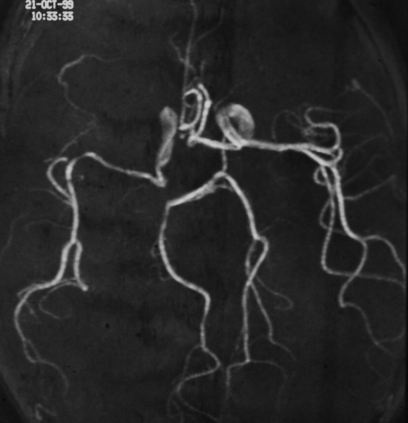
  • Fig 2.
    • Download figure
    • Open in new tab
    • Download powerpoint
    Fig 2.

    MR angiography of the patient at 2 years old shows a hypoplastic right siphon and middle cerebral artery, with a stenotic Sylvian bifurcation.
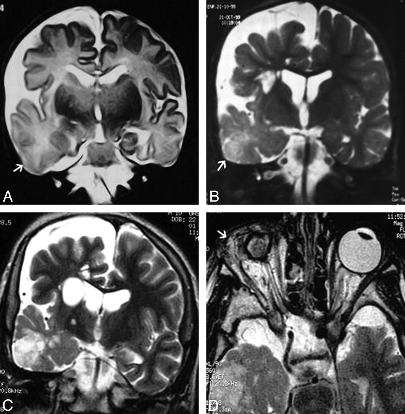
  • Fig 3.
    • Download figure
    • Open in new tab
    • Download powerpoint
    Fig 3.

    A, Coronal T2-weighted image of the patient at 20 days old shows a right cerebral hemiatrophy with cortical thinning and mild parenchymal change in the temporal lobe (arrow). On the same side, macrocrania and dilated liquoral spaces are present. B, Coronal T2-weighted image of the patient at 2 years old shows hypomyelination and an old infarct (arrow) in the right temporal lobe. Next to the ventricle, confluent and enlarged VRSs are recognizable. C, MR image of the patient at 10 years old. Coronal T2-weighted image shows, on the right side, stable cerebral hemiatrophy, a giant VRS next to the ventricle, and old ischemic changes within the hypomyelinated temporal lobe. D, High-resolution axial T2-weighted image shows a scleroatrophic right eyeball with a hypoplastic optic nerve. Lipodermoid tissue in the lacrimal fossa is present (arrow).

PreviousNext
Back to top

In this issue

American Journal of Neuroradiology: 31 (5)
American Journal of Neuroradiology
Vol. 31, Issue 5
1 May 2010
  • Table of Contents
  • Index by author
Advertisement
Print
Download PDF
Email Article
Cite this article
0 Responses
Respond to this article
Share
Bookmark this article
Schimmelpenning Syndrome: A Kind of Craniofacial Epidermal Nevus Associated with Cerebral and Ocular MR Imaging Abnormalities
C. Amato, M. Elia, C. Schepis
American Journal of Neuroradiology May 2010, 31 (5) E47-E48; DOI: 10.3174/ajnr.A2062
del.icio.us logo Twitter logo Facebook logo Mendeley logo
  • Tweet Widget
Purchase

Jump to section

  • Article
    • References
  • Figures & Data
  • Info & Metrics
  • Responses
  • References
  • PDF

Related Articles

  • No related articles found.
  • PubMed
  • Google Scholar

Cited By...

  • Schimmelpenning-Feuerstein-Mims syndrome with benign enlargement of subarachnoid space in infancy
  • Oral HRAS Mutation in Orofacial Nevus Sebaceous Syndrome (Schimmelpenning-Feuerstein-Mims-Syndrome): A Case Report With a Literature Survey
  • Crossref
  • Google Scholar

This article has not yet been cited by articles in journals that are participating in Crossref Cited-by Linking.

More in this TOC Section

  • Letter to the Editor regarding “Automated Volumetric Software in Dementia: Help or Hindrance to the Neuroradiologist?”
  • Reply:
  • Brain AVM’s Nidus: What if We Hadn’t Understood Anything?
Show more LETTERS

Similar Articles

Advertisement

Indexed Content

  • Current Issue
  • Accepted Manuscripts
  • Article Preview
  • Past Issues
  • Editorials
  • Editor's Choice
  • Fellows' Journal Club
  • Letters to the Editor
  • Video Articles

Cases

  • Case Collection
  • Archive - Case of the Week
  • Archive - Case of the Month
  • Archive - Classic Case

More from AJNR

  • Trainee Corner
  • Imaging Protocols
  • MRI Safety Corner
  • Book Reviews

Multimedia

  • AJNR Podcasts
  • AJNR Scantastics

Resources

  • Turnaround Time
  • Submit a Manuscript
  • Submit a Video Article
  • Submit an eLetter to the Editor/Response
  • Manuscript Submission Guidelines
  • Statistical Tips
  • Fast Publishing of Accepted Manuscripts
  • Graphical Abstract Preparation
  • Imaging Protocol Submission
  • Evidence-Based Medicine Level Guide
  • Publishing Checklists
  • Author Policies
  • Become a Reviewer/Academy of Reviewers
  • News and Updates

About Us

  • About AJNR
  • Editorial Board
  • Editorial Board Alumni
  • Alerts
  • Permissions
  • Not an AJNR Subscriber? Join Now
  • Advertise with Us
  • Librarian Resources
  • Feedback
  • Terms and Conditions
  • AJNR Editorial Board Alumni

American Society of Neuroradiology

  • Not an ASNR Member? Join Now

© 2025 by the American Society of Neuroradiology All rights, including for text and data mining, AI training, and similar technologies, are reserved.
Print ISSN: 0195-6108 Online ISSN: 1936-959X

Powered by HighWire
Email this Article

Thank you for your interest in spreading the word on American Journal of Neuroradiology.

NOTE: We only request your email address so that the person you are recommending the page to knows that you wanted them to see it, and that it is not junk mail. We do not capture any email address.

Enter multiple addresses on separate lines or separate them with commas.
Schimmelpenning Syndrome: A Kind of Craniofacial Epidermal Nevus Associated with Cerebral and Ocular MR Imaging Abnormalities
(Your Name) has sent you a message from American Journal of Neuroradiology
(Your Name) thought you would like to see the American Journal of Neuroradiology web site.
CAPTCHA
This question is for testing whether or not you are a human visitor and to prevent automated spam submissions.
Cite this article
C. Amato, M. Elia, C. Schepis
Schimmelpenning Syndrome: A Kind of Craniofacial Epidermal Nevus Associated with Cerebral and Ocular MR Imaging Abnormalities
American Journal of Neuroradiology May 2010, 31 (5) E47-E48; DOI: 10.3174/ajnr.A2062

Citation Manager Formats

  • BibTeX
  • Bookends
  • EasyBib
  • EndNote (tagged)
  • EndNote 8 (xml)
  • Medlars
  • Mendeley
  • Papers
  • RefWorks Tagged
  • Ref Manager
  • RIS
  • Zotero

We use cookies on this site to enhance your user experience. By clicking any link on this page you are giving your consent for us to set cookies.