Advertisement

Main menu

  • Home
  • Content
    • Current Issue
    • Accepted Manuscripts
    • Article Preview
    • Past Issue Archive
    • Video Articles
    • AJNR Case Collection
    • Case of the Week Archive
    • Case of the Month Archive
    • Classic Case Archive
  • Special Collections
    • AJNR Awards
    • Low-Field MRI
    • Alzheimer Disease
    • ASNR Foundation Special Collection
    • Photon-Counting CT
    • View All
  • Multimedia
    • AJNR Podcasts
    • AJNR SCANtastic
    • Trainee Corner
    • MRI Safety Corner
    • Imaging Protocols
  • For Authors
    • Submit a Manuscript
    • Submit a Video Article
    • Submit an eLetter to the Editor/Response
    • Manuscript Submission Guidelines
    • Statistical Tips
    • Fast Publishing of Accepted Manuscripts
    • Graphical Abstract Preparation
    • Imaging Protocol Submission
    • Author Policies
  • About Us
    • About AJNR
    • Editorial Board
    • Editorial Board Alumni
  • More
    • Become a Reviewer/Academy of Reviewers
    • Subscribers
    • Permissions
    • Alerts
    • Feedback
    • Advertisers
    • ASNR Home

User menu

  • Alerts
  • Log in

Search

  • Advanced search
American Journal of Neuroradiology

American Journal of Neuroradiology

ASHNR American Society of Functional Neuroradiology ASHNR American Society of Pediatric Neuroradiology ASSR
  • Alerts
  • Log in

Advanced Search

  • Home
  • Content
    • Current Issue
    • Accepted Manuscripts
    • Article Preview
    • Past Issue Archive
    • Video Articles
    • AJNR Case Collection
    • Case of the Week Archive
    • Case of the Month Archive
    • Classic Case Archive
  • Special Collections
    • AJNR Awards
    • Low-Field MRI
    • Alzheimer Disease
    • ASNR Foundation Special Collection
    • Photon-Counting CT
    • View All
  • Multimedia
    • AJNR Podcasts
    • AJNR SCANtastic
    • Trainee Corner
    • MRI Safety Corner
    • Imaging Protocols
  • For Authors
    • Submit a Manuscript
    • Submit a Video Article
    • Submit an eLetter to the Editor/Response
    • Manuscript Submission Guidelines
    • Statistical Tips
    • Fast Publishing of Accepted Manuscripts
    • Graphical Abstract Preparation
    • Imaging Protocol Submission
    • Author Policies
  • About Us
    • About AJNR
    • Editorial Board
    • Editorial Board Alumni
  • More
    • Become a Reviewer/Academy of Reviewers
    • Subscribers
    • Permissions
    • Alerts
    • Feedback
    • Advertisers
    • ASNR Home
  • Follow AJNR on Twitter
  • Visit AJNR on Facebook
  • Follow AJNR on Instagram
  • Join AJNR on LinkedIn
  • RSS Feeds

AJNR Awards, New Junior Editors, and more. Read the latest AJNR updates

Research ArticleBrain

Quantification of Cerebrovascular Reactivity by Blood Oxygen Level–Dependent MR Imaging and Correlation with Conventional Angiography in Patients with Moyamoya Disease

C. Heyn, J. Poublanc, A. Crawley, D. Mandell, J.S. Han, M. Tymianski, K. terBrugge, J.A. Fisher and D.J. Mikulis
American Journal of Neuroradiology May 2010, 31 (5) 862-867; DOI: https://doi.org/10.3174/ajnr.A1922
C. Heyn
  • Find this author on Google Scholar
  • Find this author on PubMed
  • Search for this author on this site
J. Poublanc
  • Find this author on Google Scholar
  • Find this author on PubMed
  • Search for this author on this site
A. Crawley
  • Find this author on Google Scholar
  • Find this author on PubMed
  • Search for this author on this site
D. Mandell
  • Find this author on Google Scholar
  • Find this author on PubMed
  • Search for this author on this site
J.S. Han
  • Find this author on Google Scholar
  • Find this author on PubMed
  • Search for this author on this site
M. Tymianski
  • Find this author on Google Scholar
  • Find this author on PubMed
  • Search for this author on this site
K. terBrugge
  • Find this author on Google Scholar
  • Find this author on PubMed
  • Search for this author on this site
J.A. Fisher
  • Find this author on Google Scholar
  • Find this author on PubMed
  • Search for this author on this site
D.J. Mikulis
  • Find this author on Google Scholar
  • Find this author on PubMed
  • Search for this author on this site
  • Article
  • Figures & Data
  • Info & Metrics
  • Responses
  • References
  • PDF
Loading

Article Figures & Data

Figures

  • Tables
  • Fig 1.
    • Download figure
    • Open in new tab
    • Download powerpoint
    Fig 1.

    A 48-year-old woman who presented with headache. A, There is high-grade stenosis of the main trunk of the right MCA with “puff of smoke” vessels in the region of the stenotic trunk. B, The left ICA and branches are normal. Angiography indicates a Suzuki grade II right hemisphere and a Suzuki grade 0 left hemisphere. The arrows indicate surface collaterals extending from the right ACA to the right MCA territory. C, The CVR map shows absence of reactivity to the CO2 stimulus (no color) in the right parietal lobe, corresponding to the region of the angiographic collaterals.
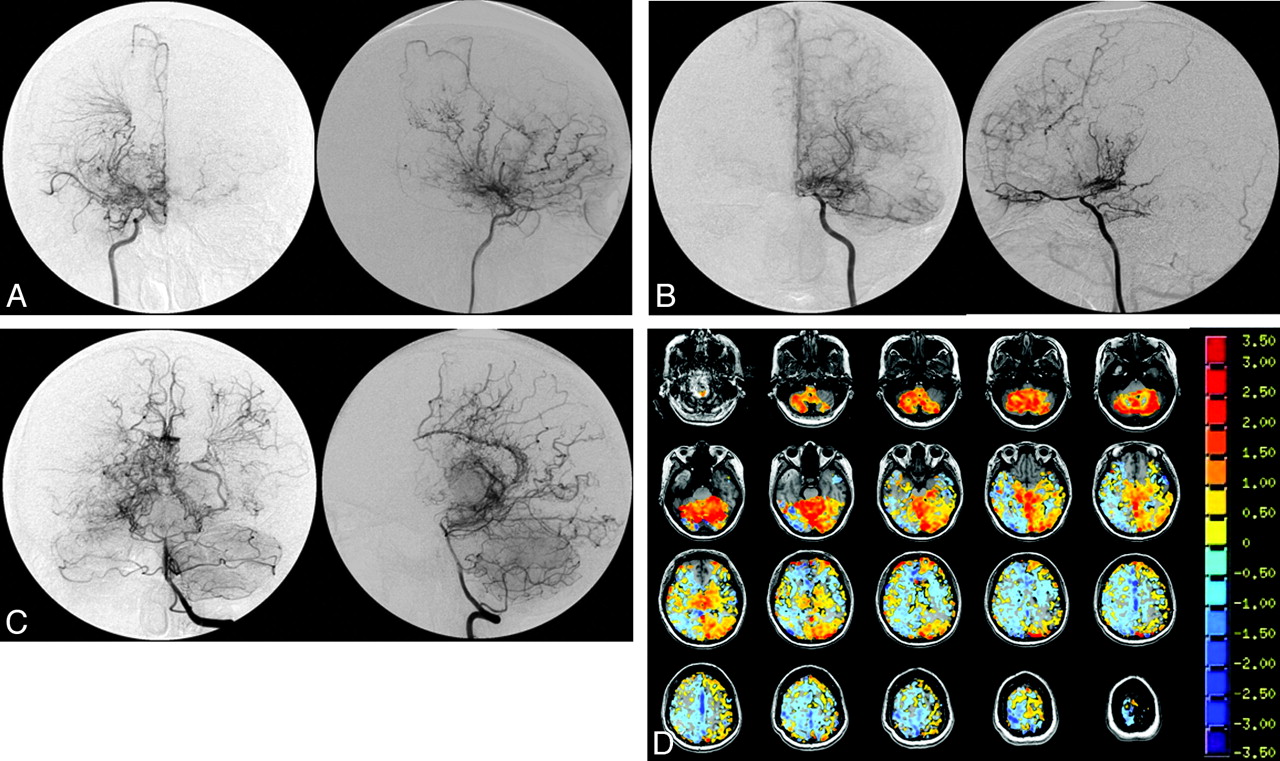
  • Fig 2.
    • Download figure
    • Open in new tab
    • Download powerpoint
    Fig 2.

    A 35-year-old woman who presented with TIA consisting of left hemiparesis. A−C, The angiograms indicate severe stenosis/occlusion of the distal ICAs bilaterally, with extensive Moyamoya vessels consistent with Suzuki grade IV bilaterally. The left vertebral injection (C) shows extensive pial collateralization from the posterior circulation to the ACA and MCA territories. However, the right hemisphere is not as well supplied as the left. D, This finding is supported by the CVR map, which shows a greater extent of steal phenomena (blue pixels) in the right hemisphere compared with the left. Notice the preservation of reactivity in the relatively well-supplied cerebellum.

  • Fig 3.
    • Download figure
    • Open in new tab
    • Download powerpoint
    Fig 3.

    Relationship between Mean CVRcombined versus disease severity as measured by the modified Suzuki score. A, Mean CVRcombined versus the Suzuki score for the MCA territory. B, Mean CVRcombined versus the Suzuki score for the ACA territory.
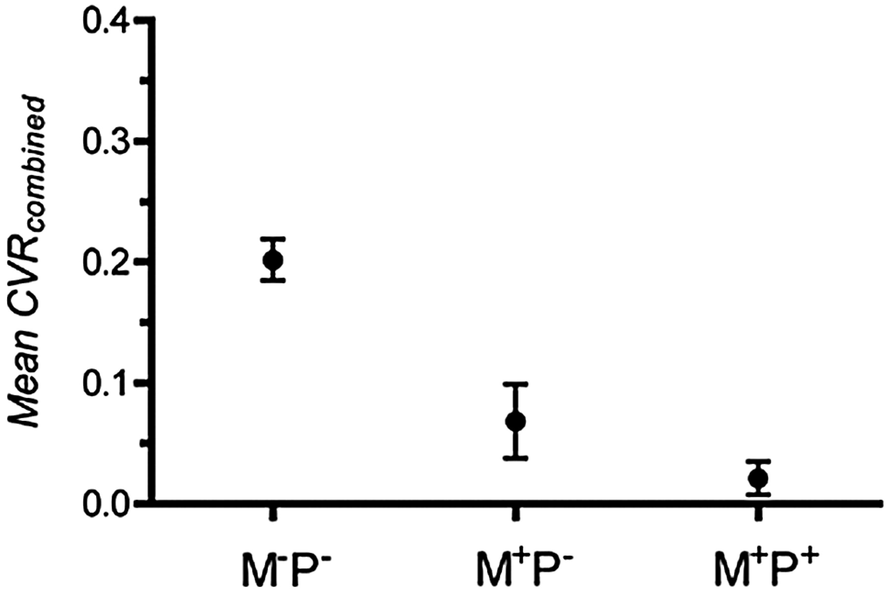
  • Fig 4.
    • Download figure
    • Open in new tab
    • Download powerpoint
    Fig 4.

    Correlation between Mean CVRcombined for MCA and ACA territories and the presence of pial or Moyamoya collaterals. M-P- indicates the absence of Moyamoya or pial collaterals, M+P− indicates the presence of Moyamoya collaterals but the absence of pial collaterals, and M+P+ indicates the presence of Moyamoya and pial collaterals.

  • Fig 5.
    • Download figure
    • Open in new tab
    • Download powerpoint
    Fig 5.

    Relationship between Mean CVRpos, Mean CVRcombined, Mean CVRneg, and the degree of vascular steal (fneg) for all ACA, MCA, and PCA territories. Closed circles represent vascular territories with a Suzuki score of 0, and open circles, Suzuki >0. A, Mean CVRpos versus fneg. B, Mean CVRcombined versus fneg. An exponential decay has been fitted to the data. The dotted lines indicate the 95% confidence band. C, Mean CVRneg versus fneg.

Tables

  • Figures
  • Clinical and angiographic characteristics of 11 patientsa

    Case No.Age (yr)SexClinical PresentationSuzukiCollaterals ACACollaterals MCACollaterals PCA
    LeftRightLeftRightLeftRightLeftRight
    148FHeadache0III000200
    236MHeadache0III000200
    345MTIA (left weakness)IIIV220200
    439FTIA (right weakness)II0002000
    533FHeadacheIIII211100
    635FTIA (left weakness)IVIV221111
    744MRight facial, arm numbness, slurred speechIV0002000
    841MTIA (right weakness)IVIII012200
    910FAsymptomaticIV0102000
    1011FTIA (right hand weakness)IVIV222200
    1133FAsymptomaticIIIII222201
    • a 0 indicates no collaterals (M−P−); 1, Moyamoya collaterals only (M+P−); 2, pial and Moyamoya collaterals (M+P+).

PreviousNext
Back to top

In this issue

American Journal of Neuroradiology: 31 (5)
American Journal of Neuroradiology
Vol. 31, Issue 5
1 May 2010
  • Table of Contents
  • Index by author
Advertisement
Print
Download PDF
Email Article

Thank you for your interest in spreading the word on American Journal of Neuroradiology.

NOTE: We only request your email address so that the person you are recommending the page to knows that you wanted them to see it, and that it is not junk mail. We do not capture any email address.

Enter multiple addresses on separate lines or separate them with commas.
Quantification of Cerebrovascular Reactivity by Blood Oxygen Level–Dependent MR Imaging and Correlation with Conventional Angiography in Patients with Moyamoya Disease
(Your Name) has sent you a message from American Journal of Neuroradiology
(Your Name) thought you would like to see the American Journal of Neuroradiology web site.
CAPTCHA
This question is for testing whether or not you are a human visitor and to prevent automated spam submissions.
Cite this article
C. Heyn, J. Poublanc, A. Crawley, D. Mandell, J.S. Han, M. Tymianski, K. terBrugge, J.A. Fisher, D.J. Mikulis
Quantification of Cerebrovascular Reactivity by Blood Oxygen Level–Dependent MR Imaging and Correlation with Conventional Angiography in Patients with Moyamoya Disease
American Journal of Neuroradiology May 2010, 31 (5) 862-867; DOI: 10.3174/ajnr.A1922

Citation Manager Formats

  • BibTeX
  • Bookends
  • EasyBib
  • EndNote (tagged)
  • EndNote 8 (xml)
  • Medlars
  • Mendeley
  • Papers
  • RefWorks Tagged
  • Ref Manager
  • RIS
  • Zotero
0 Responses
Respond to this article
Share
Bookmark this article
Quantification of Cerebrovascular Reactivity by Blood Oxygen Level–Dependent MR Imaging and Correlation with Conventional Angiography in Patients with Moyamoya Disease
C. Heyn, J. Poublanc, A. Crawley, D. Mandell, J.S. Han, M. Tymianski, K. terBrugge, J.A. Fisher, D.J. Mikulis
American Journal of Neuroradiology May 2010, 31 (5) 862-867; DOI: 10.3174/ajnr.A1922
del.icio.us logo Twitter logo Facebook logo Mendeley logo
  • Tweet Widget
  • Facebook Like
  • Google Plus One
Purchase

Jump to section

  • Article
    • Abstract
    • Abbreviations
    • Materials and Methods
    • Results
    • Discussion
    • Conclusions
    • Footnotes
    • References
  • Figures & Data
  • Info & Metrics
  • Responses
  • References
  • PDF

Related Articles

  • No related articles found.
  • PubMed
  • Google Scholar

Cited By...

  • Predicting Ischemic Risk Using Blood Oxygen Level-Dependent MRI in Children with Moyamoya
  • Breath-Hold Blood Oxygen Level-Dependent MRI: A Tool for the Assessment of Cerebrovascular Reserve in Children with Moyamoya Disease
  • Cerebrovascular Reactivity Mapping: An Evolving Standard for Clinical Functional Imaging
  • Novel MRI Approaches for Assessing Cerebral Hemodynamics in Ischemic Cerebrovascular Disease
  • Impact of Extracranial-Intracranial Bypass on Cerebrovascular Reactivity and Clinical Outcome in Patients With Symptomatic Moyamoya Vasculopathy
  • Neurosurgical Advances in the Treatment of Moyamoya Disease
  • Crossref (62)
  • Google Scholar

This article has been cited by the following articles in journals that are participating in Crossref Cited-by Linking.

  • Reliable quantification of BOLD fMRI cerebrovascular reactivity despite poor breath-hold performance
    Molly G. Bright, Kevin Murphy
    NeuroImage 2013 83
  • Low Frequency Systemic Hemodynamic “Noise” in Resting State BOLD fMRI: Characteristics, Causes, Implications, Mitigation Strategies, and Applications
    Yunjie Tong, Lia M. Hocke, Blaise B. Frederick
    Frontiers in Neuroscience 2019 13
  • Neurosurgical Advances in the Treatment of Moyamoya Disease
    Paritosh Pandey, Gary K. Steinberg
    Stroke 2011 42 11
  • Cerebrovascular Reactivity Mapping: An Evolving Standard for Clinical Functional Imaging
    J. J. Pillai, D. J. Mikulis
    American Journal of Neuroradiology 2015 36 1
  • Cerebrovascular reactivity mapping in patients with low grade gliomas undergoing presurgical sensorimotor mapping with BOLD fMRI
    Domenico Zacà, Jorge Jovicich, Sreenivasan R. Nadar, James T. Voyvodic, Jay J. Pillai
    Journal of Magnetic Resonance Imaging 2014 40 2
  • Impaired peri-nidal cerebrovascular reserve in seizure patients with brain arteriovenous malformations
    Jorn Fierstra, John Conklin, Timo Krings, Marat Slessarev, Jay S. Han, Joseph A. Fisher, Karel terBrugge, M. Christopher Wallace, Michael Tymianski, David J. Mikulis
    Brain 2011 134 1
  • Impact of Extracranial–Intracranial Bypass on Cerebrovascular Reactivity and Clinical Outcome in Patients With Symptomatic Moyamoya Vasculopathy
    Jay S. Han, Amal Abou-Hamden, Daniel M. Mandell, Julien Poublanc, Adrian P. Crawley, Joseph A. Fisher, David J. Mikulis, Michael Tymianski
    Stroke 2011 42 11
  • Investigating the non-linearity of the BOLD cerebrovascular reactivity response to targeted hypo/hypercapnia at 7T
    Alex A. Bhogal, Jeroen C.W. Siero, Joseph A. Fisher, Martijn Froeling, Peter Luijten, Marielle Philippens, Hans Hoogduin
    NeuroImage 2014 98
  • Relationships between hypercarbic reactivity, cerebral blood flow, and arterial circulation times in patients with moyamoya disease
    Manus J. Donahue, Michael Ayad, Ryan Moore, Matthias van Osch, Robert Singer, Paul Clemmons, Megan Strother
    Journal of Magnetic Resonance Imaging 2013 38 5
  • Measuring vascular reactivity with breath‐holds after stroke: A method to aid interpretation of group‐level BOLD signal changes in longitudinal fMRI studies
    Fatemeh Geranmayeh, Richard J.S. Wise, Robert Leech, Kevin Murphy
    Human Brain Mapping 2015 36 5

More in this TOC Section

  • Multimodal CT Provides Improved Performance for Lacunar Infarct Detection
  • Optimal MRI Sequence for Identifying Occlusion Location in Acute Stroke: Which Value of Time-Resolved Contrast-Enhanced MRA?
  • Evaluating the Effects of White Matter Multiple Sclerosis Lesions on the Volume Estimation of 6 Brain Tissue Segmentation Methods
Show more BRAIN

Similar Articles

Advertisement

Indexed Content

  • Current Issue
  • Accepted Manuscripts
  • Article Preview
  • Past Issues
  • Editorials
  • Editor's Choice
  • Fellows' Journal Club
  • Letters to the Editor
  • Video Articles

Cases

  • Case Collection
  • Archive - Case of the Week
  • Archive - Case of the Month
  • Archive - Classic Case

More from AJNR

  • Trainee Corner
  • Imaging Protocols
  • MRI Safety Corner
  • Book Reviews

Multimedia

  • AJNR Podcasts
  • AJNR Scantastics

Resources

  • Turnaround Time
  • Submit a Manuscript
  • Submit a Video Article
  • Submit an eLetter to the Editor/Response
  • Manuscript Submission Guidelines
  • Statistical Tips
  • Fast Publishing of Accepted Manuscripts
  • Graphical Abstract Preparation
  • Imaging Protocol Submission
  • Evidence-Based Medicine Level Guide
  • Publishing Checklists
  • Author Policies
  • Become a Reviewer/Academy of Reviewers
  • News and Updates

About Us

  • About AJNR
  • Editorial Board
  • Editorial Board Alumni
  • Alerts
  • Permissions
  • Not an AJNR Subscriber? Join Now
  • Advertise with Us
  • Librarian Resources
  • Feedback
  • Terms and Conditions
  • AJNR Editorial Board Alumni

American Society of Neuroradiology

  • Not an ASNR Member? Join Now

© 2025 by the American Society of Neuroradiology All rights, including for text and data mining, AI training, and similar technologies, are reserved.
Print ISSN: 0195-6108 Online ISSN: 1936-959X

Powered by HighWire