Advertisement
  • Main menu
  • User menu
  • Search
  • English ▼
    • English
    • Afrikaans
    • Albanian
    • Amharic
    • Arabic
    • Armenian
    • Azerbaijani
    • Basque
    • Belarusian
    • Bengali
    • Bosnian
    • Bulgarian
    • Catalan
    • Cebuano
    • Chichewa
    • Chinese (Simplified)
    • Chinese (Traditional)
    • Corsican
    • Croatian
    • Czech
    • Danish
    • Dutch
    • Esperanto
    • Estonian
    • Filipino
    • Finnish
    • French
    • Frisian
    • Galician
    • Georgian
    • German
    • Greek
    • Gujarati
    • Haitian Creole
    • Hausa
    • Hawaiian
    • Hebrew
    • Hindi
    • Hmong
    • Hungarian
    • Icelandic
    • Igbo
    • Indonesian
    • Irish
    • Italian
    • Japanese
    • Javanese
    • Kannada
    • Kazakh
    • Khmer
    • Korean
    • Kurdish (Kurmanji)
    • Kyrgyz
    • Lao
    • Latin
    • Latvian
    • Lithuanian
    • Luxembourgish
    • Macedonian
    • Malagasy
    • Malay
    • Malayalam
    • Maltese
    • Maori
    • Marathi
    • Mongolian
    • Myanmar (Burmese)
    • Nepali
    • Norwegian
    • Pashto
    • Persian
    • Polish
    • Portuguese
    • Punjabi
    • Romanian
    • Russian
    • Samoan
    • Scottish Gaelic
    • Serbian
    • Sesotho
    • Shona
    • Sindhi
    • Sinhala
    • Slovak
    • Slovenian
    • Somali
    • Spanish
    • Sudanese
    • Swahili
    • Swedish
    • Tajik
    • Tamil
    • Telugu
    • Thai
    • Turkish
    • Ukrainian
    • Urdu
    • Uzbek
    • Vietnamese
    • Welsh
    • Xhosa
    • Yiddish
    • Yoruba
    • Zulu

Main menu

  • Home
  • Content
    • Current Issue
    • Accepted Manuscripts
    • Article Preview
    • Past Issue Archive
    • Video Articles
    • AJNR Case Collection
    • Case of the Week Archive
    • Case of the Month Archive
    • Classic Case Archive
  • Special Collections
    • AJNR Awards
    • Low-Field MRI
    • Alzheimer Disease
    • ASNR Foundation Special Collection
    • Photon-Counting CT
    • View All
  • Multimedia
    • AJNR Podcasts
    • AJNR SCANtastic
    • Trainee Corner
    • MRI Safety Corner
    • Imaging Protocols
  • For Authors
    • Submit a Manuscript
    • Submit a Video Article
    • Submit an eLetter to the Editor/Response
    • Manuscript Submission Guidelines
    • Statistical Tips
    • Fast Publishing of Accepted Manuscripts
    • Graphical Abstract Preparation
    • Imaging Protocol Submission
    • Author Policies
  • About Us
    • About AJNR
    • Editorial Board
    • Editorial Board Alumni
  • More
    • Become a Reviewer/Academy of Reviewers
    • Subscribers
    • Permissions
    • Alerts
    • Feedback
    • Advertisers
    • ASNR Home

User menu

  • Alerts
  • Log in

Search

  • Advanced search
American Journal of Neuroradiology

American Journal of Neuroradiology

ASHNR American Society of Functional Neuroradiology ASHNR American Society of Pediatric Neuroradiology ASSR
  • Alerts
  • Log in

Advanced Search

English ▼
  • English
  • Afrikaans
  • Albanian
  • Amharic
  • Arabic
  • Armenian
  • Azerbaijani
  • Basque
  • Belarusian
  • Bengali
  • Bosnian
  • Bulgarian
  • Catalan
  • Cebuano
  • Chichewa
  • Chinese (Simplified)
  • Chinese (Traditional)
  • Corsican
  • Croatian
  • Czech
  • Danish
  • Dutch
  • Esperanto
  • Estonian
  • Filipino
  • Finnish
  • French
  • Frisian
  • Galician
  • Georgian
  • German
  • Greek
  • Gujarati
  • Haitian Creole
  • Hausa
  • Hawaiian
  • Hebrew
  • Hindi
  • Hmong
  • Hungarian
  • Icelandic
  • Igbo
  • Indonesian
  • Irish
  • Italian
  • Japanese
  • Javanese
  • Kannada
  • Kazakh
  • Khmer
  • Korean
  • Kurdish (Kurmanji)
  • Kyrgyz
  • Lao
  • Latin
  • Latvian
  • Lithuanian
  • Luxembourgish
  • Macedonian
  • Malagasy
  • Malay
  • Malayalam
  • Maltese
  • Maori
  • Marathi
  • Mongolian
  • Myanmar (Burmese)
  • Nepali
  • Norwegian
  • Pashto
  • Persian
  • Polish
  • Portuguese
  • Punjabi
  • Romanian
  • Russian
  • Samoan
  • Scottish Gaelic
  • Serbian
  • Sesotho
  • Shona
  • Sindhi
  • Sinhala
  • Slovak
  • Slovenian
  • Somali
  • Spanish
  • Sudanese
  • Swahili
  • Swedish
  • Tajik
  • Tamil
  • Telugu
  • Thai
  • Turkish
  • Ukrainian
  • Urdu
  • Uzbek
  • Vietnamese
  • Welsh
  • Xhosa
  • Yiddish
  • Yoruba
  • Zulu
  • Home
  • Content
    • Current Issue
    • Accepted Manuscripts
    • Article Preview
    • Past Issue Archive
    • Video Articles
    • AJNR Case Collection
    • Case of the Week Archive
    • Case of the Month Archive
    • Classic Case Archive
  • Special Collections
    • AJNR Awards
    • Low-Field MRI
    • Alzheimer Disease
    • ASNR Foundation Special Collection
    • Photon-Counting CT
    • View All
  • Multimedia
    • AJNR Podcasts
    • AJNR SCANtastic
    • Trainee Corner
    • MRI Safety Corner
    • Imaging Protocols
  • For Authors
    • Submit a Manuscript
    • Submit a Video Article
    • Submit an eLetter to the Editor/Response
    • Manuscript Submission Guidelines
    • Statistical Tips
    • Fast Publishing of Accepted Manuscripts
    • Graphical Abstract Preparation
    • Imaging Protocol Submission
    • Author Policies
  • About Us
    • About AJNR
    • Editorial Board
    • Editorial Board Alumni
  • More
    • Become a Reviewer/Academy of Reviewers
    • Subscribers
    • Permissions
    • Alerts
    • Feedback
    • Advertisers
    • ASNR Home
  • Follow AJNR on Twitter
  • Visit AJNR on Facebook
  • Follow AJNR on Instagram
  • Join AJNR on LinkedIn
  • RSS Feeds

AJNR Awards, New Junior Editors, and more. Read the latest AJNR updates

Research ArticleBrain
Open Access

Pitfalls in the Use of Voxel-Based Morphometry as a Biomarker: Examples from Huntington Disease

S.M.D. Henley, G.R. Ridgway, R.I. Scahill, S. Klöppel, S.J. Tabrizi, N.C. Fox, J. Kassubek and for the EHDN Imaging Working Group
American Journal of Neuroradiology April 2010, 31 (4) 711-719; DOI: https://doi.org/10.3174/ajnr.A1939
S.M.D. Henley
  • Find this author on Google Scholar
  • Find this author on PubMed
  • Search for this author on this site
G.R. Ridgway
  • Find this author on Google Scholar
  • Find this author on PubMed
  • Search for this author on this site
R.I. Scahill
  • Find this author on Google Scholar
  • Find this author on PubMed
  • Search for this author on this site
S. Klöppel
  • Find this author on Google Scholar
  • Find this author on PubMed
  • Search for this author on this site
S.J. Tabrizi
  • Find this author on Google Scholar
  • Find this author on PubMed
  • Search for this author on this site
N.C. Fox
  • Find this author on Google Scholar
  • Find this author on PubMed
  • Search for this author on this site
J. Kassubek
  • Find this author on Google Scholar
  • Find this author on PubMed
  • Search for this author on this site
  • Article
  • Figures & Data
  • Supplemental
  • Info & Metrics
  • Responses
  • References
  • PDF
Loading

Article Figures & Data

Figures

  • Tables
  • Fig 1.
    • Download figure
    • Open in new tab
    • Download powerpoint
    Fig 1.

    Effect of type and level of statistical correction. All SPMs show the same contrast: regions in which the early HD group has reduced GM volume relative to controls (this is true throughout the article unless otherwise stated). SPMs are smoothed at 4-mm FWHM. The 3 SPMs in the top panel show various levels of FWE correction, and the 3 SPMs below show various levels of uncorrected SPMs. The color bar shows the t value and is applicable to all figures in this article.

  • Fig 2.
    • Download figure
    • Open in new tab
    • Download powerpoint
    Fig 2.

    Effect of using modulated or unmodulated data. Both SPMs show the same contrast of early HD versus controls, corrected at FWE P < .05, smoothed at 4-mm FWHM.

  • Fig 3.
    • Download figure
    • Open in new tab
    • Download powerpoint
    Fig 3.

    Effect of smoothing kernel size. All SPMs show early HD versus controls, corrected at FWE P < .05. The SPMs are smoothed at 4-, 6-, and 8-mm FWHM.
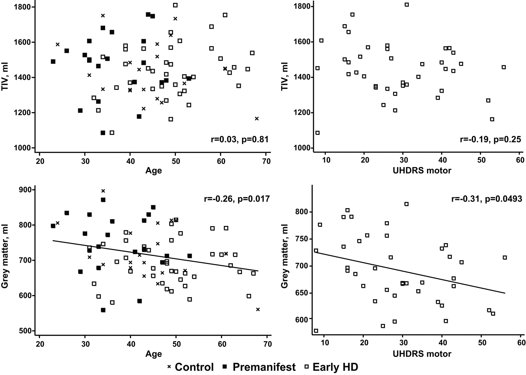
  • Fig 4.
    • Download figure
    • Open in new tab
    • Download powerpoint
    Fig 4.

    Graphs demonstrate how TIV and total GM volume vary with age and motor score (an index of HD severity). The top 2 graphs show that the relationship between TIV and both age and motor score is small and not statistically significant. The bottom 2 graphs show that total GM volume decreases with age (r = −0.26, P = .017) and motor score (r = −0.31, P = .0493).
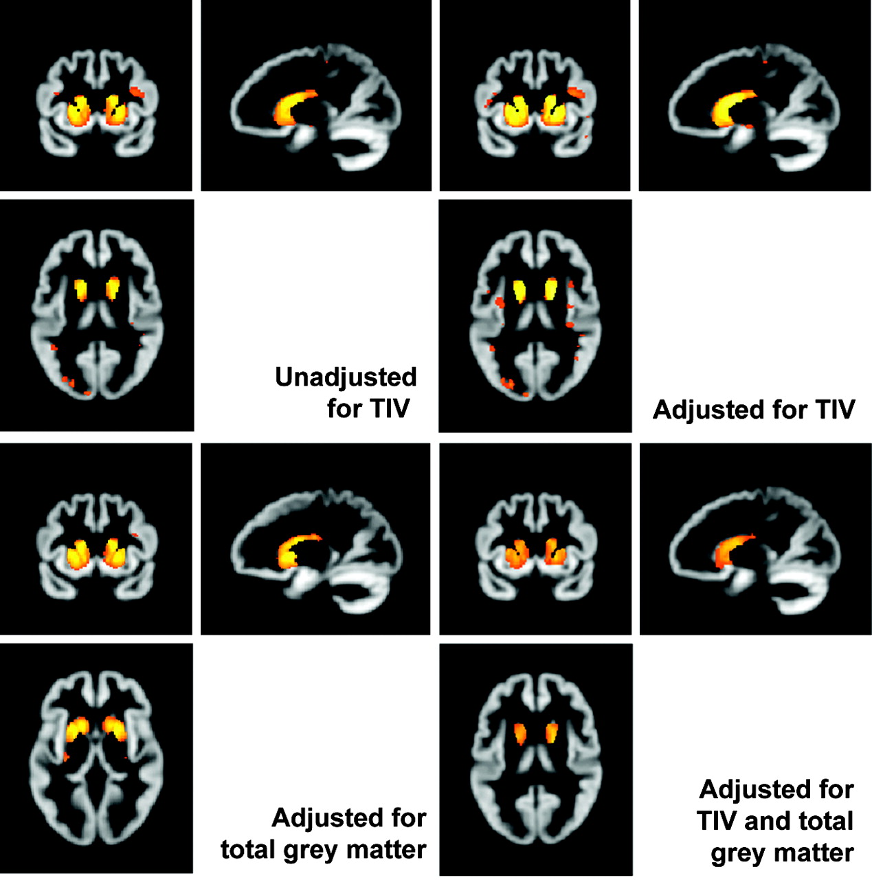
  • Fig 5.
    • Download figure
    • Open in new tab
    • Download powerpoint
    Fig 5.

    Effects of adjusting for TIV with and without including total GM volume. All SPMs show early HD versus controls, corrected at FWE P < .05, smoothed at 4-mm FWHM. The top row shows the effect of including or excluding TIV as a covariate. The bottom row shows the effect of adjusting for total GM volume with and without TIV.
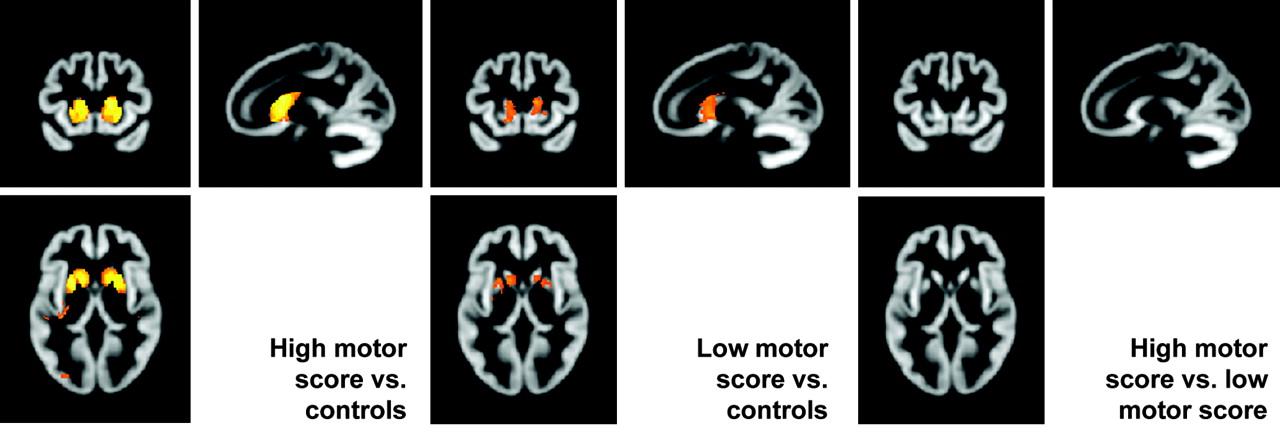
  • Fig 6.
    • Download figure
    • Open in new tab
    • Download powerpoint
    Fig 6.

    Subgroup analyses. The left SPM shows regions in which a group of high motor scorers have reduced GM volume relative to matched controls. The center SPM shows regions in which a group of low motor scorers have reduced GM volume relative to controls. The right SPM shows the results when the high and low motor scorer groups are compared directly.

Tables

  • Figures
    • View popup
    Table 1:

    Demographic dataa

    ControlPremanifestEarly HD
    (n = 20)(n = 21)(n = 40)
    Gender (M:F)7:1310:1120:20
    Age (yr)44.9 (10.5)37.2 (7.9)48.5 (9.6)
    CAG repeat lengthNA42.2 (1.8), range, 40–4543.7 (2.4), range, 40–50
    Predicted years to onsetbNA18.2 (7.1), range, 9–35NA
    Disease duration (yr since onset)NANA4.1 (2.6)
    UHDRS motorc1.1 (0.9)3.6 (4.0)28.9 (12.6)
    UHDRS independenced100 (0)100 (0)90.4 (9.6)
    UHDRS TFCe13 (0)13 (0)10.9 (1.8)
    • a Data are mean (SD) with the exception of gender and handedness.

    • b Onset was defined as a 60% chance of showing motor signs (a greater chance of showing signs than not, as described in Feigin et al30) and was predicted using the equation of Langbehn et al.31

    • c UHDRS motor is out of 124; higher score indicates more severely impaired.

    • d Independence is a percentage; higher score indicates better function.

    • e TFC is out of 13; higher score indicates better function.

    • View popup
    Table 2:

    Summary of processing methods used by other groupsa

    StudySPM VersionNormalizationSegmentationMod.Smoothing FWHM (mm)Correction
    Thieben et al3299Study-specific GM template, patients and controlsUnspecifiedYes10SPM uncorrected, p < .005; reported results mostly small-volume-corrected
    Ho et al3399Study-specific GM template, all controls onlyStudy-specific GM template, all controls onlyYes12SPM and reported results uncorrected, P < .0001; cluster 10 voxels
    Kassubek et al1899Study-specific template, whole-brain or GM unspecified; subjects unspecifiedUnspecifiedNo6SPM and reported results FWE P < .001; clusters 54 voxels
    Kassubek et al3499Study-specific template, 50:50 patients:controls; whole-brain or GM unspecifiedStudy-specific template, 50:50 patients:controlsYes6SPM and reported results FWE P < .001
    Peinemann et al2699Study-specific template, whole-brain or GM only unspecified; subjects unspecifiedUnspecifiedNo6SPM and reported results FWE P < .05
    Douaud et al352Study-specific symmetric GM template, 50:50 patients:controls, from original and mid-plane-reflected imagesStudy-specific GM template, 50:50 patients:controls, from original and symmetric imagesYes8SPM and reported results FDR P < .01
    Barrios et al27Not specifiedStandard whole-brain templateNot specifiedNo4SPM and reported results uncorrected; P < .01, clusters >10 mm3
    Gavazzi et al362Study-specific GM template, subjects unspecifiedStudy-specific GM template, subjects unspecifiedYes10SPM and reported results corrected; P < .01 (type unspecified)
    Jech et al372Study-specific GM template, all patients; no controls in studyStudy-specific GM template, all patients; no controls in studyYes10SPM uncorrected P < .001; reported results uncorrected in striatum or rolandic area, P < .001, elsewhere, FDR P < .05
    Kipps et al382Study-specific template, patients and controls, exact makeup unspecifiedNot specifiedYes8Uncorrected, P < .05
    Mühlau et al392Study-specific prior probability maps, subjects and whether used for normalization as well as segmentation unspecifiedYes8SPM and reported results FWE P <.05, extent P <.05, clusters P < .001
    Mühlau et al252Study-specific prior probability maps, subjects and whether used for normalization as well as segmentation unspecifiedYes8SPM and reported results FWE P < .05, clusters P < .05
    Ruocco et al402Study-specific GM template, healthy volunteers otherwise unused in studyStudy-specific GM template, healthy volunteers otherwise unused in studyYes10SPM and reported results FDR P < .05
    Wolf et al412Study-specific whole-brain template, 50:50 patients:controlsStudy-specific GM templates, 50:50 patients:controlsYes8SPM not shown; reported results FWE P < .001
    Henley et al72Standard GM templateStandard GM templateYes8SPM and reported results small-volume corrected FDR P < .05
    Wolf et al422Study-specific whole-brain template, 50:50 patients:controlsStudy-specific GM templates, 50:50 patients:controlsYes8SPM and reported results FWE P < .001
    • a Studies are listed by year and then author.

PreviousNext
Back to top

In this issue

American Journal of Neuroradiology: 31 (4)
American Journal of Neuroradiology
Vol. 31, Issue 4
1 Apr 2010
  • Table of Contents
  • Index by author
Advertisement
Print
Download PDF
Email Article
Cite this article
0 Responses
Respond to this article
Share
Bookmark this article
Pitfalls in the Use of Voxel-Based Morphometry as a Biomarker: Examples from Huntington Disease
S.M.D. Henley, G.R. Ridgway, R.I. Scahill, S. Klöppel, S.J. Tabrizi, N.C. Fox, J. Kassubek, for the EHDN Imaging Working Group
American Journal of Neuroradiology Apr 2010, 31 (4) 711-719; DOI: 10.3174/ajnr.A1939
del.icio.us logo Twitter logo Facebook logo Mendeley logo
  • Tweet Widget
Purchase

Jump to section

  • Article
    • Abstract
    • Abbreviations
    • Materials and Methods
    • Results
    • Discussion
    • Conclusions
    • Acknowledgments
    • Appendix
    • Footnotes
    • References
  • Figures & Data
  • Supplemental
  • Info & Metrics
  • Responses
  • References
  • PDF

Related Articles

  • PubMed
  • Google Scholar

Cited By...

  • Widespread Focal Cortical Alterations in Autism Spectrum Disorder with Intellectual Disability Detected by Threshold-Free Cluster Enhancement
  • Grey matter volume increase following electroconvulsive therapy in patients with late life depression: a longitudinal MRI study
  • Brain Structure and Function in Patients after Metal-on-Metal Hip Resurfacing
  • The Impact of Lesion In-Painting and Registration Methods on Voxel-Based Morphometry in Detecting Regional Cerebral Gray Matter Atrophy in Multiple Sclerosis
  • Fully Automated Atlas-Based MR Imaging Volumetry in Huntington Disease, Compared with Manual Volumetry
  • Voxelwise Analysis of Diffusion Tensor Imaging and Structural MR Imaging in Patients with the m.3243A>G Mutation in Mitochondrial DNA
  • Crossref
  • Google Scholar

This article has not yet been cited by articles in journals that are participating in Crossref Cited-by Linking.

More in this TOC Section

  • Quiet PROPELLER MRI Techniques Match the Quality of Conventional PROPELLER Brain Imaging Techniques
  • Predictors of Reperfusion in Patients with Acute Ischemic Stroke
  • Enhanced Axonal Metabolism during Early Natalizumab Treatment in Relapsing-Remitting Multiple Sclerosis
Show more Brain

Similar Articles

Advertisement

Indexed Content

  • Current Issue
  • Accepted Manuscripts
  • Article Preview
  • Past Issues
  • Editorials
  • Editor's Choice
  • Fellows' Journal Club
  • Letters to the Editor
  • Video Articles

Cases

  • Case Collection
  • Archive - Case of the Week
  • Archive - Case of the Month
  • Archive - Classic Case

More from AJNR

  • Trainee Corner
  • Imaging Protocols
  • MRI Safety Corner
  • Book Reviews

Multimedia

  • AJNR Podcasts
  • AJNR Scantastics

Resources

  • Turnaround Time
  • Submit a Manuscript
  • Submit a Video Article
  • Submit an eLetter to the Editor/Response
  • Manuscript Submission Guidelines
  • Statistical Tips
  • Fast Publishing of Accepted Manuscripts
  • Graphical Abstract Preparation
  • Imaging Protocol Submission
  • Evidence-Based Medicine Level Guide
  • Publishing Checklists
  • Author Policies
  • Become a Reviewer/Academy of Reviewers
  • News and Updates

About Us

  • About AJNR
  • Editorial Board
  • Editorial Board Alumni
  • Alerts
  • Permissions
  • Not an AJNR Subscriber? Join Now
  • Advertise with Us
  • Librarian Resources
  • Feedback
  • Terms and Conditions
  • AJNR Editorial Board Alumni

American Society of Neuroradiology

  • Not an ASNR Member? Join Now

© 2025 by the American Society of Neuroradiology All rights, including for text and data mining, AI training, and similar technologies, are reserved.
Print ISSN: 0195-6108 Online ISSN: 1936-959X

Powered by HighWire
Email this Article

Thank you for your interest in spreading the word on American Journal of Neuroradiology.

NOTE: We only request your email address so that the person you are recommending the page to knows that you wanted them to see it, and that it is not junk mail. We do not capture any email address.

Enter multiple addresses on separate lines or separate them with commas.
Pitfalls in the Use of Voxel-Based Morphometry as a Biomarker: Examples from Huntington Disease
(Your Name) has sent you a message from American Journal of Neuroradiology
(Your Name) thought you would like to see the American Journal of Neuroradiology web site.
CAPTCHA
This question is for testing whether or not you are a human visitor and to prevent automated spam submissions.
Cite this article
S.M.D. Henley, G.R. Ridgway, R.I. Scahill, S. Klöppel, S.J. Tabrizi, N.C. Fox, J. Kassubek, for the EHDN Imaging Working Group
Pitfalls in the Use of Voxel-Based Morphometry as a Biomarker: Examples from Huntington Disease
American Journal of Neuroradiology Apr 2010, 31 (4) 711-719; DOI: 10.3174/ajnr.A1939

Citation Manager Formats

  • BibTeX
  • Bookends
  • EasyBib
  • EndNote (tagged)
  • EndNote 8 (xml)
  • Medlars
  • Mendeley
  • Papers
  • RefWorks Tagged
  • Ref Manager
  • RIS
  • Zotero

We use cookies on this site to enhance your user experience. By clicking any link on this page you are giving your consent for us to set cookies.