Advertisement

Main menu

  • Home
  • Content
    • Current Issue
    • Accepted Manuscripts
    • Article Preview
    • Past Issue Archive
    • Video Articles
    • AJNR Case Collection
    • Case of the Week Archive
    • Case of the Month Archive
    • Classic Case Archive
  • Special Collections
    • AJNR Awards
    • Low-Field MRI
    • Alzheimer Disease
    • ASNR Foundation Special Collection
    • Photon-Counting CT
    • View All
  • Multimedia
    • AJNR Podcasts
    • AJNR SCANtastic
    • Trainee Corner
    • MRI Safety Corner
    • Imaging Protocols
  • For Authors
    • Submit a Manuscript
    • Submit a Video Article
    • Submit an eLetter to the Editor/Response
    • Manuscript Submission Guidelines
    • Statistical Tips
    • Fast Publishing of Accepted Manuscripts
    • Graphical Abstract Preparation
    • Imaging Protocol Submission
    • Author Policies
  • About Us
    • About AJNR
    • Editorial Board
    • Editorial Board Alumni
  • More
    • Become a Reviewer/Academy of Reviewers
    • Subscribers
    • Permissions
    • Alerts
    • Feedback
    • Advertisers
    • ASNR Home

User menu

  • Alerts
  • Log in

Search

  • Advanced search
American Journal of Neuroradiology

American Journal of Neuroradiology

ASHNR American Society of Functional Neuroradiology ASHNR American Society of Pediatric Neuroradiology ASSR
  • Alerts
  • Log in

Advanced Search

  • Home
  • Content
    • Current Issue
    • Accepted Manuscripts
    • Article Preview
    • Past Issue Archive
    • Video Articles
    • AJNR Case Collection
    • Case of the Week Archive
    • Case of the Month Archive
    • Classic Case Archive
  • Special Collections
    • AJNR Awards
    • Low-Field MRI
    • Alzheimer Disease
    • ASNR Foundation Special Collection
    • Photon-Counting CT
    • View All
  • Multimedia
    • AJNR Podcasts
    • AJNR SCANtastic
    • Trainee Corner
    • MRI Safety Corner
    • Imaging Protocols
  • For Authors
    • Submit a Manuscript
    • Submit a Video Article
    • Submit an eLetter to the Editor/Response
    • Manuscript Submission Guidelines
    • Statistical Tips
    • Fast Publishing of Accepted Manuscripts
    • Graphical Abstract Preparation
    • Imaging Protocol Submission
    • Author Policies
  • About Us
    • About AJNR
    • Editorial Board
    • Editorial Board Alumni
  • More
    • Become a Reviewer/Academy of Reviewers
    • Subscribers
    • Permissions
    • Alerts
    • Feedback
    • Advertisers
    • ASNR Home
  • Follow AJNR on Twitter
  • Visit AJNR on Facebook
  • Follow AJNR on Instagram
  • Join AJNR on LinkedIn
  • RSS Feeds

AJNR Awards, New Junior Editors, and more. Read the latest AJNR updates

Review ArticleReview Articles
Open Access

Fluorodeoxyglucose–Positron-Emission Tomography Imaging of Head and Neck Squamous Cell Cancer

R.M. Subramaniam, M. Truong, P. Peller, O. Sakai and G. Mercier
American Journal of Neuroradiology April 2010, 31 (4) 598-604; DOI: https://doi.org/10.3174/ajnr.A1760
R.M. Subramaniam
  • Find this author on Google Scholar
  • Find this author on PubMed
  • Search for this author on this site
M. Truong
  • Find this author on Google Scholar
  • Find this author on PubMed
  • Search for this author on this site
P. Peller
  • Find this author on Google Scholar
  • Find this author on PubMed
  • Search for this author on this site
O. Sakai
  • Find this author on Google Scholar
  • Find this author on PubMed
  • Search for this author on this site
G. Mercier
  • Find this author on Google Scholar
  • Find this author on PubMed
  • Search for this author on this site
  • Article
  • Figures & Data
  • Info & Metrics
  • Responses
  • References
  • PDF
Loading

Article Figures & Data

Figures

  • Fig 1.
    • Download figure
    • Open in new tab
    • Download powerpoint
    Fig 1.

    Surgical planning and metabolic staging. A 50-year-old man with T4N2bM0 SCC of the right tonsil and base of the tongue. The contrast-enhanced CT component of the PET/DCT provides the high-resolution imaging necessary for surgical planning (red arrows), with the tumor involving the right tonsil and tongue base. The PET component of the PET/DCT provides the metabolic information about the primary tumor and locoregional and distant metastatic disease. The patient underwent resection with pectoralis flap reconstruction and concurrent chemoradiation. A and B, Contrast-enhanced CT of PET/CT. C−E, Fused PET/CT images. FDG uptake provides the primary tumor and metastatic lymph node metabolism (yellow arrows).

  • Fig 2.
    • Download figure
    • Open in new tab
    • Download powerpoint
    Fig 2.

    Detection of unknown primary tumor. A 58-year-old man who presented with a right neck mass. A neck CT shows multiple enlarged necrotic right level II, III, and IV lymph nodes. FNA of the right level III node showed a metastatic poorly differentiated carcinoma with extensive necrosis. PET/CT showed a 1.0 × 1.2 cm hypermetabolic (gold arrow) and enhancing (red arrow) lesion at the right base of the tongue, large nodes in the right levels II/III, an 8-mm node at level IV, and no left-sided nodes or distant metastases. Direct laryngoscopy with biopsy and cervical esophagoscopy showed no lesions, but palpation showed a 1-cm firm right base of tongue lesion not crossing the midline, which was pathologically proved to be SCC. PET/CT is useful in identifying the unknown primary tumor in approximately one-third of patients after physical examination, anatomic cross-sectional imaging, and endoscopy. A−C, Fused PET/CT images. D−F, Contrast-enhanced CT of the PET/CT.

  • Fig 3.
    • Download figure
    • Open in new tab
    • Download powerpoint
    Fig 3.

    Failure to detect an unknown primary tumor. A 50-year-old man who underwent a dental extraction in which the oral surgeon incidentally identified a right-sided neck mass that was tender to touch. FNA revealed moderate to poorly differentiated SCC. PET/DCT demonstrates FDG hypermetabolic (gold arrow) and enhancing (red arrow) lymphadenopathy in the right levels I-III and fails to reveal a primary lesion. Same-day rigid esophagoscopy, direct laryngoscopy, nasal endoscopy with biopsy of the base of the tongue bilaterally, and tonsillectomy showed no tumor. The patient was treated in all potential mucosal primary sites and the bilateral neck with IMRT to a dose of 69.96 Gy in 33 fractions with concurrent cisplatin. A, C, and D, Fused PET/CT images. B, Contrast-enhanced CT of the PET/CT.

  • Fig 4.
    • Download figure
    • Open in new tab
    • Download powerpoint
    Fig 4.

    Perineural spread. A 65-year-old woman diagnosed with T4N0M0 SCC of the skin of the left upper lip and perineural spread along infraorbital nerve and VI to the skull base. PET/DCT demonstrates a hypermetabolic and enhancing mass in the left upper lip extending along the infraorbital nerve and at the skull base (gold arrows). MR imaging demonstrates these findings (red arrows) with higher resolution. The perineural spread is better depicted on the MR imaging than PET/CT due to higher resolution and small-volume disease along the nerve. A, B, and D, PET/DCT fused images. C, Contrast-enhanced coronal CT scan. E and G, Coronal T1 pregadolinium images. F and H, Postgadolinium coronal T1 images.

  • Fig 5.
    • Download figure
    • Open in new tab
    • Download powerpoint
    Fig 5.

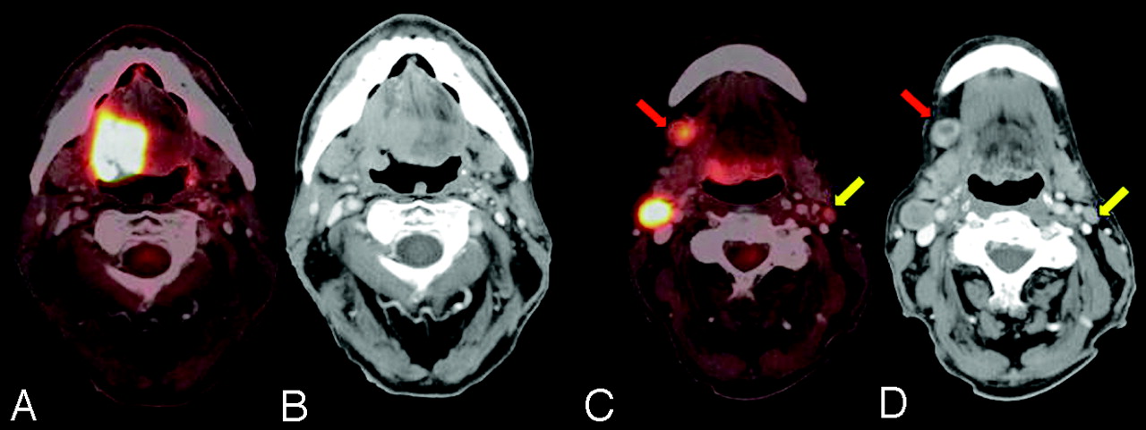
    Staging. A 62-year-old man with a stage IVa (T2 N2cM0) SCC of the tongue with a central necrotic right IB (red arrow) and right IIA (yellow arrow) FDG hypermetabolic lymphadenopathy. The left IIA lymph node (<10 mm) demonstrates FDG hypermetabolism, proved pathologically to be metastatic. Even smaller lymph nodes with apparent mild FDG uptake due to partial volume may harbor metastatic deposits.

  • Fig 6.
    • Download figure
    • Open in new tab
    • Download powerpoint
    Fig 6.

    Radiation therapy planning. A, Planning CT GTV in red and PTV in blue. Green and aqua contours represent elective nodal volumes. B, PET. C, Rigid fusion of planning CT and PET for radiation planning. The PET/CT scan was obtained in the treatment position with rigid immobilization in a thermoplastic head and neck mask.

  • Fig 7.
    • Download figure
    • Open in new tab
    • Download powerpoint
    Fig 7.

    Therapy assessment, complete response. A 39-year-old male patient with a stage IVa (T4N3M0) SCC of the right tonsil who underwent induction chemotherapy. A postinduction therapy PET/DCT demonstrates complete metabolic resolution of the primary tumor and nodal metastases. A and E, Maximum intensity projection views. B−D and F−H, Fused PET/CT images.

  • Fig 8.
    • Download figure
    • Open in new tab
    • Download powerpoint
    Fig 8.

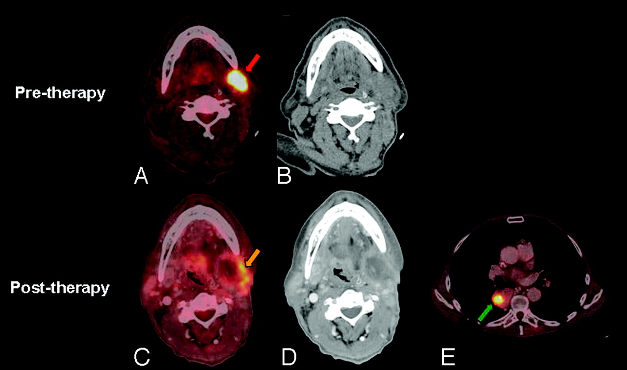
    Therapy assessment, partial response. A 67-year-old male patient with a history of recurrent SCC of the oral tongue, SCC of the midesophagus, and SCC of the left soft palate (T2N0M0) status postresection with negative margins, who presented with a recurrent left neck mass. The pretherapy PET/CT demonstrates FDG hypermetabolic left level I nodal recurrence (red arrow), and the patient completed chemoradiotherapy to the recurrent tumor in his left neck with IMRT to a dose of 70 Gy. A 12-week posttherapy PET/DCT scan demonstrates residual activity (gold arrow) in the periphery and central necrosis of the left neck nodal mass, representing partial response and a new recurrence of the esophageal malignancy (green arrow).

PreviousNext
Back to top

In this issue

American Journal of Neuroradiology: 31 (4)
American Journal of Neuroradiology
Vol. 31, Issue 4
1 Apr 2010
  • Table of Contents
  • Index by author
Advertisement
Print
Download PDF
Email Article

Thank you for your interest in spreading the word on American Journal of Neuroradiology.

NOTE: We only request your email address so that the person you are recommending the page to knows that you wanted them to see it, and that it is not junk mail. We do not capture any email address.

Enter multiple addresses on separate lines or separate them with commas.
Fluorodeoxyglucose–Positron-Emission Tomography Imaging of Head and Neck Squamous Cell Cancer
(Your Name) has sent you a message from American Journal of Neuroradiology
(Your Name) thought you would like to see the American Journal of Neuroradiology web site.
CAPTCHA
This question is for testing whether or not you are a human visitor and to prevent automated spam submissions.
Cite this article
R.M. Subramaniam, M. Truong, P. Peller, O. Sakai, G. Mercier
Fluorodeoxyglucose–Positron-Emission Tomography Imaging of Head and Neck Squamous Cell Cancer
American Journal of Neuroradiology Apr 2010, 31 (4) 598-604; DOI: 10.3174/ajnr.A1760

Citation Manager Formats

  • BibTeX
  • Bookends
  • EasyBib
  • EndNote (tagged)
  • EndNote 8 (xml)
  • Medlars
  • Mendeley
  • Papers
  • RefWorks Tagged
  • Ref Manager
  • RIS
  • Zotero
0 Responses
Respond to this article
Share
Bookmark this article
Fluorodeoxyglucose–Positron-Emission Tomography Imaging of Head and Neck Squamous Cell Cancer
R.M. Subramaniam, M. Truong, P. Peller, O. Sakai, G. Mercier
American Journal of Neuroradiology Apr 2010, 31 (4) 598-604; DOI: 10.3174/ajnr.A1760
del.icio.us logo Twitter logo Facebook logo Mendeley logo
  • Tweet Widget
  • Facebook Like
  • Google Plus One
Purchase

Jump to section

  • Article
    • Abstract
    • Abbreviations
    • PET/CT Protocols
    • Detection of Unknown Primary Tumor
    • Staging
    • Radiation-Therapy Planning
    • Evaluation of Therapy Response
    • Evaluation of Recurrence and Surveillance
    • References
  • Figures & Data
  • Info & Metrics
  • Responses
  • References
  • PDF

Related Articles

  • No related articles found.
  • PubMed
  • Google Scholar

Cited By...

  • An Imagers Guide to Perineural Tumor Spread in Head and Neck Cancers: Radiologic Footprints on 18F-FDG PET, with CT and MRI Correlates
  • Tumor Metabolic Features Identified by 18F-FDG PET Correlate with Gene Networks of Immune Cell Microenvironment in Head and Neck Cancer
  • Restricting carbohydrates to fight head and neck cancer--is this realistic?
  • PET/CT Imaging and Human Papilloma Virus-Positive Oropharyngeal Squamous Cell Cancer: Evolving Clinical Imaging Paradigm
  • Addition of 18F-FDG PET/CT to Clinical Assessment Predicts Overall Survival in HNSCC: A Retrospective Analysis with Follow-up for 12 Years
  • 18F-FDG Metabolic Tumor Volume and Total Glycolytic Activity of Oral Cavity and Oropharyngeal Squamous Cell Cancer: Adding Value to Clinical Staging
  • Biologic Imaging of Head and Neck Cancer: The Present and the Future
  • Crossref (94)
  • Google Scholar

This article has been cited by the following articles in journals that are participating in Crossref Cited-by Linking.

  • Cancer metabolism, stemness and tumor recurrence
    Joseph M. Curry, Madalina Tuluc, Diana Whitaker-Menezes, Julie A. Ames, Archana Anantharaman, Aileen Butera, Benjamin Leiby, David Cognetti, Federica Sotgia, Michael P. Lisanti, Ubaldo E. Martinez-Outschoorn
    Cell Cycle 2013 12 9
  • 18F-FDG Metabolic Tumor Volume and Total Glycolytic Activity of Oral Cavity and Oropharyngeal Squamous Cell Cancer: Adding Value to Clinical Staging
    Elizabeth H. Dibble, Ana C. Lara Alvarez, Minh-Tam Truong, Gustavo Mercier, Earl F. Cook, Rathan M. Subramaniam
    Journal of Nuclear Medicine 2012 53 5
  • FDG-PET/CT pitfalls in oncological head and neck imaging
    Bela S. Purohit, Angeliki Ailianou, Nicolas Dulguerov, Christoph D. Becker, Osman Ratib, Minerva Becker
    Insights into Imaging 2014 5 5
  • Clinical Practice in PET/CT for the Management of Head and Neck Squamous Cell Cancer
    Reema Goel, William Moore, Baran Sumer, Saad Khan, David Sher, Rathan M. Subramaniam
    American Journal of Roentgenology 2017 209 2
  • Posttreatment CT and MR Imaging in Head and Neck Cancer: What the Radiologist Needs to Know
    Naoko Saito, Rohini N. Nadgir, Mitsuhiko Nakahira, Masahiro Takahashi, Akira Uchino, Fumiko Kimura, Minh Tam Truong, Osamu Sakai
    RadioGraphics 2012 32 5
  • Perineural Spread in Head and Neck Malignancies: Clinical Significance and Evaluation with18F-FDG PET/CT
    Fabio M. Paes, Adam D. Singer, Adam N. Checkver, Ricardo A. Palmquist, Gabriela De La Vega, Charif Sidani
    RadioGraphics 2013 33 6
  • Diagnostic Accuracy of Follow-Up FDG PET or PET/CT in Patients With Head and Neck Cancer After Definitive Treatment: A Systematic Review and Meta-Analysis
    Sara Sheikhbahaei, Mehdi Taghipour, Rubina Ahmad, Carole Fakhry, Ana P. Kiess, Christine H. Chung, Rathan M. Subramaniam
    American Journal of Roentgenology 2015 205 3
  • Imaging in head and neck squamous cell carcinoma: the potential role of PET/MRI
    Minerva Becker, Habib Zaidi
    The British Journal of Radiology 2014 87 1036
  • PET/CT of Cancer Patients: Part 1, Pancreatic Neoplasms
    Elizabeth H. Dibble, Dimitrios Karantanis, Gustavo Mercier, Patrick J. Peller, Lisa A. Kachnic, Rathan M. Subramaniam
    American Journal of Roentgenology 2012 199 5
  • FDG PET/CT Imaging of Oropharyngeal Squamous Cell Carcinoma
    Abdel K. Tahari, Krishna C. Alluri, Harry Quon, Wayne Koch, Richard L. Wahl, Rathan M. Subramaniam
    Clinical Nuclear Medicine 2014 39 3

More in this TOC Section

  • An Atlas of Neonatal Neurovascular Imaging Anatomy as Depicted with Microvascular Imaging: The Intracranial Arteries
  • An Atlas of Neonatal Neurovascular Imaging Anatomy as Depicted with Microvascular Imaging: The Intracranial Veins
  • Clinical Translation of Hyperpolarized 13C Metabolic Probes for Glioma Imaging
Show more Review articles

Similar Articles

Advertisement

Indexed Content

  • Current Issue
  • Accepted Manuscripts
  • Article Preview
  • Past Issues
  • Editorials
  • Editor's Choice
  • Fellows' Journal Club
  • Letters to the Editor
  • Video Articles

Cases

  • Case Collection
  • Archive - Case of the Week
  • Archive - Case of the Month
  • Archive - Classic Case

More from AJNR

  • Trainee Corner
  • Imaging Protocols
  • MRI Safety Corner
  • Book Reviews

Multimedia

  • AJNR Podcasts
  • AJNR Scantastics

Resources

  • Turnaround Time
  • Submit a Manuscript
  • Submit a Video Article
  • Submit an eLetter to the Editor/Response
  • Manuscript Submission Guidelines
  • Statistical Tips
  • Fast Publishing of Accepted Manuscripts
  • Graphical Abstract Preparation
  • Imaging Protocol Submission
  • Evidence-Based Medicine Level Guide
  • Publishing Checklists
  • Author Policies
  • Become a Reviewer/Academy of Reviewers
  • News and Updates

About Us

  • About AJNR
  • Editorial Board
  • Editorial Board Alumni
  • Alerts
  • Permissions
  • Not an AJNR Subscriber? Join Now
  • Advertise with Us
  • Librarian Resources
  • Feedback
  • Terms and Conditions
  • AJNR Editorial Board Alumni

American Society of Neuroradiology

  • Not an ASNR Member? Join Now

© 2025 by the American Society of Neuroradiology All rights, including for text and data mining, AI training, and similar technologies, are reserved.
Print ISSN: 0195-6108 Online ISSN: 1936-959X

Powered by HighWire