Advertisement

Main menu

  • Home
  • Content
    • Current Issue
    • Accepted Manuscripts
    • Article Preview
    • Past Issue Archive
    • Video Articles
    • AJNR Case Collection
    • Case of the Week Archive
    • Case of the Month Archive
    • Classic Case Archive
  • Special Collections
    • AJNR Awards
    • Low-Field MRI
    • Alzheimer Disease
    • ASNR Foundation Special Collection
    • Photon-Counting CT
    • View All
  • Multimedia
    • AJNR Podcasts
    • AJNR SCANtastic
    • Trainee Corner
    • MRI Safety Corner
    • Imaging Protocols
  • For Authors
    • Submit a Manuscript
    • Submit a Video Article
    • Submit an eLetter to the Editor/Response
    • Manuscript Submission Guidelines
    • Statistical Tips
    • Fast Publishing of Accepted Manuscripts
    • Graphical Abstract Preparation
    • Imaging Protocol Submission
    • Author Policies
  • About Us
    • About AJNR
    • Editorial Board
    • Editorial Board Alumni
  • More
    • Become a Reviewer/Academy of Reviewers
    • Subscribers
    • Permissions
    • Alerts
    • Feedback
    • Advertisers
    • ASNR Home

User menu

  • Alerts
  • Log in

Search

  • Advanced search
American Journal of Neuroradiology

American Journal of Neuroradiology

ASHNR American Society of Functional Neuroradiology ASHNR American Society of Pediatric Neuroradiology ASSR
  • Alerts
  • Log in

Advanced Search

  • Home
  • Content
    • Current Issue
    • Accepted Manuscripts
    • Article Preview
    • Past Issue Archive
    • Video Articles
    • AJNR Case Collection
    • Case of the Week Archive
    • Case of the Month Archive
    • Classic Case Archive
  • Special Collections
    • AJNR Awards
    • Low-Field MRI
    • Alzheimer Disease
    • ASNR Foundation Special Collection
    • Photon-Counting CT
    • View All
  • Multimedia
    • AJNR Podcasts
    • AJNR SCANtastic
    • Trainee Corner
    • MRI Safety Corner
    • Imaging Protocols
  • For Authors
    • Submit a Manuscript
    • Submit a Video Article
    • Submit an eLetter to the Editor/Response
    • Manuscript Submission Guidelines
    • Statistical Tips
    • Fast Publishing of Accepted Manuscripts
    • Graphical Abstract Preparation
    • Imaging Protocol Submission
    • Author Policies
  • About Us
    • About AJNR
    • Editorial Board
    • Editorial Board Alumni
  • More
    • Become a Reviewer/Academy of Reviewers
    • Subscribers
    • Permissions
    • Alerts
    • Feedback
    • Advertisers
    • ASNR Home
  • Follow AJNR on Twitter
  • Visit AJNR on Facebook
  • Follow AJNR on Instagram
  • Join AJNR on LinkedIn
  • RSS Feeds

AJNR Awards, New Junior Editors, and more. Read the latest AJNR updates

LetterLetter

Unusual Extensive T1 Hyperintense Signals on MR Imaging in Neurocysticercosis

P. Lalitha and B. Reddy
American Journal of Neuroradiology March 2010, 31 (3) E33; DOI: https://doi.org/10.3174/ajnr.A1956
P. Lalitha
  • Find this author on Google Scholar
  • Find this author on PubMed
  • Search for this author on this site
B. Reddy
  • Find this author on Google Scholar
  • Find this author on PubMed
  • Search for this author on this site
  • Article
  • Figures & Data
  • Info & Metrics
  • Responses
  • References
  • PDF
Loading

Neurocysticercosis (NCC) is a common cause of morbidity in many developing countries and a common cause of seizure disorder in children and adults. The 4 stages of NCC are vesicular, vesicular colloidal, granular nodular, and nodular calcified.1 The appearance on MR imaging would depend on the stage of the disease. Most commonly, NCC presents as isohypointense lesions on T1-weighted MR imaging. To our knowledge, extensive hyperintense signals on T1-weighted images have not been described for NCC.

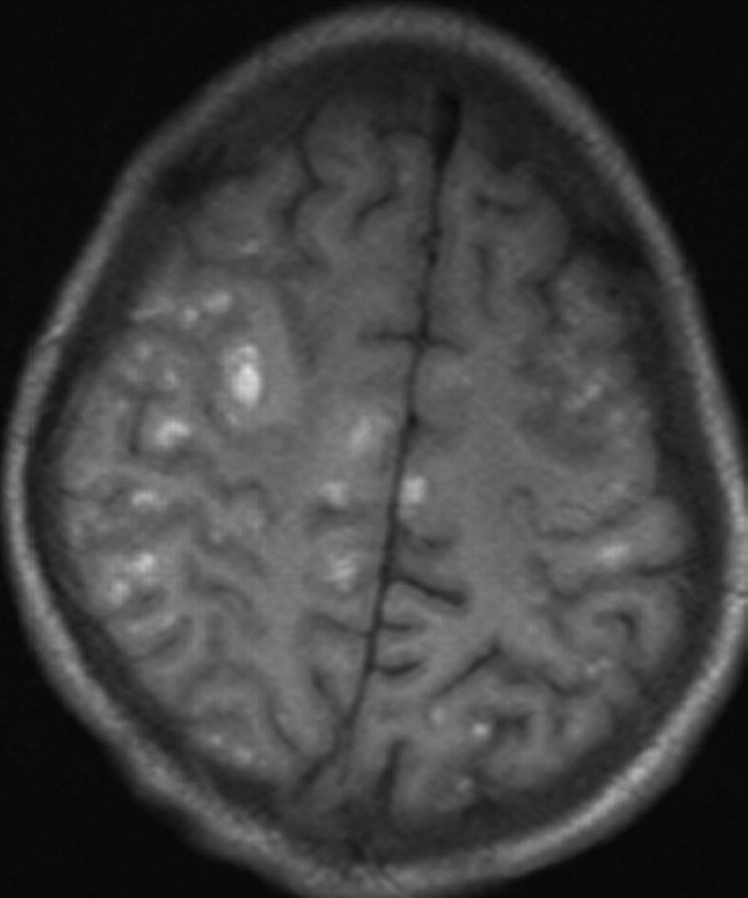
A young man 22 years of age presented with headache on and off for 5–6 months. There was no history of seizures, which was also very unusual in his case. Plain and contrast MR imaging of the brain was performed. T1-weighted images revealed multiple hyperintense foci of varying sizes, in bilateral cerebral, cerebellar hemispheres and the brain stem (Fig 1). The T1 hyperintense signals were thicker in the right cerebellar hemisphere (Fig 2). Multiple other lesions were revealed on gradient images, which were not demonstrated on routine MR images. These multiple extensive T1 hyperintense signals were confirmed to be calcifications on CT, which was performed subsequent to the MR imaging. Few of the T1 hyperintense lesions showed mild enhancement on contrast administration.

Fig 1.
  • Download figure
  • Open in new tab
  • Download powerpoint
Fig 1.

Plain T1-weighted image reveals small hyperintense foci in the bilateral cerebral hemispheres.

Fig 2.
  • Download figure
  • Open in new tab
  • Download powerpoint
Fig 2.

Plain T1-weighted image reveals hyperintense signals in both cerebellar hemispheres, thicker on the right side.

The MR imaging appearance and contrast characteristics would depend on the stage of the parasitic cyst. The unusual appearance of such extensive T1 hyperintense signals, as seen in our patient, has not been described earlier and is probably due to calcifications, which are known to appear hyperintense on T1-weighted MR imaging.2,3 The reason for calcifications demonstrating T1 hyperintense signals has not been clearly elucidated.2 These signals in calcifications are probably due to shortening of the T1 relaxation time, due to the paramagnetic effect of calcium on adjacent water.2 The degree of T1 shortening is larger with a larger area of calcification. Calcifications may also appear hyperintense on the T1-weighted images if they contain other paramagnetic elements like manganese or iron.2 Such extensive T1 hyperintense calcifications with blooming on gradient images must not be mistaken for multiple bleeds.

References

  1. 1.↵
    1. Osborne AG
    . Diagnostic Neuroradiology: The Requisites. St Louis: Mosby; 2003:709–12
  2. 2.↵
    1. Tawil MI,
    2. Wilson JP,
    3. Wright NB
    . Intracranial lithography? Br J Radiol 2002:75:563–64
    FREE Full Text
  3. 3.↵
    1. Dell LA,
    2. Brown MS,
    3. Orrison WW,
    4. et al
    . Physiologic intracranial calcification with hyperintensity on MR imaging: case report and experimental model. AJNR Am J Neuroradiol 1988;9:1145–48
    Abstract/FREE Full Text
  • Copyright © American Society of Neuroradiology
PreviousNext
Back to top

In this issue

American Journal of Neuroradiology: 31 (3)
American Journal of Neuroradiology
Vol. 31, Issue 3
1 Mar 2010
  • Table of Contents
  • Index by author
Advertisement
Print
Download PDF
Email Article

Thank you for your interest in spreading the word on American Journal of Neuroradiology.

NOTE: We only request your email address so that the person you are recommending the page to knows that you wanted them to see it, and that it is not junk mail. We do not capture any email address.

Enter multiple addresses on separate lines or separate them with commas.
Unusual Extensive T1 Hyperintense Signals on MR Imaging in Neurocysticercosis
(Your Name) has sent you a message from American Journal of Neuroradiology
(Your Name) thought you would like to see the American Journal of Neuroradiology web site.
CAPTCHA
This question is for testing whether or not you are a human visitor and to prevent automated spam submissions.
Cite this article
P. Lalitha, B. Reddy
Unusual Extensive T1 Hyperintense Signals on MR Imaging in Neurocysticercosis
American Journal of Neuroradiology Mar 2010, 31 (3) E33; DOI: 10.3174/ajnr.A1956

Citation Manager Formats

  • BibTeX
  • Bookends
  • EasyBib
  • EndNote (tagged)
  • EndNote 8 (xml)
  • Medlars
  • Mendeley
  • Papers
  • RefWorks Tagged
  • Ref Manager
  • RIS
  • Zotero
0 Responses
Respond to this article
Share
Bookmark this article
Unusual Extensive T1 Hyperintense Signals on MR Imaging in Neurocysticercosis
P. Lalitha, B. Reddy
American Journal of Neuroradiology Mar 2010, 31 (3) E33; DOI: 10.3174/ajnr.A1956
del.icio.us logo Twitter logo Facebook logo Mendeley logo
  • Tweet Widget
  • Facebook Like
  • Google Plus One
Purchase

Jump to section

  • Article
    • References
  • Figures & Data
  • Info & Metrics
  • Responses
  • References
  • PDF

Related Articles

  • No related articles found.
  • PubMed
  • Google Scholar

Cited By...

  • Atypical T1 Hyperintense Neurocysticercosis Masquerading As Cystic Brain Metastases
  • Crossref (2)
  • Google Scholar

This article has been cited by the following articles in journals that are participating in Crossref Cited-by Linking.

  • Parasitic Diseases of the Central Nervous System
    Ahmed Abdel Khalek Abdel Razek, Arvemas Watcharakorn, Mauricio Castillo
    Neuroimaging Clinics of North America 2011 21 4
  • Atypical T1 Hyperintense Neurocysticercosis Masquerading As Cystic Brain Metastases
    Abhishek Mahajan, Mehul Patel, Nilesh Sable, Meenakshi H. Thakur
    Journal of Global Oncology 2017 3 5

More in this TOC Section

  • Letter to the Editor regarding “Automated Volumetric Software in Dementia: Help or Hindrance to the Neuroradiologist?”
  • Reply:
  • Brain AVM’s Nidus: What if We Hadn’t Understood Anything?
Show more LETTERS

Similar Articles

Advertisement

Indexed Content

  • Current Issue
  • Accepted Manuscripts
  • Article Preview
  • Past Issues
  • Editorials
  • Editor's Choice
  • Fellows' Journal Club
  • Letters to the Editor
  • Video Articles

Cases

  • Case Collection
  • Archive - Case of the Week
  • Archive - Case of the Month
  • Archive - Classic Case

More from AJNR

  • Trainee Corner
  • Imaging Protocols
  • MRI Safety Corner

Multimedia

  • AJNR Podcasts
  • AJNR Scantastics

Resources

  • Turnaround Time
  • Submit a Manuscript
  • Submit a Video Article
  • Submit an eLetter to the Editor/Response
  • Manuscript Submission Guidelines
  • Statistical Tips
  • Fast Publishing of Accepted Manuscripts
  • Graphical Abstract Preparation
  • Imaging Protocol Submission
  • Evidence-Based Medicine Level Guide
  • Publishing Checklists
  • Author Policies
  • Become a Reviewer/Academy of Reviewers
  • News and Updates

About Us

  • About AJNR
  • Editorial Board
  • Editorial Board Alumni
  • Alerts
  • Permissions
  • Not an AJNR Subscriber? Join Now
  • Advertise with Us
  • Librarian Resources
  • Feedback
  • Terms and Conditions
  • AJNR Editorial Board Alumni

American Society of Neuroradiology

  • Not an ASNR Member? Join Now

© 2025 by the American Society of Neuroradiology All rights, including for text and data mining, AI training, and similar technologies, are reserved.
Print ISSN: 0195-6108 Online ISSN: 1936-959X

Powered by HighWire