
Article Figures & Data
Figures
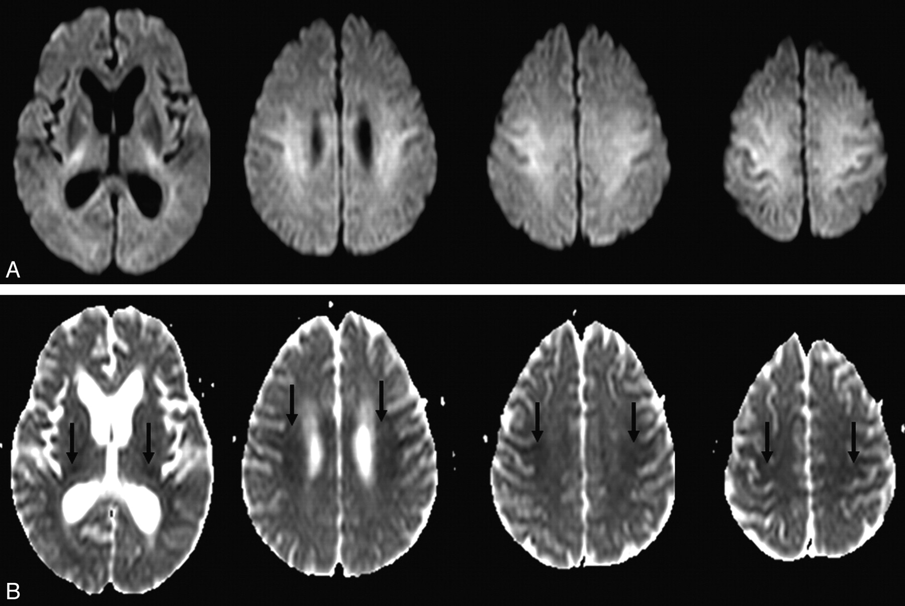
- Fig 1.
A 63-year-old woman (patient 4) was found unconscious. DWI on admission shows hyperintense lesions in the IC, CR, and CS (A) with reduced ADC values (arrows, B). These lesions are bilateral and symmetric.
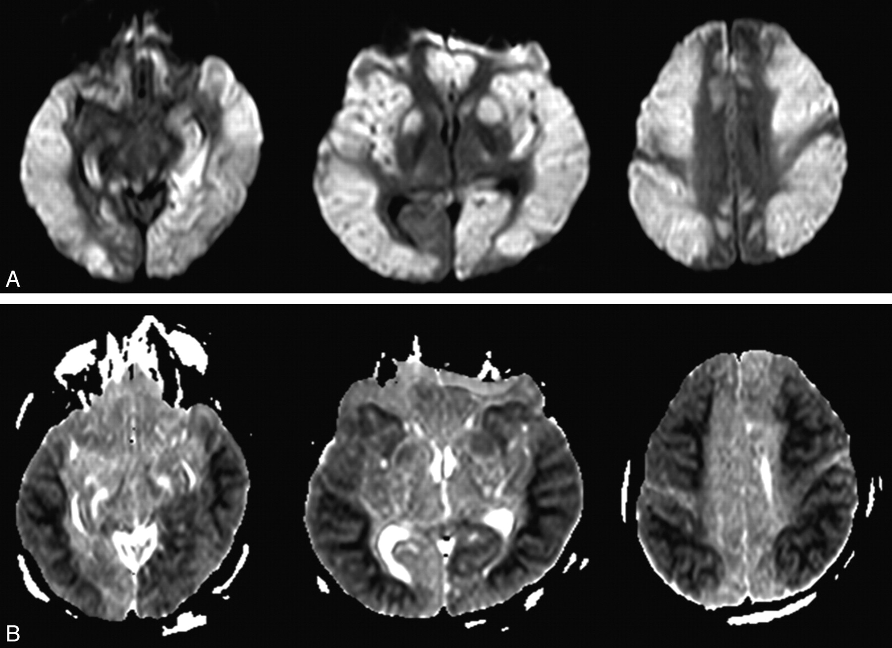
- Fig 2.
A 45-year-old man (patient 11) was found in a coma. A, DWI shows bilaterally asymmetric confluent hyperintense lesions in the frontal, parietal, insular, temporal, occipital cortices, and BG. B, On ADC maps, these lesions show low SI, but the WM was spared.
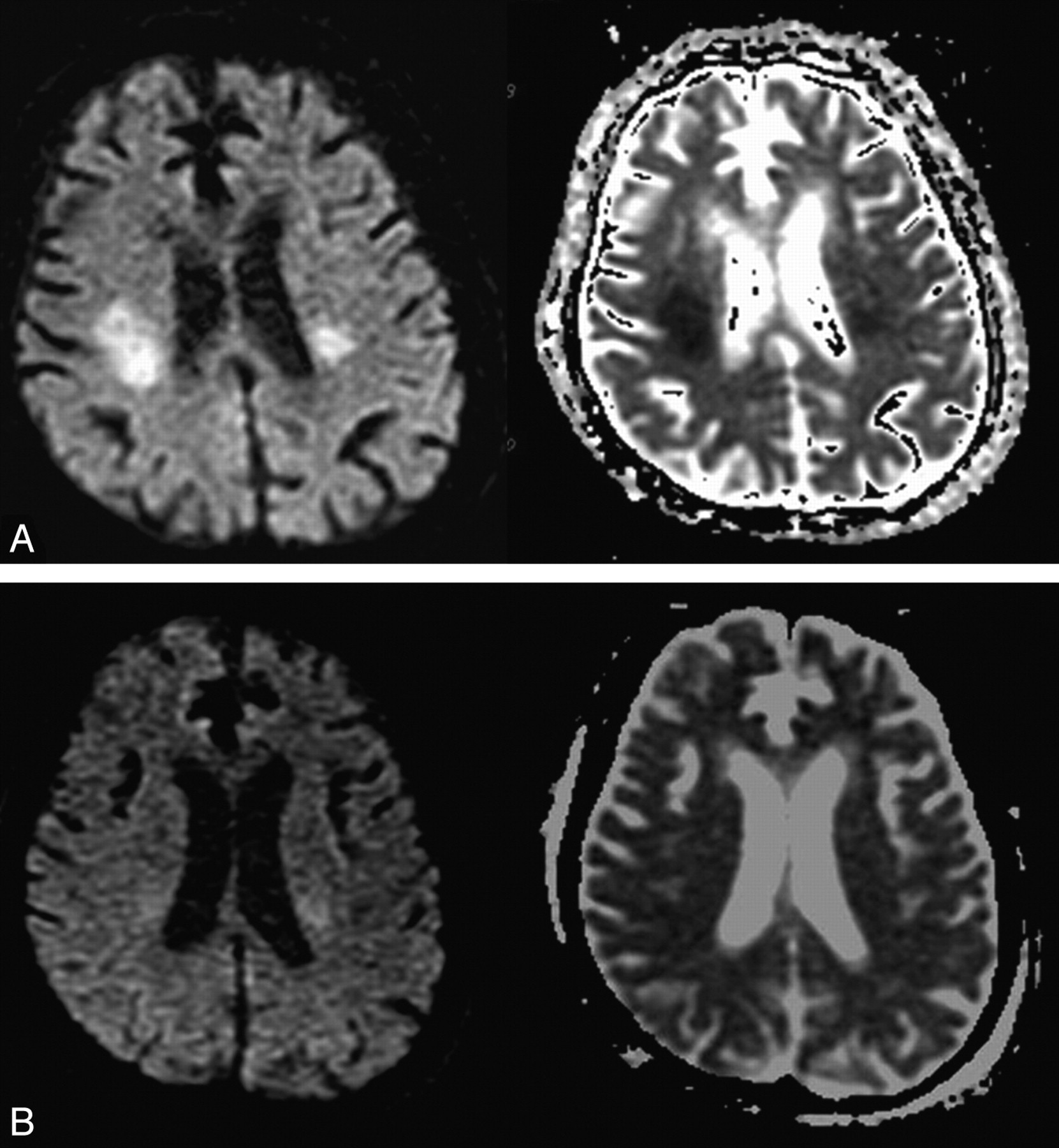
- Fig 3.
A 75-year-old woman (patient 7) was found in a confused mental state. A, Initial DWI on admission shows hyperintense lesions in the IC and CR, with reduced ADC values. B, Follow-up DWI obtained 4 days after symptom improvement shows that the hyperintense lesions have disappeared, with normalization of ADC values.


Tables
Patient No. WM Gray matter Hippocampus Bilaterality CS CR IC CC FL PA TL OC IN BG 1 + + + – – + – – – – – + 2 + + – – + – – – – – – + 3 – – – – + + – + – – + + 4 + + + – + + – – – – – + 5 + + + – + – – – – – – + 6 + + + – – – – – – – + –b 7 + – + – + – – – – – – + 8 + – – – + – – – – – – + 9 + + + – – – – – – – – + 10 + + – – – – – – – – + + 11 – – – – + + + + + + + + a + indicates the presence of a lesion at each anatomic location; –, no abnormality at each anatomic location on MR image.
b Unilateral involvement because there was an old infarction at the right MCA territory.
Patient No. MR Imaging Findings Follow-Up MR Imaging Interval between Initial and Follow-Up (Day) SI on DWI ADC Value of Lesion (10−6 mm2/s) ADC of the Non-Involved Area (10−6 mm2/s) 1 High 542 ± 31.37 946 ± 52.50 Reversible 11 2 High 416 ± 33.69 837 ± 117.90 No 3 High 432 ± 55.79 794 ± 52.50 No 4 High 578 ± 40.93 914 ± 45.04 No 5 High 496 ± 33.31 784 ± 43.93 Reversible 18 6 High 413 ± 26.15 882 ± 95.70 Reversible 19 7 High 458 ± 35.21 786 ± 61.04 Reversible 6 8 High 486 ± 28.62 892 ± 50.33 Reversible 4 9 High 514 ± 32.54 788 ± 81.04 Reversible 8 10 High 354 ± 42.32 759 ± 43.42 No 11 High 248 ± 70.13 833 ± 84.22 No a ADC values are presented as the mean ± SD.
In this issue


Jump to section
Related Articles
Cited By...
- Diffusion Restricted Lesions in the Splenium of the Corpus Callosum
- Diffusion-Weighted MR Imaging in a Prospective Cohort of Children with Cerebral Malaria Offers Insights into Pathophysiology and Prognosis
- Forgetting to remember: hypoglycaemic encephalopathy
- Hypoglycemic encephalopathy
- Cortical abnormalities on MRI: what a neurologist should know
- Effect of Hypoglycemia on Brain Structure in People With Type 2 Diabetes: Epidemiological Analysis of the ACCORD-MIND MRI Trial
- Neuroimaging in Patients with Abnormal Blood Glucose Levels
- Diffusion-Weighted Imaging Changes Caused by Acute Hypoglycemia and Prolonged Febrile Convulsion in Childhood
- Diagnostic approach to restricted-diffusion patterns on MR imaging
- Early Diffusion MR Imaging Findings and Short-Term Outcome in Comatose Patients with Hypoglycemia
- Serum Calcium Concentration Affects Signal Changes on Diffusion-Weighted Imaging in Hypoglycemic Encephalopathy