
Article Figures & Data
Figures
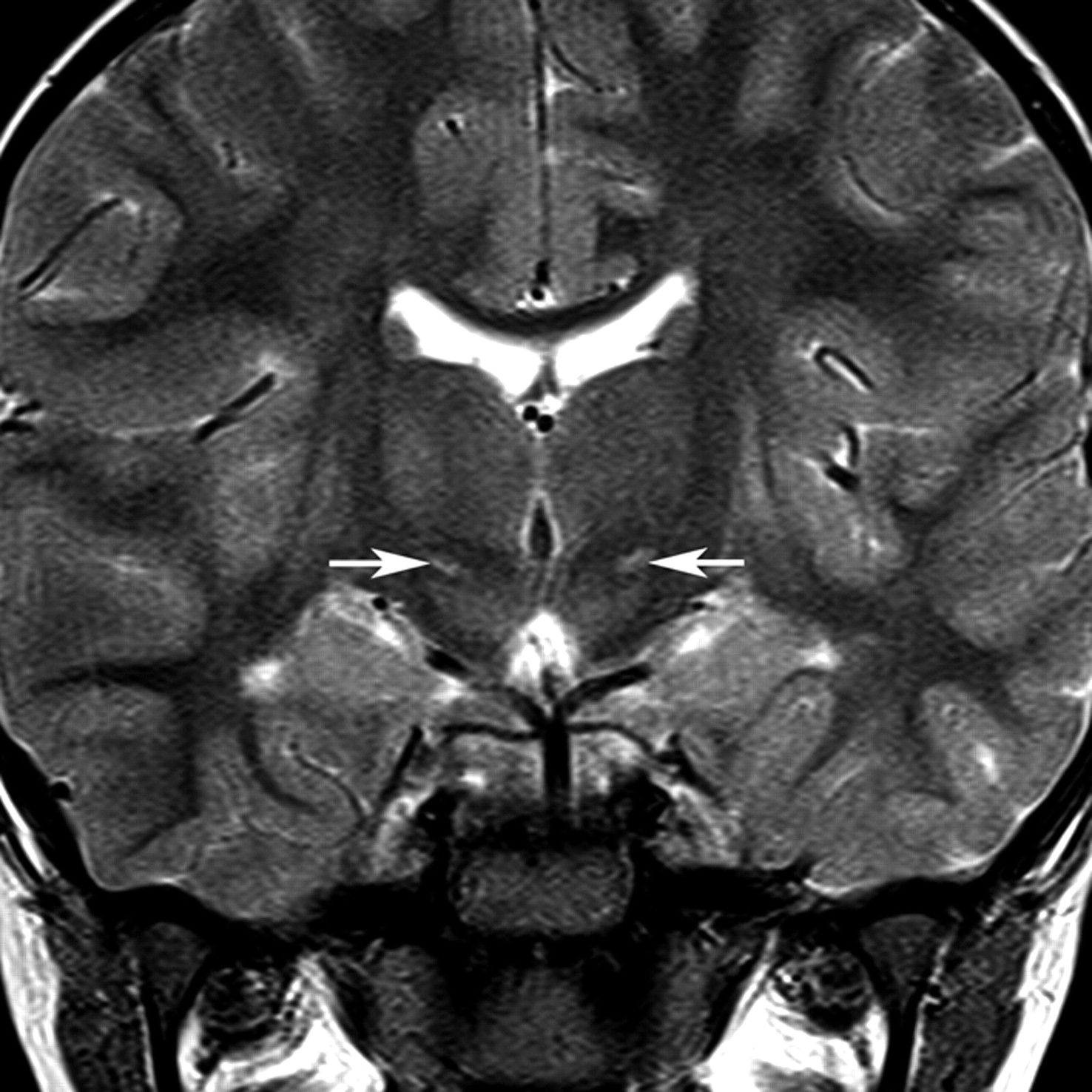
- Fig 1.
Location of the STN. The normal STN is not seen on T2-weighted images at 1.5T because of its small size and signal-intensity characteristics close to the surrounding white matter structures. This coronal T2-weighted image from a child with dyskinetic CP arising from hypoxic-ischemic injury shows the STNs (arrows) as regions of high signal intensity because of gliosis within the nuclei.
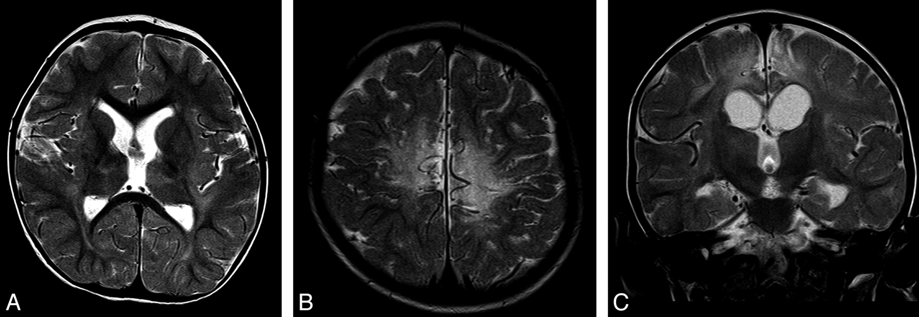
- Fig 2.
Typical appearances of HIBD changes in a child with spastic CP following acute profound HIBD. A and B, Axial T2 images show moderate PCWM signal-intensity abnormality and very mild involvement of the putamen and thalamus. C, Coronal T2 image shows no signal-intensity abnormality in the region of the STN.
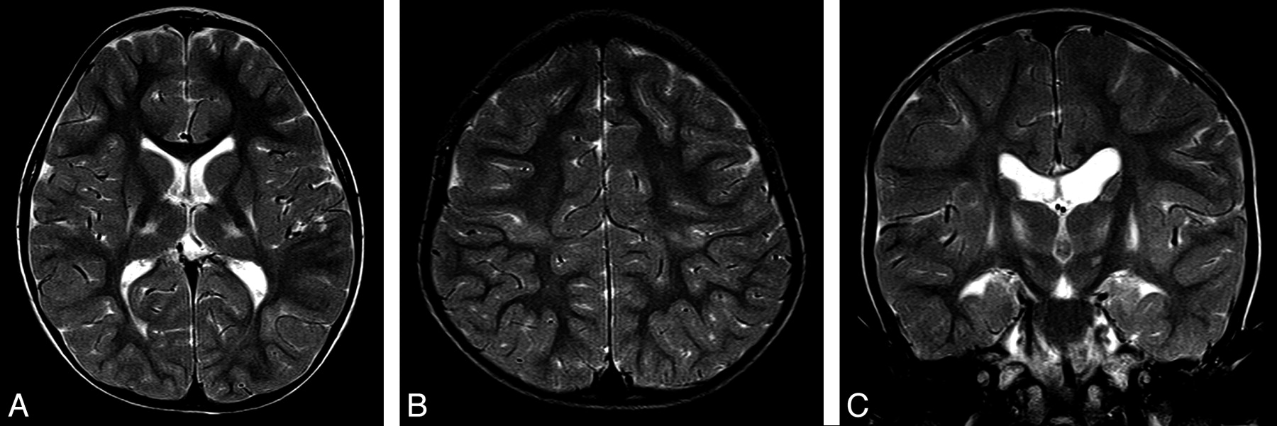
- Fig 3.
Typical appearances of HIBD changes in a child with dyskinetic CP following acute profound HIBD. A and B, Axial T2 images show mild involvement of the putamen, thalamus, and PCWM. C, Coronal T2 image shows high signal intensity in the region of the STN.
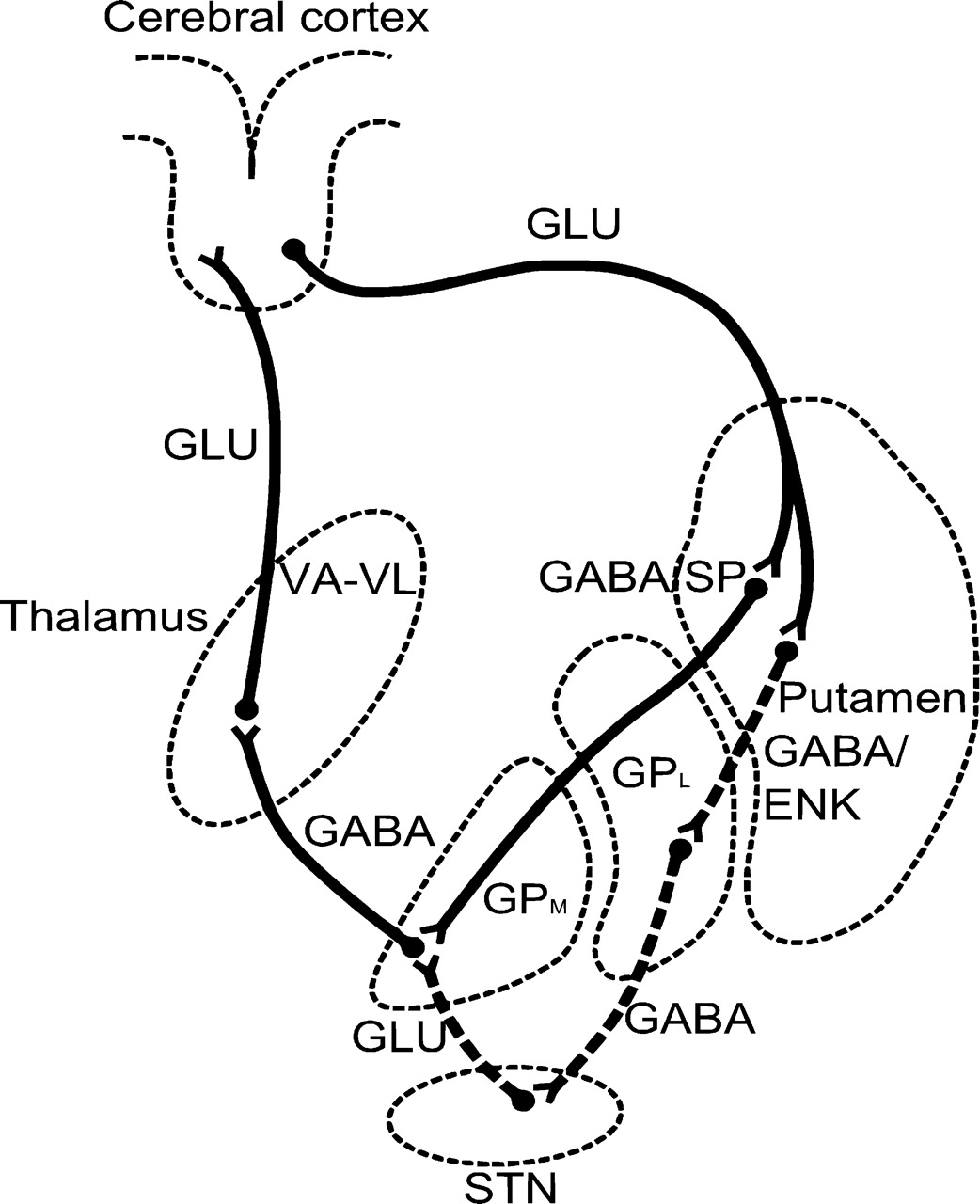
- Fig 4.
Line diagram shows the direct and indirect pathways from the putamen to the thalamus and hence to the motor cortex. Dashed lines indicate the neurons of the indirect pathway traveling via the STN (drawn based on data in Crossman12).


Tables
Variable Dyskinetic CP (n = 20) Spastic CP (n = 20) P Value Age at time of MR imaging (yr) 4.8 ± 3.7 6.0 ± 3.9 .35 Gestational age (wk) 40.0 ± 1.3 39.1 ± 2.0 .12 Birth weight (kg) 3.3 ± 0.5 3.3 ± 0.6 .78 Apgar score (5-minute) 3 (1–7) 4 (0–6) .27 Cord blood pH at birth 6.92 ± 0.15 6.83 ± 0.20 .18 Head circumference (percentile) 53.5 ± 26.0 35.4 ± 20.5 .02b Age at onset of seizures (hr) 3 (1–14) 3.5 (1–18) .17 Putaminal injury score (0–3) Left 2 (1–3) 1.5 (0–3) .72 Right 1 (0–2) 1 (0–3) .78 Thalamic injury score (0–3) Left 1 (1–3) 1.5 (0–3) .84 Right 1 (1–3) 1 (0–3) .64 PCWM severity score (0–3) Left 1 (0–2) 2 (1–3) .01b Right 1 (0–2) 2 (0–3 .07 Total injury score (0–18) 8.0 (4–14) 9.5 (4–18) .38 STN injury (No.) (%) 15 (75) 6 (30) .01b Caudate injury (No.) (%) 2 (10) 8 (40) .06 GP injury (No.) (%) 2 (10) 6 (30) .24 a Groups were compared by the Student t-test. All scores and age at onset of seizures are expressed as medians with ranges in parentheses and are compared by the Wilcoxon rank-sum test. The proportion of patients in each group with STN, caudate, and GP injuries was evaluated by the Fisher exact test.
b Statistically significant, P < .05.
Predictor Variable LRT P Value Odds Ratioa 95% CI PCWM left (0–3) 8.2 .004 0.2 0.1–0.7 STN injury 11.1 .001 18.6 2.4–141.0 Head circumferenceb 6.7 .010 3.0 1.2–7.3 a Odds ratios and 95% CIs are with respect to dyskinetic CP and can be reciprocated to estimate the odds of spastic CP. Cox and Snell model R2 = 63%, indicating good fit to the data.
b Based on percentiles (10th, 25th, 50th, 75th, 90th) for head circumference with the 10th as the reference category.
HC (percentile) STN Injury No STN Injury PCWM Injury Score, Left PCWM Injury Score, Left 0 1 2 3 0 1 2 3 10th 93 70 30 8 40 11 2 1 25th 97 87 55 19 66 26 6 2 50th 98 95 78 50 85 51 16 4 75th 99 98 90 66 94 75 35 9 90th 99 99 97 85 98 90 62 23 a Values represent percentage probabilities of dyskinetic CP according to the combination of the 3 predictors in the logistic regression model. Children with dyskinetic CP have a larger HC, more commonly have STN injury compared with those with spastic CP, and have lower left-sided PCWM scores. The probability of spastic CP can be determined by taking 100 minus the value in the table.


