
Article Figures & Data
Figures
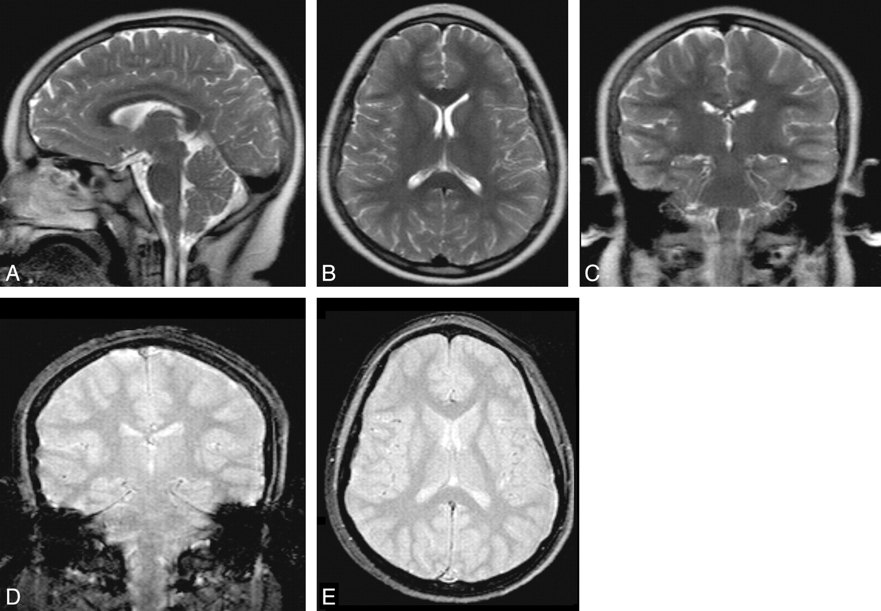
- Fig 1.
Rapid nonsedated pediatric brain MR imaging protocol. The upper row contains TSE-T2-weighted MR images. The bottom row has fast SS-GRE MR images. The total imaging time was approximately 150 seconds.
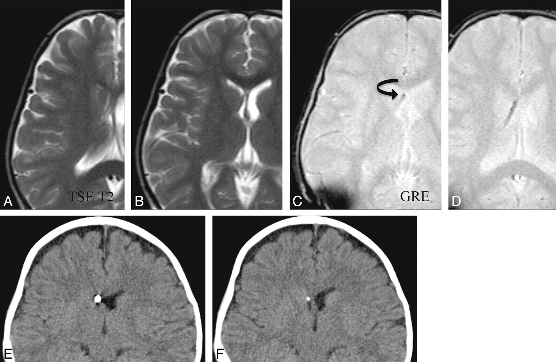
- Fig 2.
A representative sample in which SS-GRE is better than TSE-T2 for localizing the ventricular catheter tip. The upper row has TSE-T2-weighted MR images; and the lower row, SS-GRE MR images. The catheter is inserted from a right frontal approach and travels posterolaterally to the left. On TSE-T2-weighted MR images, the exact delineation of the catheter is not certain. On SS-GRE MR images, the entirety of the catheter can be identified with its tip ending in the left posterior periventricular white matter (curved arrow).
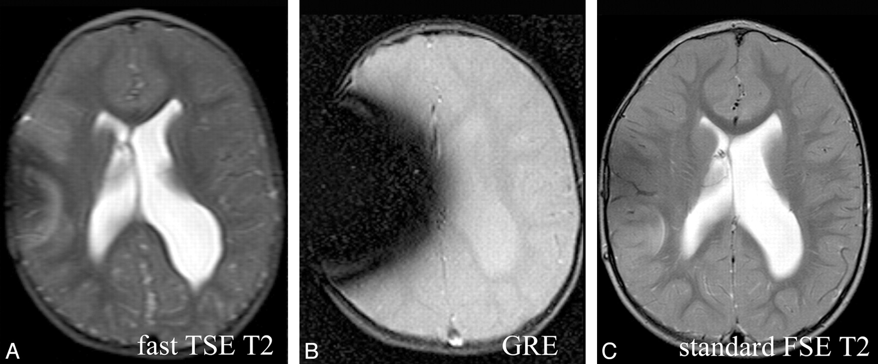
- Fig 3.
Comparison of fast SS-GRE MR images with standard T2-weighted MR images for localization of the ventricular catheter tip. On the standard T2-weighted MR image and the SS-GRE MR image, the catheter tip is easily identified in the right frontal white matter (curved arrows). The tip is less conspicuous on the faster TSE T2-weighted MR image.
- Fig 4.
Comparison of fast SS-GRE MR images with CT for localization of the ventricular catheter tip. The catheter tip is visible within the right frontal horn on CT as well as on SS-GRE MR images (curved arrow). On fast TSE-T2-weighted MR images, the catheter tip is not as readily identified.
- Fig 5.
Artifacts secondary to the shunt reservoir. The SS-GRE MR image is the most affected by these artifacts. There are lesser degrees of susceptibility on the fast TSE-T2-weighted and standard T2-weighted MR images, respectively.


