Advertisement

Main menu

  • Home
  • Content
    • Current Issue
    • Accepted Manuscripts
    • Article Preview
    • Past Issue Archive
    • Video Articles
    • AJNR Case Collection
    • Case of the Week Archive
    • Case of the Month Archive
    • Classic Case Archive
  • Special Collections
    • AJNR Awards
    • Low-Field MRI
    • Alzheimer Disease
    • ASNR Foundation Special Collection
    • Photon-Counting CT
    • View All
  • Multimedia
    • AJNR Podcasts
    • AJNR SCANtastic
    • Trainee Corner
    • MRI Safety Corner
    • Imaging Protocols
  • For Authors
    • Submit a Manuscript
    • Submit a Video Article
    • Submit an eLetter to the Editor/Response
    • Manuscript Submission Guidelines
    • Statistical Tips
    • Fast Publishing of Accepted Manuscripts
    • Graphical Abstract Preparation
    • Imaging Protocol Submission
    • Author Policies
  • About Us
    • About AJNR
    • Editorial Board
    • Editorial Board Alumni
  • More
    • Become a Reviewer/Academy of Reviewers
    • Subscribers
    • Permissions
    • Alerts
    • Feedback
    • Advertisers
    • ASNR Home

User menu

  • Alerts
  • Log in

Search

  • Advanced search
American Journal of Neuroradiology

American Journal of Neuroradiology

ASHNR American Society of Functional Neuroradiology ASHNR American Society of Pediatric Neuroradiology ASSR
  • Alerts
  • Log in

Advanced Search

  • Home
  • Content
    • Current Issue
    • Accepted Manuscripts
    • Article Preview
    • Past Issue Archive
    • Video Articles
    • AJNR Case Collection
    • Case of the Week Archive
    • Case of the Month Archive
    • Classic Case Archive
  • Special Collections
    • AJNR Awards
    • Low-Field MRI
    • Alzheimer Disease
    • ASNR Foundation Special Collection
    • Photon-Counting CT
    • View All
  • Multimedia
    • AJNR Podcasts
    • AJNR SCANtastic
    • Trainee Corner
    • MRI Safety Corner
    • Imaging Protocols
  • For Authors
    • Submit a Manuscript
    • Submit a Video Article
    • Submit an eLetter to the Editor/Response
    • Manuscript Submission Guidelines
    • Statistical Tips
    • Fast Publishing of Accepted Manuscripts
    • Graphical Abstract Preparation
    • Imaging Protocol Submission
    • Author Policies
  • About Us
    • About AJNR
    • Editorial Board
    • Editorial Board Alumni
  • More
    • Become a Reviewer/Academy of Reviewers
    • Subscribers
    • Permissions
    • Alerts
    • Feedback
    • Advertisers
    • ASNR Home
  • Follow AJNR on Twitter
  • Visit AJNR on Facebook
  • Follow AJNR on Instagram
  • Join AJNR on LinkedIn
  • RSS Feeds

AJNR Awards, New Junior Editors, and more. Read the latest AJNR updates

Review ArticleReview Articles
Open Access

MR Imaging Findings in Brachial Plexopathy with Thoracic Outlet Syndrome

A. Aralasmak, K. Karaali, C. Cevikol, H. Uysal and U. Senol
American Journal of Neuroradiology March 2010, 31 (3) 410-417; DOI: https://doi.org/10.3174/ajnr.A1700
A. Aralasmak
  • Find this author on Google Scholar
  • Find this author on PubMed
  • Search for this author on this site
K. Karaali
  • Find this author on Google Scholar
  • Find this author on PubMed
  • Search for this author on this site
C. Cevikol
  • Find this author on Google Scholar
  • Find this author on PubMed
  • Search for this author on this site
H. Uysal
  • Find this author on Google Scholar
  • Find this author on PubMed
  • Search for this author on this site
U. Senol
  • Find this author on Google Scholar
  • Find this author on PubMed
  • Search for this author on this site
  • Article
  • Figures & Data
  • Info & Metrics
  • Responses
  • References
  • PDF
Loading

Article Figures & Data

Figures

  • Tables
  • Fig 1.
    • Download figure
    • Open in new tab
    • Download powerpoint
    Fig 1.

    A, Coronal drawing demonstrates the basic anatomy of the BPL. B–D, Oblique sagittal drawings (B–D from medial to lateral) demonstrate 3 parts of the BPL. The supraclavicular plexus is composed of roots and trunks. Roots are seen at the interscalene triangle between the anterior and middle scalene muscles. The subclavian artery forms the floor of the interscalene triangle (B). Roots then form the trunks at the lateral border of the middle scalene muscles. The retroclavicular plexus is composed of divisions situated in the costoclavicular space between the first rib and clavicula, and the BPL is seen in the superior and posterior aspect of the subclavian artery (C). The infraclavicular plexus is composed of cords and terminal branches located in the retropectoralis minor space. The BPL is situated in the posterior and superior aspect of axillary artery (D). The subclavian artery and vein take the name of axillary artery and vein at the lateral border of first rib.

  • Fig 2.
    • Download figure
    • Open in new tab
    • Download powerpoint
    Fig 2.

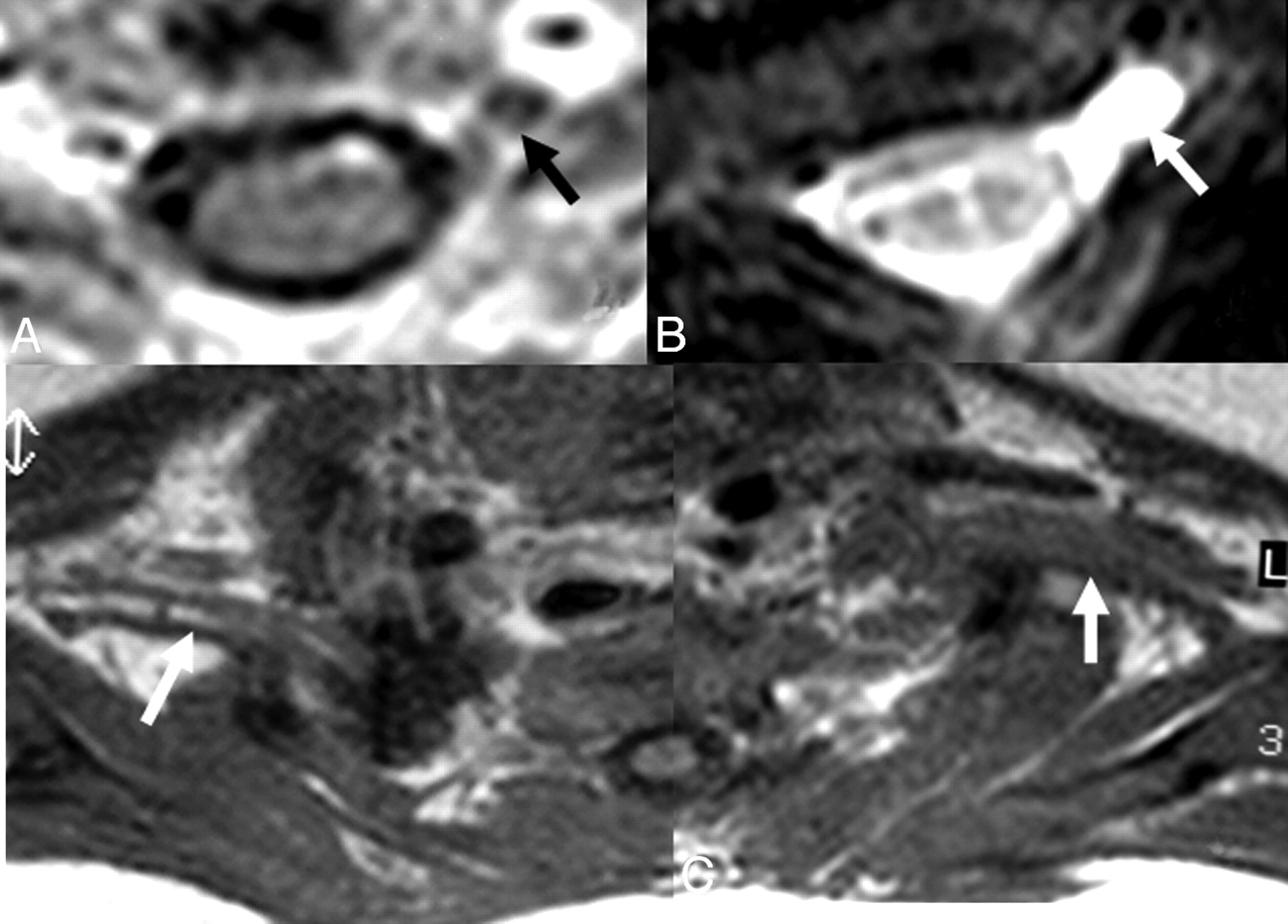
    A and B, In a 1-month-old infant with a history of difficult birth, contrast-enhanced axial T1-weighted (A) and axial MR myelography (B) views show avulsion of the left C5 anterior root with contrast enhancement at its root stump (A) and a pseudomeningocele (arrows), consistent with preganglionic injury. C, Distal to the injury, the BPL on the left appears thickened and irregular on axial T1WI images compared with the BPL of the normal right side (arrows).

  • Fig 3.
    • Download figure
    • Open in new tab
    • Download powerpoint
    Fig 3.

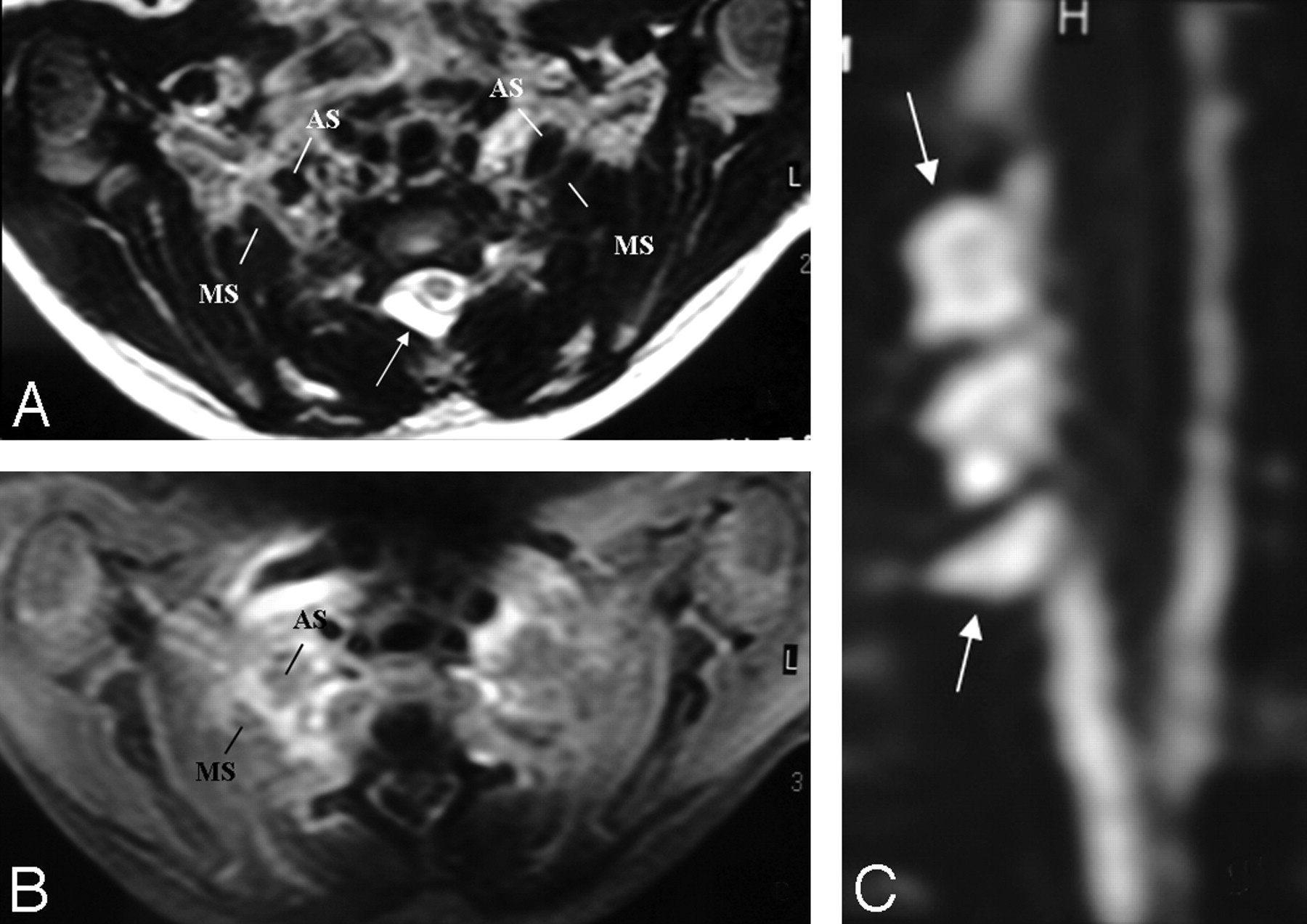
    A and B, There is asymmetric thickening, T2 hyperintensity, and contrast enhancement of the right BPL compared with that of the left side, better seen between the anterior and middle scalene muscles. The pseudomeningocele is noted at the right posterolateral aspect of the central canal on axial T2-weighted TSE image (arrow, A). C, However, a coronal reformatted image from MR myelography shows the roots intact within the pseudomeningocele sac (arrows), suggesting only a traction injury. There is neither asymmetric enhancement nor T2 signal-intensity change of the BPL-innervated muscles or paraspinal muscles to support preganglionic injury.

  • Fig 4.
    • Download figure
    • Open in new tab
    • Download powerpoint
    Fig 4.

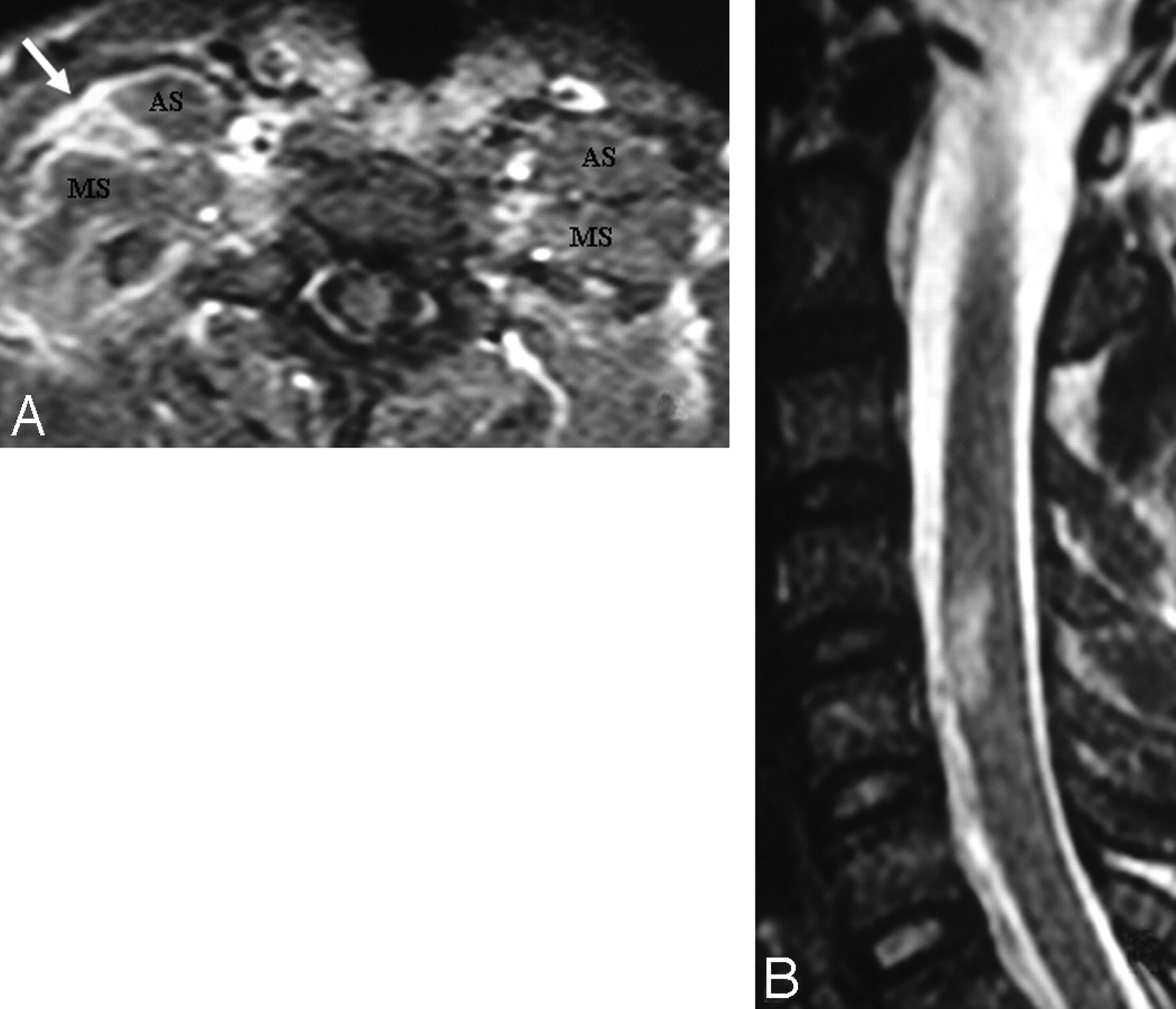
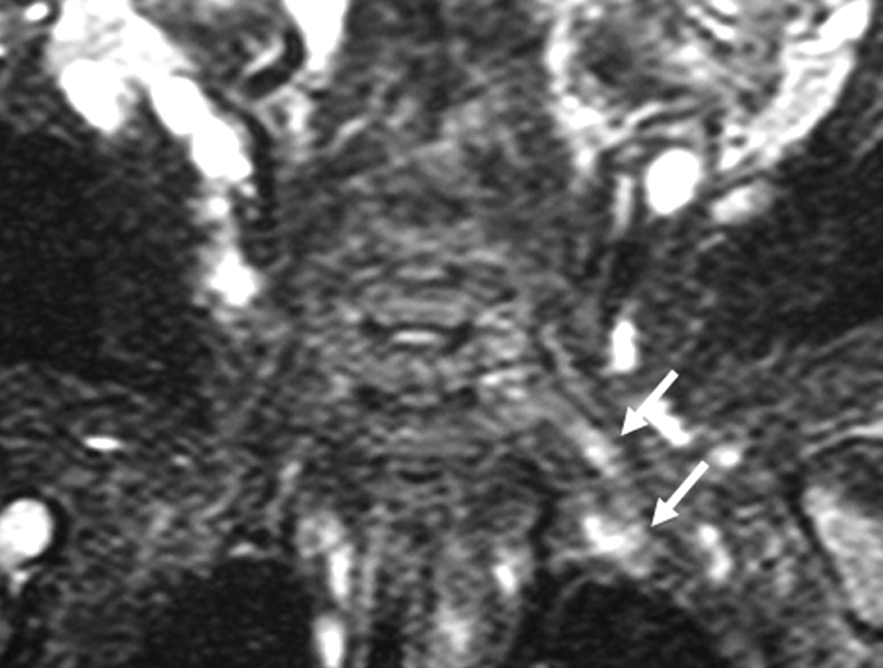
    A, In a patient with a right-sided scapula fracture and monoparesis following trauma, the right BPL (arrow) is thickened and asymmetrically enhancing at the truncus level between the anterior and middle interscalene muscles. There is no associated root avulsion or pseudomeningocele, suggesting only traction injury at first glance. B, However, the presence of an expansile spinal cord lesion at the C4-C5 level could signal the possibility of preganglionic injury.

  • Fig 5.
    • Download figure
    • Open in new tab
    • Download powerpoint
    Fig 5.

    In another infant with a left-sided BPL paralysis following birth trauma, there is only hyperintensity of the left BPL on the coronal STIR T2-weighted image (arrows) without thickening on T1-weighted and T2-weighted TSE images (not shown). There is no associated root avulsion and pseudomeningocele. Findings are suggestive of traction injury.

  • Fig 6.
    • Download figure
    • Open in new tab
    • Download powerpoint
    Fig 6.

    A Pancoast tumor surrounds the subclavian artery (arrow) circumferentially. The subclavian vein is not seen as separated. This finding means that it is either compressed or invaded. The BPL posterosuperior aspect of the subclavian artery is invaded as well.

  • Fig 7.
    • Download figure
    • Open in new tab
    • Download powerpoint
    Fig 7.

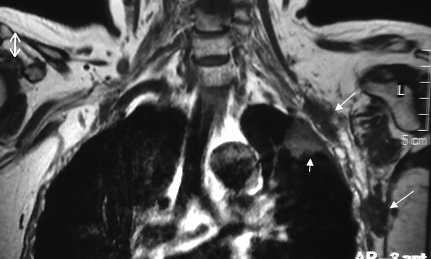
    Precontrast coronal T1-weighted view shows metastatic nodules (long arrows) from breast cancer in the vicinity of the left BPL and another metastatic mass in the upper lobe of the left lung (small arrow).

  • Fig 8.
    • Download figure
    • Open in new tab
    • Download powerpoint
    Fig 8.

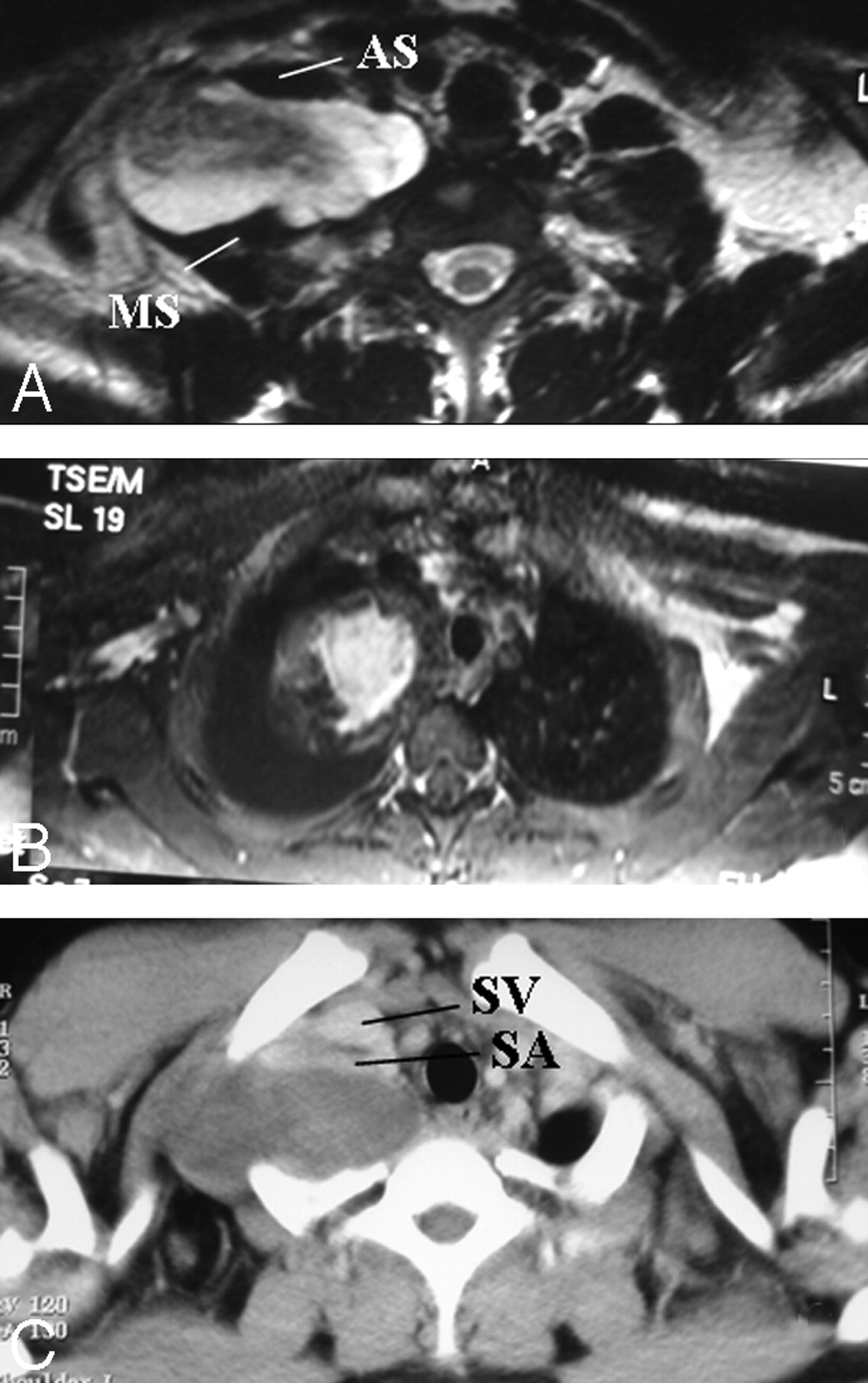
    A and B, Pathologically proved neurofibroma elongating between the anterior and middle scalene muscles. The mass lesion is fusiform, longitudinally oriented along the BPL (A), with central homogeneous enhancement (B). A target sign with central hypointensity and peripheral hyperintensity on T2WI (A) and central homogeneous enhancement (B) are suggestive of neurofibroma. C, The subclavian vein and artery are extended at the anterior aspect of the mass on the axial postcontrast CT view. There is diaphragm paralysis on the right side compatible with phrenic nerve involvement as well (not shown).

  • Fig 9.
    • Download figure
    • Open in new tab
    • Download powerpoint
    Fig 9.

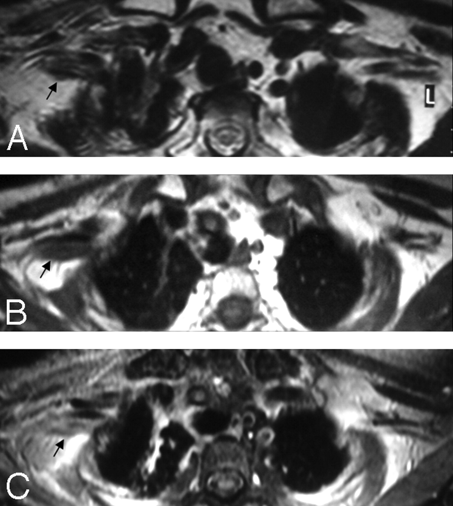
    A, In a patient with a history of breast cancer surgery and radiation treatment with right-arm weakness, axial T2WI shows diffuse thickening of the right PBL with no associated hyperintensity (arrow). B and C, Pre- (B) and postcontrast (C) axial T1WIs show minimal contrast enhancement of the thickened right BPL fibers with no associated nodularity (arrows). Findings are suggestive of radiation fibrosis rather than metastasis.

  • Fig 10.
    • Download figure
    • Open in new tab
    • Download powerpoint
    Fig 10.

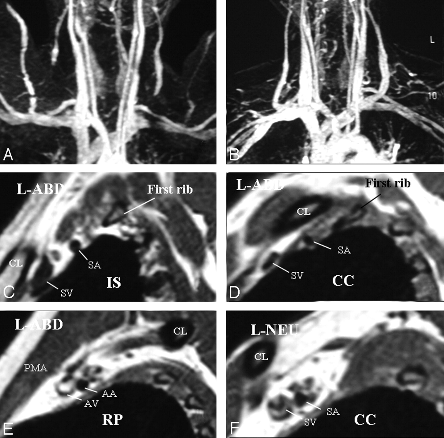
    A, Left-sided neurogenic, arterial, and venous TOS with compression in the costoclavicular space in a patient with bluish discoloration of the left hand. MRA and MRV in abduction show impingement on the left subclavian artery and vein. B, In a neutral position, impingements on the vessels are all resolved. C–E, Sagittal views through the left BPL in abduction show narrowing of the subclavian artery and vein in the costoclavicular space and a normal caliber of the vessels in the interscalene triangle and retropectoralis minor spaces. In abduction, the left BPL is also squeezed in the costoclavicular space, suggesting neurogenic TOS additionally. F, In the neutral position, there is neither narrowing of the vessels nor any impingement on the BPL nerves in the costoclavicular space of the left side.

  • Fig 11.
    • Download figure
    • Open in new tab
    • Download powerpoint
    Fig 11.

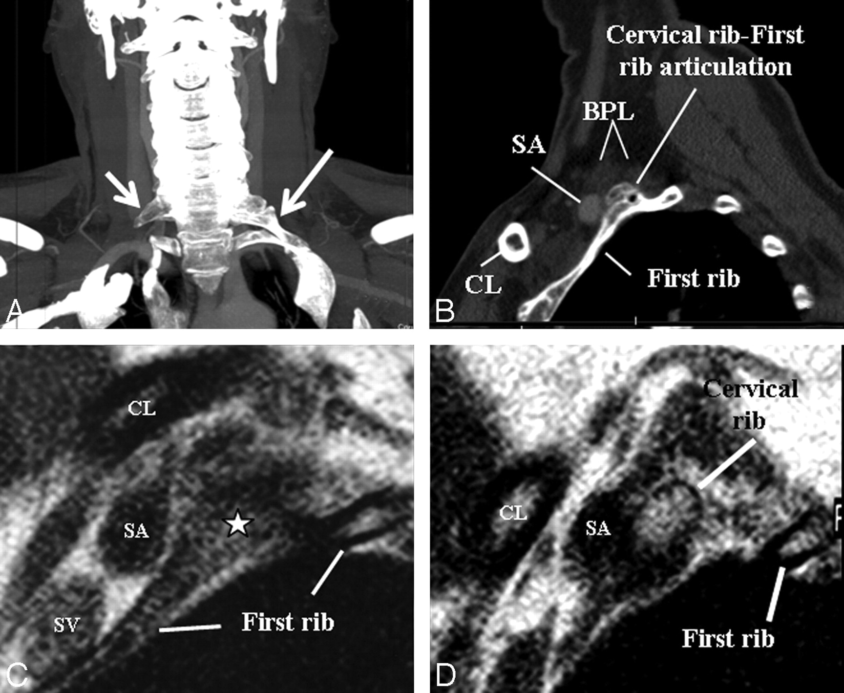
    A, An elongated C7 transverse process on the right (small arrow) and a cervical rib (long arrow) articulating with first thoracic rib on the left on a maximal-intensity-projection view of a neck CT of a patient presenting with cervical pain and bluish discoloration of both hands, more so on the left. MRA and MRV of the neck vessels (not shown) in abduction did not show any stenosis excluding vascular (arterial or venous) TOS. B–D, Sagittal CT (B) and sagittal MR imaging (C and D from lateral to medial) views in a neutral position on the left side demonstrate cervical rib−first rib articulation (star) and the cervical rib contacting the BPL fibers in the costoclavicular space, responsible for neurogenic TOS. The patient's symptoms on the right are due to the elongated C7 transverse process impinging the BPL within the interscalene triangle (not shown).

Tables

  • Figures
  • MR imaging sequences obtained in brachial plexopathy with/without TOS

    MR Imaging SequencesAssociated Anatomic Structures or Pathologic Conditions
    Sagittal TSE T2WI through cervical spineSpinal cord lesion (edema, hemorrhage, avulsion, myelomalacia, syrinx, tumor, etc)
    Precontrast axiala T1WIBPL (thickening, nodularity)
    Precontrast coronalb T1WIBPL, vertebrae, long C7 transverse process, cervical rib
    Axiala 2D TSE T2WIBPL (thickening, nodularity, signal changes better seen between anterior and middle scalene muscles), radiculopathy, diskopathy, foraminal invasion, spinal cord lesions, large-sized pseudomeningocele, muscle denervation
    Coronalb STIR T2WIBPL (any signal changes not detectable on 2D TSE T2WI, especially in traction injuries and brachial plexitis), muscle denervation in traumatic injury, and brachial plexitis
    Axiala 3D TSE heavily T2WI (MR myelography)Root avulsions, small-sized pseudomeningocele, which can be missed on 2D TSE T2WI.
    Postcontrast fat-saturated axiala T1WIBPL (contrast enhancement), contrast enhancement of root stump or intradural roots or denervated muscles in preganglionic injuries
    Postcontrast coronalb T1WIBPL (contrast enhancement)
    Sagittalc,d T1WI from the symptomatic side in abductionCompression on BPL and subclavian vessels (positional, cervical rib, long C7 transverse process, accessory muscles, fibrous band)
    Sagittalc,d T1WI from the symptomatic side in neutral if there is compressionResolution of compression on the BPL and subclavian vessels
    MRAc and MRVc of subclavian artery and vein in abductionSubclavian artery and vein (patency, thrombosis, aneurysm, any impingement on the vessels)
    MRAc and MRVc of subclavian artery and vein next day in neutral if there is impingementResolution of impingement on the subclavian vessels
    • a Perpendicular to the long axis of the vertebrae in the coronal plane.

    • b Parallel to the long axis of the lower cervical vertebrae of C4-C7.

    • c Additional MR imaging sequences obtained when there is clinical suspicion of TOS.

    • d Perpendicular to the long axis of the BPL from the spinal cord to the medial border of the humerus.

PreviousNext
Back to top

In this issue

American Journal of Neuroradiology: 31 (3)
American Journal of Neuroradiology
Vol. 31, Issue 3
1 Mar 2010
  • Table of Contents
  • Index by author
Advertisement
Print
Download PDF
Email Article

Thank you for your interest in spreading the word on American Journal of Neuroradiology.

NOTE: We only request your email address so that the person you are recommending the page to knows that you wanted them to see it, and that it is not junk mail. We do not capture any email address.

Enter multiple addresses on separate lines or separate them with commas.
MR Imaging Findings in Brachial Plexopathy with Thoracic Outlet Syndrome
(Your Name) has sent you a message from American Journal of Neuroradiology
(Your Name) thought you would like to see the American Journal of Neuroradiology web site.
CAPTCHA
This question is for testing whether or not you are a human visitor and to prevent automated spam submissions.
Cite this article
A. Aralasmak, K. Karaali, C. Cevikol, H. Uysal, U. Senol
MR Imaging Findings in Brachial Plexopathy with Thoracic Outlet Syndrome
American Journal of Neuroradiology Mar 2010, 31 (3) 410-417; DOI: 10.3174/ajnr.A1700

Citation Manager Formats

  • BibTeX
  • Bookends
  • EasyBib
  • EndNote (tagged)
  • EndNote 8 (xml)
  • Medlars
  • Mendeley
  • Papers
  • RefWorks Tagged
  • Ref Manager
  • RIS
  • Zotero
0 Responses
Respond to this article
Share
Bookmark this article
MR Imaging Findings in Brachial Plexopathy with Thoracic Outlet Syndrome
A. Aralasmak, K. Karaali, C. Cevikol, H. Uysal, U. Senol
American Journal of Neuroradiology Mar 2010, 31 (3) 410-417; DOI: 10.3174/ajnr.A1700
del.icio.us logo Twitter logo Facebook logo Mendeley logo
  • Tweet Widget
  • Facebook Like
  • Google Plus One
Purchase

Jump to section

  • Article
    • Abstract
    • Abbreviations
    • Anatomy of the BPL
    • MR Imaging Protocols
    • Physiologic, Functional, and Other Anatomic Imaging Techniques
    • Traumatic Brachial Plexopathy
    • Mass Involving or Compressing the BPL
    • Brachial Plexitis
    • TOS (Entrapment Syndrome)
    • Vascular Insult
    • Conclusions
    • Footnotes
    • References
  • Figures & Data
  • Info & Metrics
  • Responses
  • References
  • PDF

Related Articles

  • No related articles found.
  • PubMed
  • Google Scholar

Cited By...

  • Brachial plexopathy as a complication of COVID-19
  • High-Resolution 3T MR Neurography of the Brachial Plexus and Its Branches, with Emphasis on 3D Imaging
  • Crossref (53)
  • Google Scholar

This article has been cited by the following articles in journals that are participating in Crossref Cited-by Linking.

  • High-Resolution 3T MR Neurography of the Brachial Plexus and Its Branches, with Emphasis on 3D Imaging
    A. Chhabra, G.K. Thawait, T. Soldatos, R.S. Thakkar, F. Del Grande, M. Chalian, J.A. Carrino
    American Journal of Neuroradiology 2013 34 3
  • Imaging of the Patient with Thoracic Outlet Syndrome
    Constantine A. Raptis, Sreevathsan Sridhar, Robert W. Thompson, Kathryn J. Fowler, Sanjeev Bhalla
    RadioGraphics 2016 36 4
  • MRI findings in thoracic outlet syndrome
    Ayse Aralasmak, Can Cevikol, Kamil Karaali, Utku Senol, Rasul Sharifov, Rukiye Kilicarslan, Alpay Alkan
    Skeletal Radiology 2012 41 11
  • Thoracic outlet syndrome: a review
    Eric J. Panther, Christian D. Reintgen, Robert J. Cueto, Kevin A. Hao, Harvey Chim, Joseph J. King
    Journal of Shoulder and Elbow Surgery 2022 31 11
  • MR neurography in traumatic brachial plexopathy
    Vaishali Upadhyaya, Divya N. Upadhyaya, Adarsh Kumar, Ratni B. Gujral
    European Journal of Radiology 2015 84 5
  • Diagnostic accuracy of MRI in adults with suspect brachial plexus lesions: A multicentre retrospective study with surgical findings and clinical follow-up as reference standard
    Alberto Tagliafico, Giulia Succio, Giovanni Serafini, Carlo Martinoli
    European Journal of Radiology 2012 81 10
  • MR Imaging of the Brachial Plexus
    Amelie M. Lutz, Garry Gold, Christopher Beaulieu
    Neuroimaging Clinics of North America 2014 24 1
  • Diagnostic Value of Magnetic Resonance Imaging in Thoracic Outlet Syndrome
    Vinay Kumar Singh, Luckshmana Jeyaseelan, Steve Kyriacou, Subhojit Ghosh, Marco Sinisi, Michael Fox
    Journal of Orthopaedic Surgery 2014 22 2
  • ACR Appropriateness Criteria Imaging in the Diagnosis of Thoracic Outlet Syndrome
    John M. Moriarty, Dennis F. Bandyk, Daniel F. Broderick, Rebecca S. Cornelius, Karin E. Dill, Christopher J. Francois, Marie D. Gerhard-Herman, Mark E. Ginsburg, Michael Hanley, Sanjeeva P. Kalva, Jeffrey P. Kanne, Loren H. Ketai, Bill S. Majdalany, James G. Ravenel, Christopher J. Roth, Anthony G. Saleh, Matthew P. Schenker, Tan-Lucien H. Mohammed, Frank J. Rybicki
    Journal of the American College of Radiology 2015 12 5
  • High-resolution Imaging of Neural Anatomy and Pathology of the Neck
    Jeong Hyun Lee, Kai-Lung Cheng, Young Jun Choi, Jung Hwan Baek
    Korean Journal of Radiology 2017 18 1

More in this TOC Section

  • An Atlas of Neonatal Neurovascular Imaging Anatomy as Depicted with Microvascular Imaging: The Intracranial Arteries
  • An Atlas of Neonatal Neurovascular Imaging Anatomy as Depicted with Microvascular Imaging: The Intracranial Veins
  • Clinical Translation of Hyperpolarized 13C Metabolic Probes for Glioma Imaging
Show more Review articles

Similar Articles

Advertisement

Indexed Content

  • Current Issue
  • Accepted Manuscripts
  • Article Preview
  • Past Issues
  • Editorials
  • Editor's Choice
  • Fellows' Journal Club
  • Letters to the Editor
  • Video Articles

Cases

  • Case Collection
  • Archive - Case of the Week
  • Archive - Case of the Month
  • Archive - Classic Case

More from AJNR

  • Trainee Corner
  • Imaging Protocols
  • MRI Safety Corner

Multimedia

  • AJNR Podcasts
  • AJNR Scantastics

Resources

  • Turnaround Time
  • Submit a Manuscript
  • Submit a Video Article
  • Submit an eLetter to the Editor/Response
  • Manuscript Submission Guidelines
  • Statistical Tips
  • Fast Publishing of Accepted Manuscripts
  • Graphical Abstract Preparation
  • Imaging Protocol Submission
  • Evidence-Based Medicine Level Guide
  • Publishing Checklists
  • Author Policies
  • Become a Reviewer/Academy of Reviewers
  • News and Updates

About Us

  • About AJNR
  • Editorial Board
  • Editorial Board Alumni
  • Alerts
  • Permissions
  • Not an AJNR Subscriber? Join Now
  • Advertise with Us
  • Librarian Resources
  • Feedback
  • Terms and Conditions
  • AJNR Editorial Board Alumni

American Society of Neuroradiology

  • Not an ASNR Member? Join Now

© 2025 by the American Society of Neuroradiology All rights, including for text and data mining, AI training, and similar technologies, are reserved.
Print ISSN: 0195-6108 Online ISSN: 1936-959X

Powered by HighWire