
Article Figures & Data
Figures
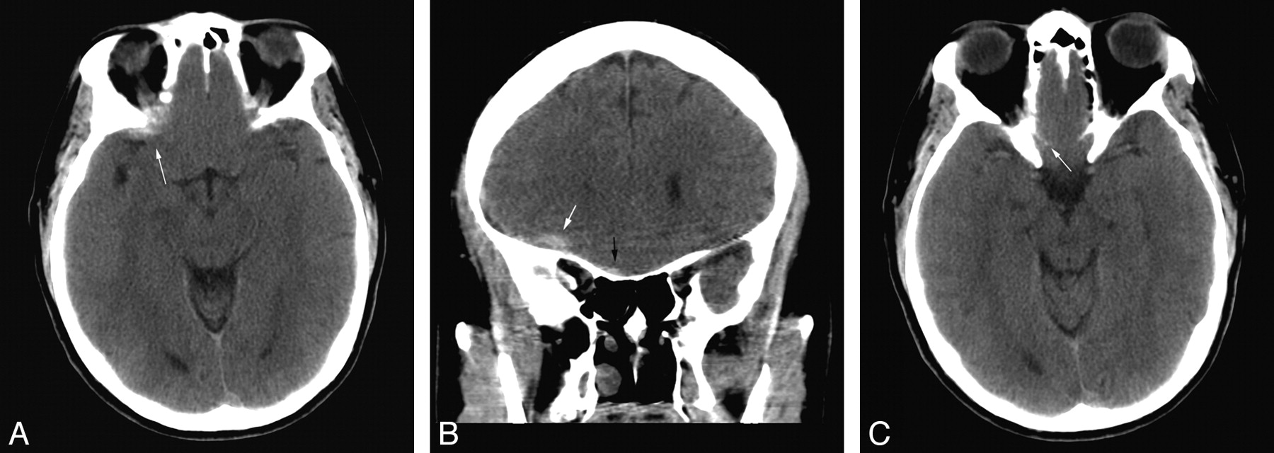
- Fig 1.
A, A small hemorrhagic contusion (white arrow) is partially obscured by volume averaging with the adjacent bone. B, The same lesion is more conspicuous and confidently identified on coronal reformations (white arrow). A small right subdural hematoma (black arrow) is noted. C, On the corresponding axial image, no abnormality can be identified in the region of the small right subdural hematoma (small white arrow).
- Fig 2.
A, Hyperattenuation (arrow) in the superficial right temporal lobe is partially obscured by volume averaging through the temporal bone. B, A clear focus of hemorrhagic contusion (arrow) is seen on the corresponding coronal image.
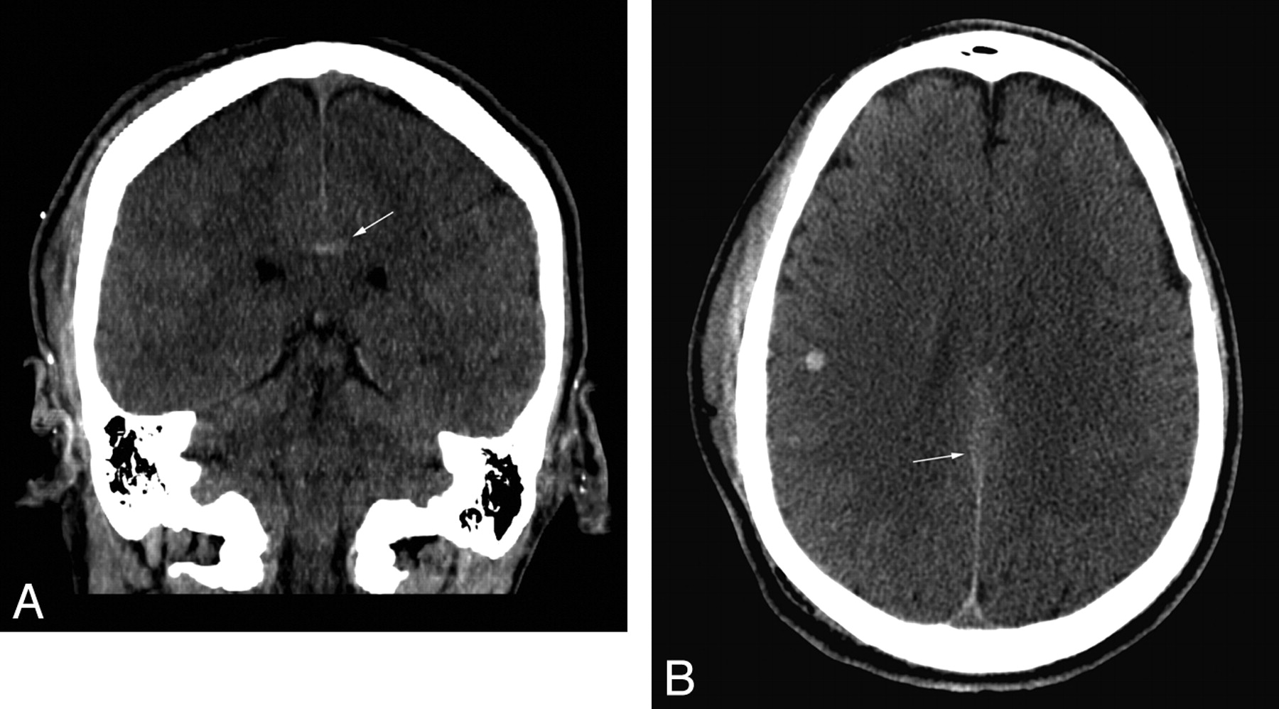
- Fig 3.
A, A small focus of parenchymal hemorrhage (arrow) is clearly seen in the corpus callosum on the coronal reformation. B, The lesion (arrow) is much more subtly apparent on the corresponding axial image.
- Fig 4.
A, Minimal thickening of the anterior falx (arrow) is not prospectively identified on initial review of the axial images. B, This is confirmed to represent a small subdural hematoma (arrow) on the corresponding coronal image.
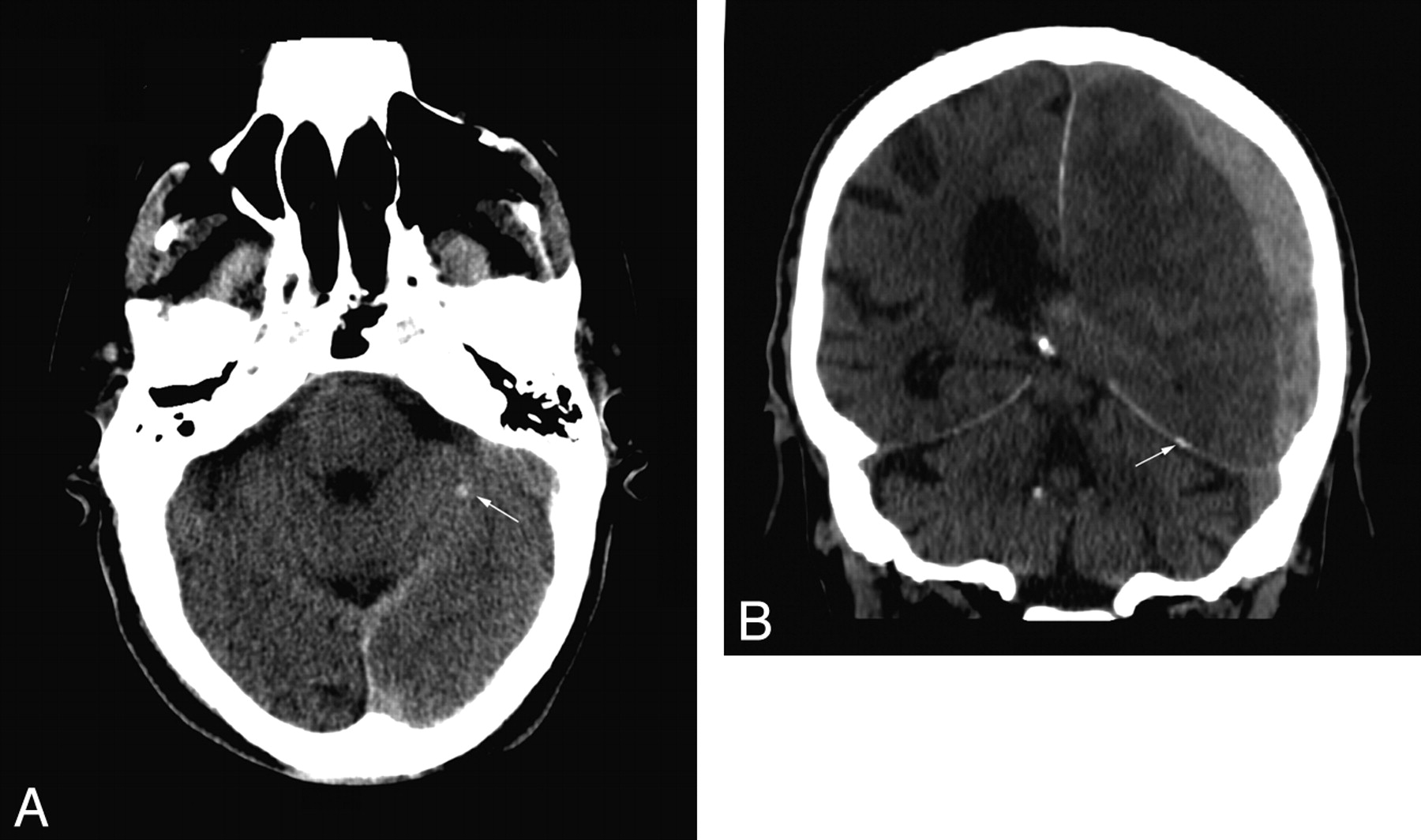
- Fig 5.
A, Hyperattenuated focus (arrow) in the left cerebellum interpreted as suspicious for parenchymal hemorrhage. B, Coronal images show that the finding represents tentorial calcification (arrow).
- Fig 6.
A, A small focus of pneumocephalus (arrow) is related to a nondisplaced fracture through the left temporal bone. B, On the axial images, the findings are obscured due to volume averaging through neighboring mastoid air cells (arrow).
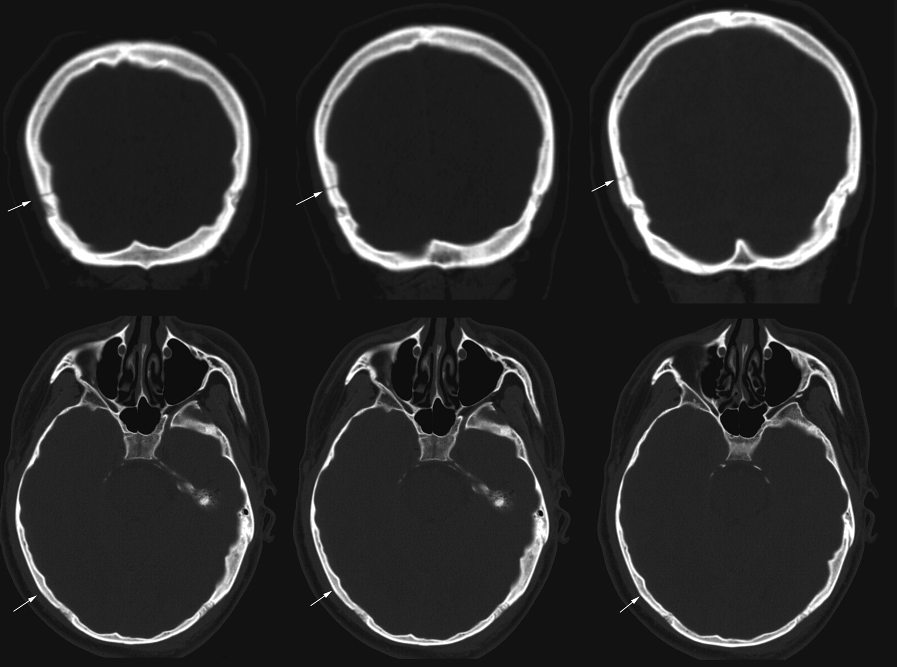
- Fig 7.
Three coronal images show a tranverse nondisplaced fracture (arrows) through the right parietal bone. The corresponding axial images show no evidence of fracture (arrows).


