Advertisement

Main menu

  • Home
  • Content
    • Current Issue
    • Accepted Manuscripts
    • Article Preview
    • Past Issue Archive
    • Video Articles
    • AJNR Case Collection
    • Case of the Week Archive
    • Case of the Month Archive
    • Classic Case Archive
  • Special Collections
    • AJNR Awards
    • Low-Field MRI
    • Alzheimer Disease
    • ASNR Foundation Special Collection
    • Photon-Counting CT
    • View All
  • Multimedia
    • AJNR Podcasts
    • AJNR SCANtastic
    • Trainee Corner
    • MRI Safety Corner
    • Imaging Protocols
  • For Authors
    • Submit a Manuscript
    • Submit a Video Article
    • Submit an eLetter to the Editor/Response
    • Manuscript Submission Guidelines
    • Statistical Tips
    • Fast Publishing of Accepted Manuscripts
    • Graphical Abstract Preparation
    • Imaging Protocol Submission
    • Author Policies
  • About Us
    • About AJNR
    • Editorial Board
    • Editorial Board Alumni
  • More
    • Become a Reviewer/Academy of Reviewers
    • Subscribers
    • Permissions
    • Alerts
    • Feedback
    • Advertisers
    • ASNR Home

User menu

  • Alerts
  • Log in

Search

  • Advanced search
American Journal of Neuroradiology

American Journal of Neuroradiology

ASHNR American Society of Functional Neuroradiology ASHNR American Society of Pediatric Neuroradiology ASSR
  • Alerts
  • Log in

Advanced Search

  • Home
  • Content
    • Current Issue
    • Accepted Manuscripts
    • Article Preview
    • Past Issue Archive
    • Video Articles
    • AJNR Case Collection
    • Case of the Week Archive
    • Case of the Month Archive
    • Classic Case Archive
  • Special Collections
    • AJNR Awards
    • Low-Field MRI
    • Alzheimer Disease
    • ASNR Foundation Special Collection
    • Photon-Counting CT
    • View All
  • Multimedia
    • AJNR Podcasts
    • AJNR SCANtastic
    • Trainee Corner
    • MRI Safety Corner
    • Imaging Protocols
  • For Authors
    • Submit a Manuscript
    • Submit a Video Article
    • Submit an eLetter to the Editor/Response
    • Manuscript Submission Guidelines
    • Statistical Tips
    • Fast Publishing of Accepted Manuscripts
    • Graphical Abstract Preparation
    • Imaging Protocol Submission
    • Author Policies
  • About Us
    • About AJNR
    • Editorial Board
    • Editorial Board Alumni
  • More
    • Become a Reviewer/Academy of Reviewers
    • Subscribers
    • Permissions
    • Alerts
    • Feedback
    • Advertisers
    • ASNR Home
  • Follow AJNR on Twitter
  • Visit AJNR on Facebook
  • Follow AJNR on Instagram
  • Join AJNR on LinkedIn
  • RSS Feeds

AJNR Awards, New Junior Editors, and more. Read the latest AJNR updates

Research ArticleNeurointerventionT

Intracranial Blood-Flow Velocity and Pressure Measurements Using an Intra-Arterial Dual-Sensor Guidewire

S.P. Ferns, J.J. Schneiders, M. Siebes, R. van den Berg, E.T. van Bavel and C.B. Majoie
American Journal of Neuroradiology February 2010, 31 (2) 324-326; DOI: https://doi.org/10.3174/ajnr.A1718
S.P. Ferns
  • Find this author on Google Scholar
  • Find this author on PubMed
  • Search for this author on this site
J.J. Schneiders
  • Find this author on Google Scholar
  • Find this author on PubMed
  • Search for this author on this site
M. Siebes
  • Find this author on Google Scholar
  • Find this author on PubMed
  • Search for this author on this site
R. van den Berg
  • Find this author on Google Scholar
  • Find this author on PubMed
  • Search for this author on this site
E.T. van Bavel
  • Find this author on Google Scholar
  • Find this author on PubMed
  • Search for this author on this site
C.B. Majoie
  • Find this author on Google Scholar
  • Find this author on PubMed
  • Search for this author on this site
  • Article
  • Figures & Data
  • Info & Metrics
  • Responses
  • References
  • PDF
Loading

Abstract

SUMMARY: Hemodynamics is thought to play a role in the growth and rupture of intracranial aneurysms. In 4 patients, we obtained local pressure and BFV by using a dual-sensor pressure and Doppler velocity wire within and in vessels surrounding unruptured aneurysms. Local BFVs can serve as boundary conditions for computational fluid dynamics, whereas pressure recordings provide direct information on the mechanical load imposed on the aneurysm. Both measurements may thus add to patient-specific rupture-risk assessment.

Abbreviations

BFV
blood flow velocity
CFD
computational fluid dynamics
ICA
internal carotid artery.

Hemodynamics is believed to play an important role in aneurysm growth and rupture.1 Hemodynamic profiles have been predicted by using computational methods, suggesting high risk of rupture in aneurysms with disturbed flow patterns, small impingement regions, and narrow jets.1,2 However, proper estimation of these flow patterns requires recording of BFVs in the vessels surrounding the aneurysms.3 In addition to flow patterns, the pressure pulse inside the aneurysm may also contribute to growth and rupture.4,5 Yet, direct recordings of local BFV are limited to MR imaging–based techniques, which, so far, have found little application in intracranial aneurysms, and local measurements of pressure profiles are fully lacking.

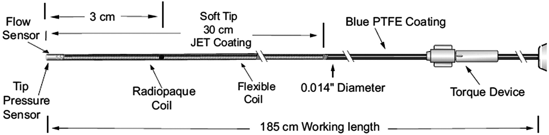
Intra-arterial BFV and pressure measurements are used on an increasingly regular basis in coronary interventions by using a dual-sensor pressure and Doppler velocity guidewire (Fig 1). Currently, the primary applications of such measurements serve to assess the hemodynamic consequences of coronary artery stenoses.6 We aimed to use this guidewire in intracranial vessels to evaluate this new use for the wire. We simultaneously acquired BFV and pressure signals in multiple locations in patients with unruptured intracranial aneurysms before endovascular treatment.

Fig 1.
  • Download figure
  • Open in new tab
  • Download powerpoint
Fig 1.

Detail of the ComboWire. Reprinted with permission of Volcano Therapeutics. PTFE indicates polytetrafluoroethylene.

Description of Techniques

Patients and Aneurysms

Four patients (52, 56, 64, and 70 years of age) scheduled to undergo endovascular coiling of an unruptured intracranial aneurysm were invited to participate in this study. Aneurysms were located at the ophthalmic artery (6 × 8 mm), the posterior communicating artery (9 × 9 and 6 × 7 mm), and the supraclinoid internal carotid artery (3 × 5 mm). The local ethics committee approved the study, and written informed consent was obtained from all participants.

Instrumentation

A 0.014-inch dual-sensor pressure and Doppler velocity wire (ComboWire; Volcano Corporation, Rancho Cordova, California) was used in combination with matching hardware (ComboMap pressure and flow system). The caliber and material of the ComboWire are identical to the standard guidewire used for endovascular treatment. Shaping of the tip for better navigation is possible but may harm its delicate sensors. To avoid potential added risk of adverse events (thrombus formation, perforation) caused by its straight tip, we did not use the ComboWire for navigation purposes. The wire tip was carefully positioned to avoid interference from the vessel wall. The velocity waveform comprises the temporal sequence of the maximum of fluid velocity within a cone-shaped beam 5 mm from the tip. Pressure measurements were considered valid when clear systole-diastole profiles were visible. Possible drift of the pressure signal intensity could be resolved by re-zeroing. All pressure and velocity signals were recorded at 200 Hz.

Flow-Velocity and Pressure Measurements

All procedures were performed with the patient under general anesthesia with full systemic heparinization on a single-plane angiographic unit (Integris Allura Neuro; Philips Medical Systems, Best, the Netherlands) following institutional protocol. The recordings also included the electrocardiogram and pressure derived from an intra-arterial pressure catheter in the radial artery.

A 5F guiding catheter was positioned in the vessel harboring the aneurysm, and the standard microcatheter was navigated beyond and within the aneurysm by using a standard microguidewire. Subsequently, the microguidewire was withdrawn and replaced by the ComboWire. All measurements were performed in the ICA, distal to the aneurysm, proximal to the aneurysm, and at the cervical level, consecutively. If aneurysm geometry and size allowed safe catheterization, measurements were also performed within the aneurysm. The exact locations were recorded with roadmap control. At each location, we recorded simultaneous BFV and pressure waveforms for a minimum of 10 consecutive cardiac cycles. All hemodynamic measurements took approximately 15 minutes and were completed before coil placement.

Data Analysis

The guidewire could not be positioned in 2 of 16 locations (12.5%) due to unfavorable geometry. The recorded raw data were extracted for further analysis. Figure 2 illustrates typical results in 1 patient: a poorly defined BFV pattern within the aneurysm, a lower maximal BFV at the cervical level compared with other locations, and stable pressure pulses (systolic minus diastolic pressure) in all locations. Averaging pressure pulses measured in the different locations resulted in a mean of 65.8 ± 9.2 mm Hg in patient 1; 65.4 ± 2.59 mm Hg in patient 2; 48.3 ± 3.1 mm Hg in patient 3; and 48.8 ± 1.5 mm Hg in patient 4.

Fig 2.
  • Download figure
  • Open in new tab
  • Download powerpoint
Fig 2.

BFV and pressure measurements using the ComboWire in the ICA at the cervical level (A), proximal to the aneurysm (B), intra-aneurysmally (C), and distal to the aneurysm (D). The graphs show matching flow velocity and pressure waveforms extracted from the ComboMap recordings. The upper part of each panel depicts pressure (mm Hg): systemic pressure measured in the radial artery (Pa) and intracranial pressure measured at the tip of the wire (Pd). The lower part of the graphs show flow velocity (centimeters per second). Sec indicates seconds.

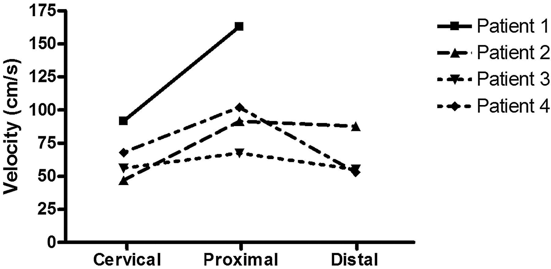
Figure 3 shows maximal BFV measured in all patients. An increase of maximal BFV proximal to the aneurysms compared with the cervical level of the ICA was seen in all patients (range, 11.5–71.4 cm/s). Measurements distal to the aneurysm were successful in 3 patients and failed in 1. Compared to proximal, BFV distal to the aneurysm remained stable in 2 patients and decreased in 1 patient.

Fig 3.
  • Download figure
  • Open in new tab
  • Download powerpoint
Fig 3.

Maximal BFV in centimeters per second measured with the ComboWire in the ICA at the cervical level (Cervical), proximal to the aneurysm (Proximal), and distal to the aneurysm (Distal). Intra-aneurysmal measurements are not plotted here because we considered them to be less accurate (see “Data Analysis”). Maximal BFV was calculated by averaging 5 consecutive peaks of the ComboMap flow signal. One measurement is lacking because the wire could not be placed distal to the aneurysm in patient 1 due to unfavorable geometry.

Discussion

We have successfully obtained simultaneous measurements of BFV and pressure in intracranial aneurysms and surrounding vessels by using a dual-sensor pressure and Doppler velocity wire (ComboWire). BFV was higher proximal to aneurysms compared with BFV at the cervical level of the ICA, consistent with the decreasing caliber of the upstream ICA. These data indicate that wall shear stress was enhanced proximal to the aneurysm. Pressure pulses were comparable between all intracranial locations within patients.

Noninvasive techniques for measuring flow velocities have been developed by using cardiac-gated 3D phase-contrast MR imaging.7 Unfortunately, prolonged MR imaging is usually not suitable in acutely ill patients with ruptured aneurysms. Benndorf et al8 have previously demonstrated the feasibility of flow-velocity measurements within giant intracranial aneurysms with a Doppler guidewire in 2 patients. We were not able to obtain clearly defined BFV measurements within aneurysms. The size and shape of the conebeam used for velocity recording makes this technique unsuitable for direct use inside the small- to medium-sized aneurysms of our study. When measuring inside an aneurysm, large spatial gradients in velocity magnitude and direction occur in the sampling volume. These lead to an envelope of maximal velocities during the heart cycle, which is difficult to interpret. We only used the ComboWire in the ICA with relatively large caliber, to diminish vessel wall interference and to obtain reliable BFV and pressure profiles. It remains to be determined whether measurements are also feasible in smaller caliber middle cerebral, anterior cerebral, and vertebrobasilar arteries.

We encountered additional difficulties using the ComboWire. Obtaining a good Doppler flow signal intensity could take several minutes because of the relatively small caliber of tortuous vessels. Wire−vessel wall interference hampered pressure measurements. The added risk to patients is negligible when the ComboWire is not used for navigation and is inserted only via the microcatheter previously deployed with a standard microguidewire. Possible risks of using the ComboWire with a straight tip for navigation may be damage of the vessel wall with possible subsequent thrombus formation and perforation of the vessel or aneurysm wall.

The capability of the ComboWire to obtain simultaneously intra-arterial pressure and BFV provides a valuable novel way for determining patient-specific hemodynamics. The simulation of fluid flows by using CFD allows the prediction of hemodynamics, but lack of accurate flow-velocity measurements has hampered the study of hemodynamics of intracranial aneurysms.1,2 For future construction of patient-specific CFD models, we are now able to provide BFV proximal and distal to the aneurysm. The pressure recordings provide additional information on the mechanical load imposed on the aneurysm. Both measurements may be vital in the individual rupture-risk assessment of patients with an unruptured aneurysm.

Acknowledgment

We thank H. Gratama van Andel for his extensive help with the measurements.

Footnotes

  • ComboWires used in this study were sponsored by Volcano Therapeutics, Europe Headquarters, Zaventem, Belgium.

References

  1. 1.↵
    1. Cebral JR,
    2. Castro MA,
    3. Burgess JE,
    4. et al
    . Characterization of cerebral aneurysms for assessing risk of rupture by using patient-specific computational hemodynamics models. AJNR Am J Neuroradiol 2005;26:2550–59
    Abstract/FREE Full Text
  2. 2.↵
    1. Castro MA,
    2. Putman CM,
    3. Cebral JR
    . Patient-specific computational fluid dynamics modeling of anterior communicating artery aneurysms: a study of the sensitivity of intra-aneurysmal flow patterns to flow conditions in the carotid arteries. AJNR Am J Neuroradiol 2006;27:2061–68
    Abstract/FREE Full Text
  3. 3.↵
    1. Venugopal P,
    2. Valentino D,
    3. Schmitt H,
    4. et al
    . Sensitivity of patient-specific numerical simulation of cerebral aneurysm hemodynamics to inflow boundary conditions. J Neurosurg 2007;106:1051–60
    CrossRefPubMed
  4. 4.↵
    1. Hashimoto T
    . Flow velocity studies in vein pouch model aneurysms. Neurol Res 1993;15:185–91
    PubMed
  5. 5.↵
    1. Chatzipromodrou I,
    2. Tricoli A,
    3. Poulikanos D,
    4. et al
    . Haemodynamics and wall remodelling of a growing cerebral aneurysm: a computational model. J Biomech 2007;40:412–26. Epub 2006 Mar 9
    CrossRefPubMed
  6. 6.↵
    1. Siebes M,
    2. Verhoeff BJ,
    3. Meeuwissen M,
    4. et al
    . Single-wire pressure and flow velocity measurement to quantify coronary stenosis hemodynamics and effects of percutaneous interventions. Circulation 2004;109:756–62
    Abstract/FREE Full Text
  7. 7.↵
    1. Meckel S,
    2. Stalder AF,
    3. Santini F,
    4. et al
    . In vivo visualization and analysis of 3-D hemodynamics in cerebral aneurysms with flow-sensitized 4-D MR imaging at 3 T. Neuroradiology 2008;50:473–84. Epub 2008 Mar 19
    CrossRefPubMed
  8. 8.↵
    1. Benndorf G,
    2. Wellnhofer E,
    3. Lanksch W,
    4. et al
    . Intraaneurysmal flow: evaluation with Doppler guidewires. AJNR Am J Neuroradiol 1996;17:1333–37
    Abstract
  • Received March 20, 2009.
  • Accepted after revision April 26, 2009.
  • Copyright © American Society of Neuroradiology
View Abstract
PreviousNext
Back to top

In this issue

American Journal of Neuroradiology: 31 (2)
American Journal of Neuroradiology
Vol. 31, Issue 2
1 Feb 2010
  • Table of Contents
  • Index by author
Advertisement
Print
Download PDF
Email Article

Thank you for your interest in spreading the word on American Journal of Neuroradiology.

NOTE: We only request your email address so that the person you are recommending the page to knows that you wanted them to see it, and that it is not junk mail. We do not capture any email address.

Enter multiple addresses on separate lines or separate them with commas.
Intracranial Blood-Flow Velocity and Pressure Measurements Using an Intra-Arterial Dual-Sensor Guidewire
(Your Name) has sent you a message from American Journal of Neuroradiology
(Your Name) thought you would like to see the American Journal of Neuroradiology web site.
CAPTCHA
This question is for testing whether or not you are a human visitor and to prevent automated spam submissions.
Cite this article
S.P. Ferns, J.J. Schneiders, M. Siebes, R. van den Berg, E.T. van Bavel, C.B. Majoie
Intracranial Blood-Flow Velocity and Pressure Measurements Using an Intra-Arterial Dual-Sensor Guidewire
American Journal of Neuroradiology Feb 2010, 31 (2) 324-326; DOI: 10.3174/ajnr.A1718

Citation Manager Formats

  • BibTeX
  • Bookends
  • EasyBib
  • EndNote (tagged)
  • EndNote 8 (xml)
  • Medlars
  • Mendeley
  • Papers
  • RefWorks Tagged
  • Ref Manager
  • RIS
  • Zotero
0 Responses
Respond to this article
Share
Bookmark this article
Intracranial Blood-Flow Velocity and Pressure Measurements Using an Intra-Arterial Dual-Sensor Guidewire
S.P. Ferns, J.J. Schneiders, M. Siebes, R. van den Berg, E.T. van Bavel, C.B. Majoie
American Journal of Neuroradiology Feb 2010, 31 (2) 324-326; DOI: 10.3174/ajnr.A1718
del.icio.us logo Twitter logo Facebook logo Mendeley logo
  • Tweet Widget
  • Facebook Like
  • Google Plus One
Purchase

Jump to section

  • Article
    • Abstract
    • Abbreviations
    • Description of Techniques
    • Discussion
    • Acknowledgment
    • Footnotes
    • References
  • Figures & Data
  • Info & Metrics
  • Responses
  • References
  • PDF

Related Articles

  • No related articles found.
  • PubMed
  • Google Scholar

Cited By...

  • Cerebral Aneurysms Treated with Flow-Diverting Stents: Computational Models with Intravascular Blood Flow Measurements
  • Crossref (19)
  • Google Scholar

This article has been cited by the following articles in journals that are participating in Crossref Cited-by Linking.

  • Computational Hemodynamics in Cerebral Aneurysms: The Effects of Modeled Versus Measured Boundary Conditions
    Alberto Marzo, Pankaj Singh, Ignacio Larrabide, Alessandro Radaelli, Stuart Coley, Matt Gwilliam, Iain D. Wilkinson, Patricia Lawford, Philippe Reymond, Umang Patel, Alejandro Frangi, D. Rod Hose
    Annals of Biomedical Engineering 2011 39 2
  • Physical Factors Effecting Cerebral Aneurysm Pathophysiology
    Chander Sadasivan, David J. Fiorella, Henry H. Woo, Baruch B. Lieber
    Annals of Biomedical Engineering 2013 41 7
  • Cerebral Aneurysms Treated with Flow-Diverting Stents: Computational Models with Intravascular Blood Flow Measurements
    M.R. Levitt, P.M. McGah, A. Aliseda, P.D. Mourad, J.D. Nerva, S.S. Vaidya, R.P. Morton, B.V. Ghodke, L.J. Kim
    American Journal of Neuroradiology 2014 35 1
  • Fully Printed, Wireless, Stretchable Implantable Biosystem toward Batteryless, Real‐Time Monitoring of Cerebral Aneurysm Hemodynamics
    Robert Herbert, Saswat Mishra, Hyo‐Ryoung Lim, Hyoungsuk Yoo, Woon‐Hong Yeo
    Advanced Science 2019 6 18
  • Fractional Flow Assessment for the Evaluation of Intracranial Atherosclerosis: A Feasibility Study
    ZhongRong Miao, David S. Liebeskind, WaiTing Lo, LiPing Liu, YueHua Pu, XinYi Leng, LiGang Song, XiaoTong Xu, BaiXue Jia, Feng Gao, DaPeng Mo, Xuan Sun, Lian Liu, Ning Ma, Bo Wang, YiLong Wang, YongJun Wang
    Interventional Neurology 2016 5 1-2
  • A Flow-Diverting Stent Is Not a Pressure-Diverting Stent
    J.J. Schneiders, E. VanBavel, C.B. Majoie, S.P. Ferns, R. van den Berg
    American Journal of Neuroradiology 2013 34 1
  • Comparison of Phase-Contrast MR Imaging and Endovascular Sonography for Intracranial Blood Flow Velocity Measurements
    J.J. Schneiders, S.P. Ferns, P. van Ooij, M. Siebes, A.J. Nederveen, R. van den Berg, J. van Lieshout, G. Jansen, E. vanBavel, C.B. Majoie
    American Journal of Neuroradiology 2012 33 9
  • Stretchable, Implantable, Nanostructured Flow-Diverter System for Quantification of Intra-aneurysmal Hemodynamics
    Connor Howe, Saswat Mishra, Yun-Soung Kim, Yanfei Chen, Sang-Ho Ye, William R. Wagner, Jae-Woong Jeong, Hun-Soo Byun, Jong-Hoon Kim, Youngjae Chun, Woon-Hong Yeo
    ACS Nano 2018 12 8
  • Non Invasive Blood Flow Features Estimation in Cerebral Arteries from Uncertain Medical Data
    R. Lal, F. Nicoud, E. Le Bars, J. Deverdun, F. Molino, V. Costalat, B. Mohammadi
    Annals of Biomedical Engineering 2017 45 11
  • Quantitative comparison of the dynamic flow waveform changes in 12 ruptured and 29 unruptured ICA–ophthalmic artery aneurysms
    Aichi Chien, James Sayre, Fernando Viñuela
    Neuroradiology 2013 55 3

More in this TOC Section

  • Contour Neurovascular System: Five Year Follow Up
  • Effect of SARS-CoV2 on Endovascular Thrombectomy
  • Flow diversion for distal circulation aneurysms
Show more NEUROINTERVENTION

Similar Articles

Advertisement

Indexed Content

  • Current Issue
  • Accepted Manuscripts
  • Article Preview
  • Past Issues
  • Editorials
  • Editor's Choice
  • Fellows' Journal Club
  • Letters to the Editor
  • Video Articles

Cases

  • Case Collection
  • Archive - Case of the Week
  • Archive - Case of the Month
  • Archive - Classic Case

More from AJNR

  • Trainee Corner
  • Imaging Protocols
  • MRI Safety Corner
  • Book Reviews

Multimedia

  • AJNR Podcasts
  • AJNR Scantastics

Resources

  • Turnaround Time
  • Submit a Manuscript
  • Submit a Video Article
  • Submit an eLetter to the Editor/Response
  • Manuscript Submission Guidelines
  • Statistical Tips
  • Fast Publishing of Accepted Manuscripts
  • Graphical Abstract Preparation
  • Imaging Protocol Submission
  • Evidence-Based Medicine Level Guide
  • Publishing Checklists
  • Author Policies
  • Become a Reviewer/Academy of Reviewers
  • News and Updates

About Us

  • About AJNR
  • Editorial Board
  • Editorial Board Alumni
  • Alerts
  • Permissions
  • Not an AJNR Subscriber? Join Now
  • Advertise with Us
  • Librarian Resources
  • Feedback
  • Terms and Conditions
  • AJNR Editorial Board Alumni

American Society of Neuroradiology

  • Not an ASNR Member? Join Now

© 2025 by the American Society of Neuroradiology All rights, including for text and data mining, AI training, and similar technologies, are reserved.
Print ISSN: 0195-6108 Online ISSN: 1936-959X

Powered by HighWire