
Article Figures & Data
Figures
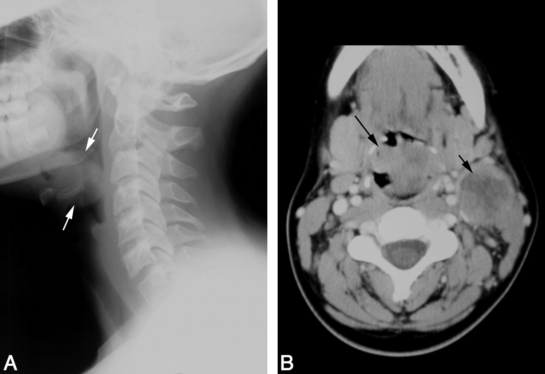
- Fig 1.
Patient 1. Carcinoma of the epiglottis; t(15;19). A, Lateral film of the neck reveals an irregular epiglottic mass (arrows). B, Contrast-enhanced neck CT scan demonstrates the irregular, midline epiglottic mass (long arrow). There is necrotic left level II adenopathy (short arrow).
- Fig 2.
Patient 2. Carcinoma of the larynx. Contrast-enhanced CT scan of the neck demonstrates an irregular and heterogeneously enhancing, off-midline submucosal mass (arrow) involving the right side of the larynx.
- Fig 3.
Patient 3. Sinonasal carcinoma. A, Coronal CT scan of the sinuses reveals a tumor (arrow) arising from the left nasal cavity. The mass causes marked thinning and lateral deviation of the medial wall of the left maxillary antrum (arrowhead) and rightward deviation of the nasal septum. B, Coronal FSEIR MR imaging demonstrates that the tumor (arrow) is hypointense compared with the trapped secretions in the left maxillary antrum (star). C, Gadolinium-enhanced, fat-suppressed coronal T1-weighted MR image shows that the tumor (arrow) enhances homogeneously.
- Fig 4.
Patient 4. Sinonasal carcinoma t(15;19). A, CT scan demonstrates a midline sinonasal tumor (arrow) with lytic bony destruction of the paranasal sinuses and hard palate (arrowheads). B, Coronal FSEIR MR demonstrates the tumor (arrow), which had a relatively short T2 relaxation time but was hyperintense relative to muscle. C, Sagittal gadolinium-enhanced, fat-suppressed T1-weighted MR image demonstrates enhancing tumor (arrow) extending into the nasopharynx. There is dural enhancement superiorly (arrowheads).


Tables
Patient results
Patient 1 Patient 2 Patient 3 Patient 4 Age (y)/Sex 13/F 14/F 15/M 12/F Symptoms 6 wk sore throat, odynophagia, voice change, neck mass 4 mos hoarseness, 1 mo odynophagia, dysphagia Nasal mass 3 wk URT symptoms and nasal mass Tumor location Epiglottis Epiglottis and glottis Sinonasal Sinonasal CT scan results 3-cm heterogeneously enhancing epiglottic mass necrotic, level II LN 3-cm heterogeneously enhancing submucosal mass 3 cm lobulated, moderately enhancing mass left nasal cavity, bony remodeling 5 × 7-cm moderately enhancing mass, nasal cavity. Bony destruction sinuses, skull base MR imaging results Isointense with cortex on FSEIR Isointense with cortex on FSEIR Isointense with cortex on FSEIR Surgery Modified radical neck dissection Biopsy Midface degloving; left Caldwell-Luc Biopsy Histopathology Undifferentiated carcinoma Well-differentiated squamous carcinoma Poorly differentiated squamous carcinoma Poorly differentiated squamous carcinoma Cytogenetics 47,XX, +8, t(15;19)(q13;p13.1). 46,XY, +der(1)t(1;5)(p12;q11.1), −5,del(22)(q11q13) T(15;19) EBV Negative Negative Negative Chemotherapy Cisplatin, 5-fluorouracil, docetaxel, methotrexate, vinblastine Cisplatin VCR/Cytoxan, Adriamycin, etoposide, carboplatin Carboplatin Radiation 69 Gy to supraglottis 70.2 Gy to larynx 64.4 Gy to nasopharynx 54.0 Gy to neck nodes 52.2 Gy to neck nodes 50.4 Gy to neck nodes Clinical course Disseminated disease. Death 10 mos after diagnosis Alive at 8 y f/u. Laryngeal scarring, stenosis. Tracheostomy Disease-free at 8 y f/u Disseminated disease. Death 3 mos VBIO, VCR after diagnosis
In this issue


Jump to section
Related Articles
Cited By...
- No citing articles found.