Advertisement
  • Main menu
  • User menu
  • Search
  • English ▼
    • English
    • Afrikaans
    • Albanian
    • Amharic
    • Arabic
    • Armenian
    • Azerbaijani
    • Basque
    • Belarusian
    • Bengali
    • Bosnian
    • Bulgarian
    • Catalan
    • Cebuano
    • Chichewa
    • Chinese (Simplified)
    • Chinese (Traditional)
    • Corsican
    • Croatian
    • Czech
    • Danish
    • Dutch
    • Esperanto
    • Estonian
    • Filipino
    • Finnish
    • French
    • Frisian
    • Galician
    • Georgian
    • German
    • Greek
    • Gujarati
    • Haitian Creole
    • Hausa
    • Hawaiian
    • Hebrew
    • Hindi
    • Hmong
    • Hungarian
    • Icelandic
    • Igbo
    • Indonesian
    • Irish
    • Italian
    • Japanese
    • Javanese
    • Kannada
    • Kazakh
    • Khmer
    • Korean
    • Kurdish (Kurmanji)
    • Kyrgyz
    • Lao
    • Latin
    • Latvian
    • Lithuanian
    • Luxembourgish
    • Macedonian
    • Malagasy
    • Malay
    • Malayalam
    • Maltese
    • Maori
    • Marathi
    • Mongolian
    • Myanmar (Burmese)
    • Nepali
    • Norwegian
    • Pashto
    • Persian
    • Polish
    • Portuguese
    • Punjabi
    • Romanian
    • Russian
    • Samoan
    • Scottish Gaelic
    • Serbian
    • Sesotho
    • Shona
    • Sindhi
    • Sinhala
    • Slovak
    • Slovenian
    • Somali
    • Spanish
    • Sudanese
    • Swahili
    • Swedish
    • Tajik
    • Tamil
    • Telugu
    • Thai
    • Turkish
    • Ukrainian
    • Urdu
    • Uzbek
    • Vietnamese
    • Welsh
    • Xhosa
    • Yiddish
    • Yoruba
    • Zulu

Main menu

  • Home
  • Content
    • Current Issue
    • Accepted Manuscripts
    • Article Preview
    • Past Issue Archive
    • Video Articles
    • AJNR Case Collection
    • Case of the Week Archive
    • Case of the Month Archive
    • Classic Case Archive
  • Special Collections
    • AJNR Awards
    • Low-Field MRI
    • Alzheimer Disease
    • ASNR Foundation Special Collection
    • Photon-Counting CT
    • View All
  • Multimedia
    • AJNR Podcasts
    • AJNR SCANtastic
    • Trainee Corner
    • MRI Safety Corner
    • Imaging Protocols
  • For Authors
    • Submit a Manuscript
    • Submit a Video Article
    • Submit an eLetter to the Editor/Response
    • Manuscript Submission Guidelines
    • Statistical Tips
    • Fast Publishing of Accepted Manuscripts
    • Graphical Abstract Preparation
    • Imaging Protocol Submission
    • Author Policies
  • About Us
    • About AJNR
    • Editorial Board
    • Editorial Board Alumni
  • More
    • Become a Reviewer/Academy of Reviewers
    • Subscribers
    • Permissions
    • Alerts
    • Feedback
    • Advertisers
    • ASNR Home

User menu

  • Alerts
  • Log in

Search

  • Advanced search
American Journal of Neuroradiology

American Journal of Neuroradiology

ASHNR American Society of Functional Neuroradiology ASHNR American Society of Pediatric Neuroradiology ASSR
  • Alerts
  • Log in

Advanced Search

English ▼
  • English
  • Afrikaans
  • Albanian
  • Amharic
  • Arabic
  • Armenian
  • Azerbaijani
  • Basque
  • Belarusian
  • Bengali
  • Bosnian
  • Bulgarian
  • Catalan
  • Cebuano
  • Chichewa
  • Chinese (Simplified)
  • Chinese (Traditional)
  • Corsican
  • Croatian
  • Czech
  • Danish
  • Dutch
  • Esperanto
  • Estonian
  • Filipino
  • Finnish
  • French
  • Frisian
  • Galician
  • Georgian
  • German
  • Greek
  • Gujarati
  • Haitian Creole
  • Hausa
  • Hawaiian
  • Hebrew
  • Hindi
  • Hmong
  • Hungarian
  • Icelandic
  • Igbo
  • Indonesian
  • Irish
  • Italian
  • Japanese
  • Javanese
  • Kannada
  • Kazakh
  • Khmer
  • Korean
  • Kurdish (Kurmanji)
  • Kyrgyz
  • Lao
  • Latin
  • Latvian
  • Lithuanian
  • Luxembourgish
  • Macedonian
  • Malagasy
  • Malay
  • Malayalam
  • Maltese
  • Maori
  • Marathi
  • Mongolian
  • Myanmar (Burmese)
  • Nepali
  • Norwegian
  • Pashto
  • Persian
  • Polish
  • Portuguese
  • Punjabi
  • Romanian
  • Russian
  • Samoan
  • Scottish Gaelic
  • Serbian
  • Sesotho
  • Shona
  • Sindhi
  • Sinhala
  • Slovak
  • Slovenian
  • Somali
  • Spanish
  • Sudanese
  • Swahili
  • Swedish
  • Tajik
  • Tamil
  • Telugu
  • Thai
  • Turkish
  • Ukrainian
  • Urdu
  • Uzbek
  • Vietnamese
  • Welsh
  • Xhosa
  • Yiddish
  • Yoruba
  • Zulu
  • Home
  • Content
    • Current Issue
    • Accepted Manuscripts
    • Article Preview
    • Past Issue Archive
    • Video Articles
    • AJNR Case Collection
    • Case of the Week Archive
    • Case of the Month Archive
    • Classic Case Archive
  • Special Collections
    • AJNR Awards
    • Low-Field MRI
    • Alzheimer Disease
    • ASNR Foundation Special Collection
    • Photon-Counting CT
    • View All
  • Multimedia
    • AJNR Podcasts
    • AJNR SCANtastic
    • Trainee Corner
    • MRI Safety Corner
    • Imaging Protocols
  • For Authors
    • Submit a Manuscript
    • Submit a Video Article
    • Submit an eLetter to the Editor/Response
    • Manuscript Submission Guidelines
    • Statistical Tips
    • Fast Publishing of Accepted Manuscripts
    • Graphical Abstract Preparation
    • Imaging Protocol Submission
    • Author Policies
  • About Us
    • About AJNR
    • Editorial Board
    • Editorial Board Alumni
  • More
    • Become a Reviewer/Academy of Reviewers
    • Subscribers
    • Permissions
    • Alerts
    • Feedback
    • Advertisers
    • ASNR Home
  • Follow AJNR on Twitter
  • Visit AJNR on Facebook
  • Follow AJNR on Instagram
  • Join AJNR on LinkedIn
  • RSS Feeds

AJNR Awards, New Junior Editors, and more. Read the latest AJNR updates

Review ArticleReview Articles
Open Access

A Compartment-Based Approach for the Imaging Evaluation of Tinnitus

S. Vattoth, R. Shah and J.K. Curé
American Journal of Neuroradiology February 2010, 31 (2) 211-218; DOI: https://doi.org/10.3174/ajnr.A1704
S. Vattoth
  • Find this author on Google Scholar
  • Find this author on PubMed
  • Search for this author on this site
R. Shah
  • Find this author on Google Scholar
  • Find this author on PubMed
  • Search for this author on this site
J.K. Curé
  • Find this author on Google Scholar
  • Find this author on PubMed
  • Search for this author on this site
  • Article
  • Figures & Data
  • Supplemental
  • Info & Metrics
  • Responses
  • References
  • PDF
Loading

Article Figures & Data

Figures

  • Fig 1.
    • Download figure
    • Open in new tab
    • Download powerpoint
    Fig 1.

    Glomus tympanicum (arrow) in a 30-year-old woman with right-sided pulsatile tinnitus. Axial CT image demonstrates lobulated soft-tissue attenuation in the middle ear overlying the cochlear promontory.

  • Fig 2.
    • Download figure
    • Open in new tab
    • Download powerpoint
    Fig 2.

    Aberrant course of the ICA in a 25-year-old man presenting with pulsatile tinnitus. A, Enhanced axial CT image demonstrates an abnormal lateral course of the right ICA through the middle ear (white arrow). Also note dehiscence of the overlying bony plate. B, Anteroposterior projection image from the MR angiogram of the same patient demonstrates decreased caliber and lateral deviation of the aberrant ICA on the “right reversed-7 sign” (black arrow).
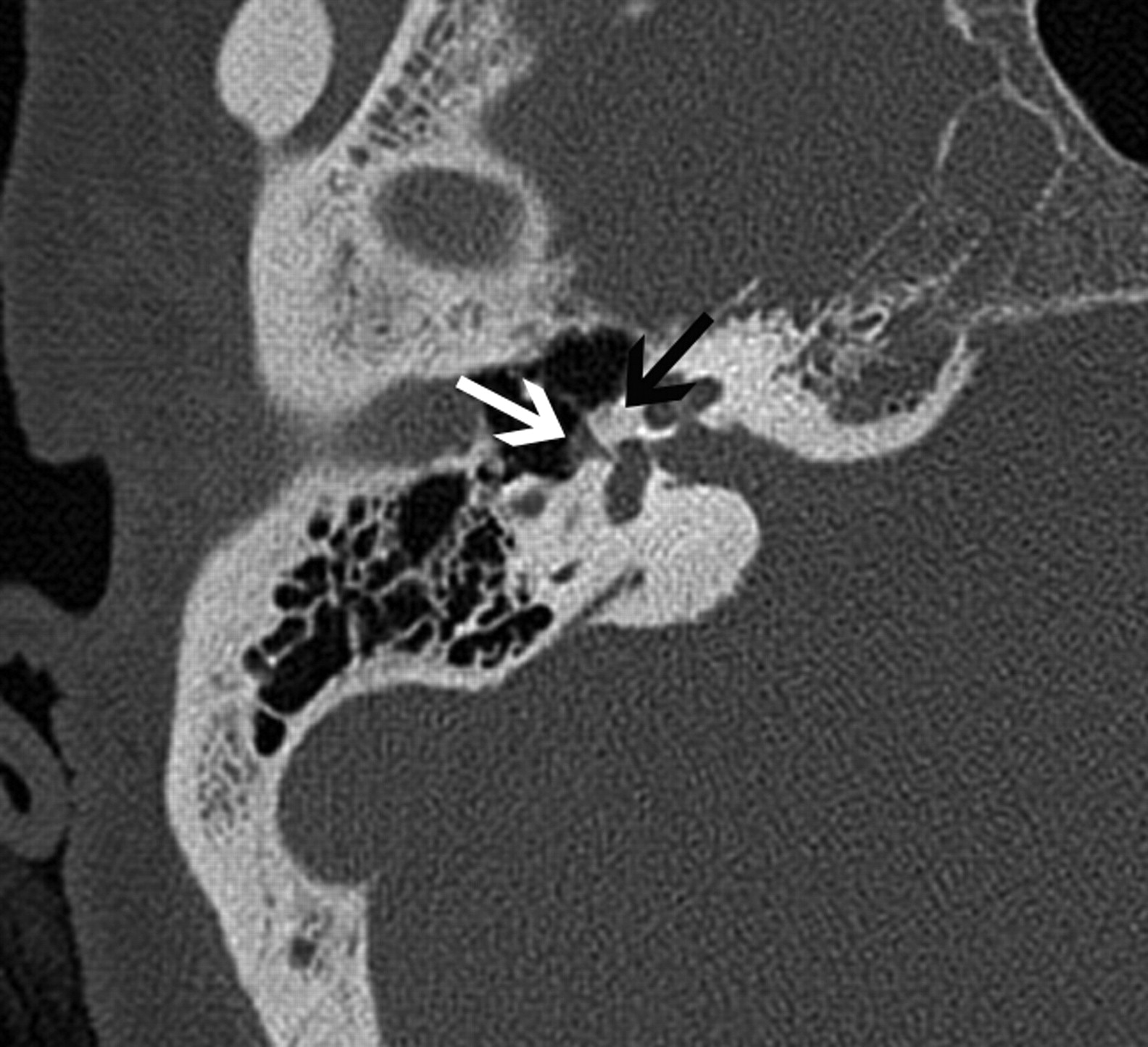
  • Fig 3.
    • Download figure
    • Open in new tab
    • Download powerpoint
    Fig 3.

    Fenestral otospongiosis in a 35-year-old woman with hearing loss and tinnitus. Axial CT image demonstrates soft-tissue attenuation in the oval window (white arrow) and abnormal lucency at the fissula antefenestram (black arrow).

  • Fig 4.
    • Download figure
    • Open in new tab
    • Download powerpoint
    Fig 4.

    A 50-year-old man with cochlear otospongiosis. Axial CT scan demonstrates abnormal lucency surrounding the left cochlea (black arrows).

  • Fig 5.
    • Download figure
    • Open in new tab
    • Download powerpoint
    Fig 5.

    Paget disease of the skull base in a 70-year-old man. Axial CT scan demonstrates diffuse expansion and sclerosis of the bones of the skull base, characteristic of Paget disease. Note the sparing of the maxillofacial bones, which, along with the age of the patient, is a helpful feature in differentiating it from fibrous dysplasia.
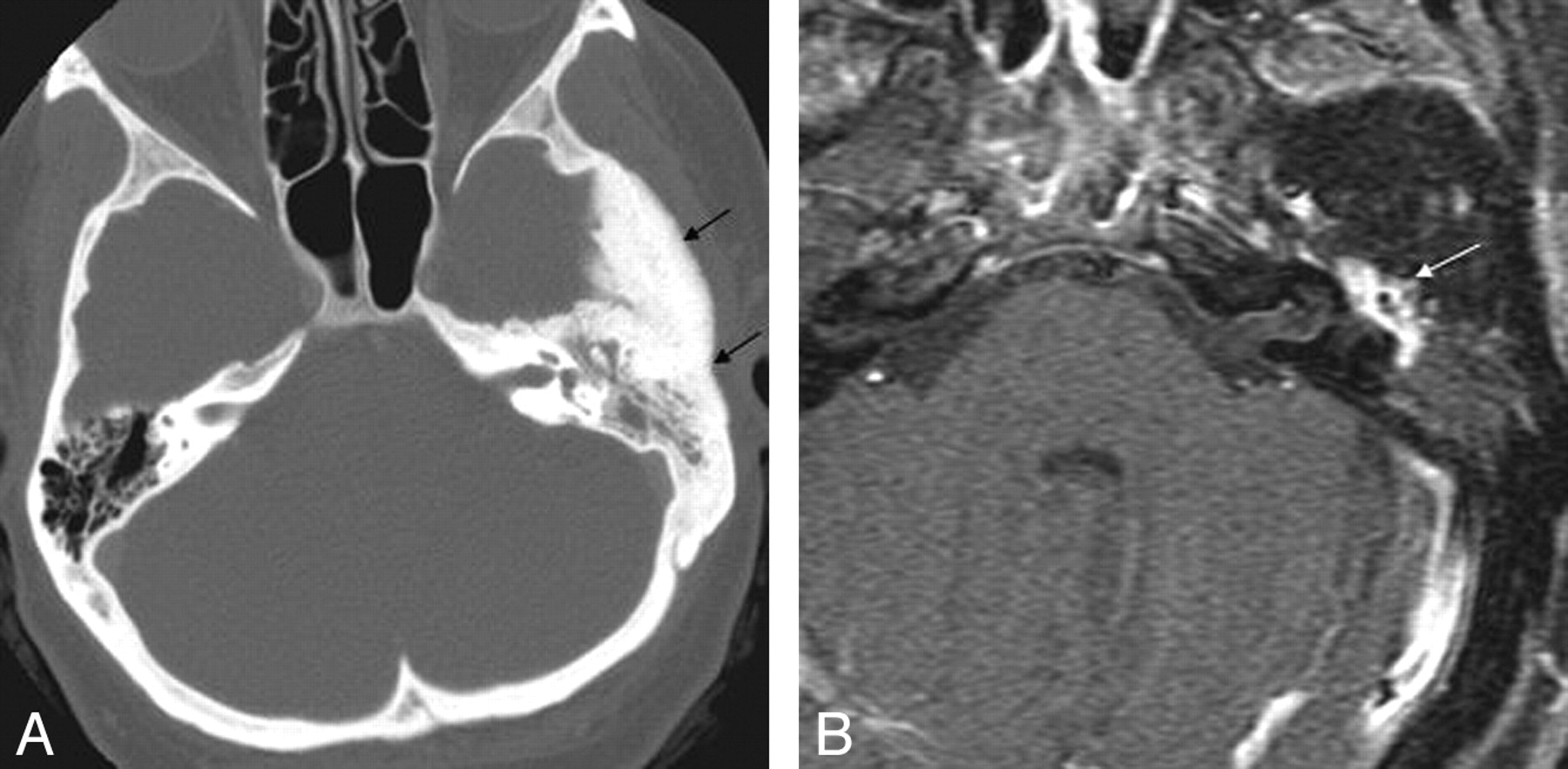
  • Fig 6.
    • Download figure
    • Open in new tab
    • Download powerpoint
    Fig 6.

    Intraosseous temporal bone meningioma in a 45-year-old woman who presented with left-sided hearing loss and tinnitus. A, Axial CT image demonstrates diffuse sclerosis of the temporal bone and soft-tissue attenuation (black arrows) in the left mastoid and middle ear cavity. B, Axial enhanced MR image in the same patient demonstrates enhancement within the left middle ear (white arrow).

  • Fig 7.
    • Download figure
    • Open in new tab
    • Download powerpoint
    Fig 7.

    A 50-year-old man with MD. Axial thin-section CT image shows decreased distance between the vertical limb of the posterior semicircular canal and the posterior edge of the temporal bone (black arrow).
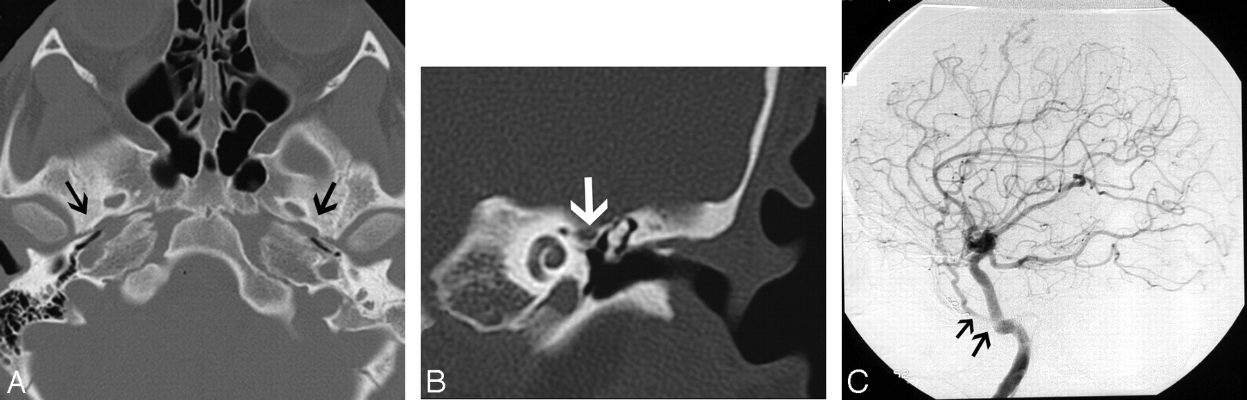
  • Fig 8.
    • Download figure
    • Open in new tab
    • Download powerpoint
    Fig 8.

    A 30-year-old man with PSA. A, Axial CT image demonstrates absence of the foramen spinosum bilaterally (black arrows). B, Coronal CT image in the same patient shows enlargement of the tympanic part of the facial nerve canal, another indirect imaging sign of PSA. C, Lateral DSA image of the internal carotid injection shows the PSA arising from the ICA (arrows).

  • Fig 9.
    • Download figure
    • Open in new tab
    • Download powerpoint
    Fig 9.

    Glomus jugulotympanicum in 45-year-old woman who presented with left-sided pulsatile tinnitus. A, Coronal CT image demonstrates soft-tissue attenuation projecting in the hypotympanum (white arrow). There is permeative osseous destruction in the left jugular foramen (black arrows), providing a clue as to the origin of the mass in the left jugular foramen. B, Enhanced coronal MR image better demonstrates the true extent of the mass within the left jugular foramen (black arrows).

  • Fig 10.
    • Download figure
    • Open in new tab
    • Download powerpoint
    Fig 10.

    Anterior inferior cerebellar artery loop in a 35-year-old man. Axial T2-weighted MR image shows a vascular loop entering the IAC (black arrow).
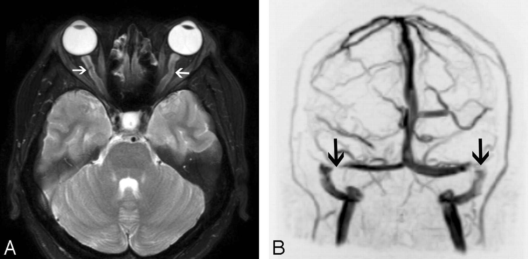
  • Fig 11.
    • Download figure
    • Open in new tab
    • Download powerpoint
    Fig 11.

    Intracranial hypertension in a 30-year-old woman presenting with headaches and tinnitus. A, Axial T2-weighted image demonstrates dilation of bilateral optic nerve sheaths (arrows) and flattening of the posterior globes. B, Coronal maximum-intensity-projection image from an MR venogram also demonstrates stenoses of bilateral transverse sinuses (arrows).
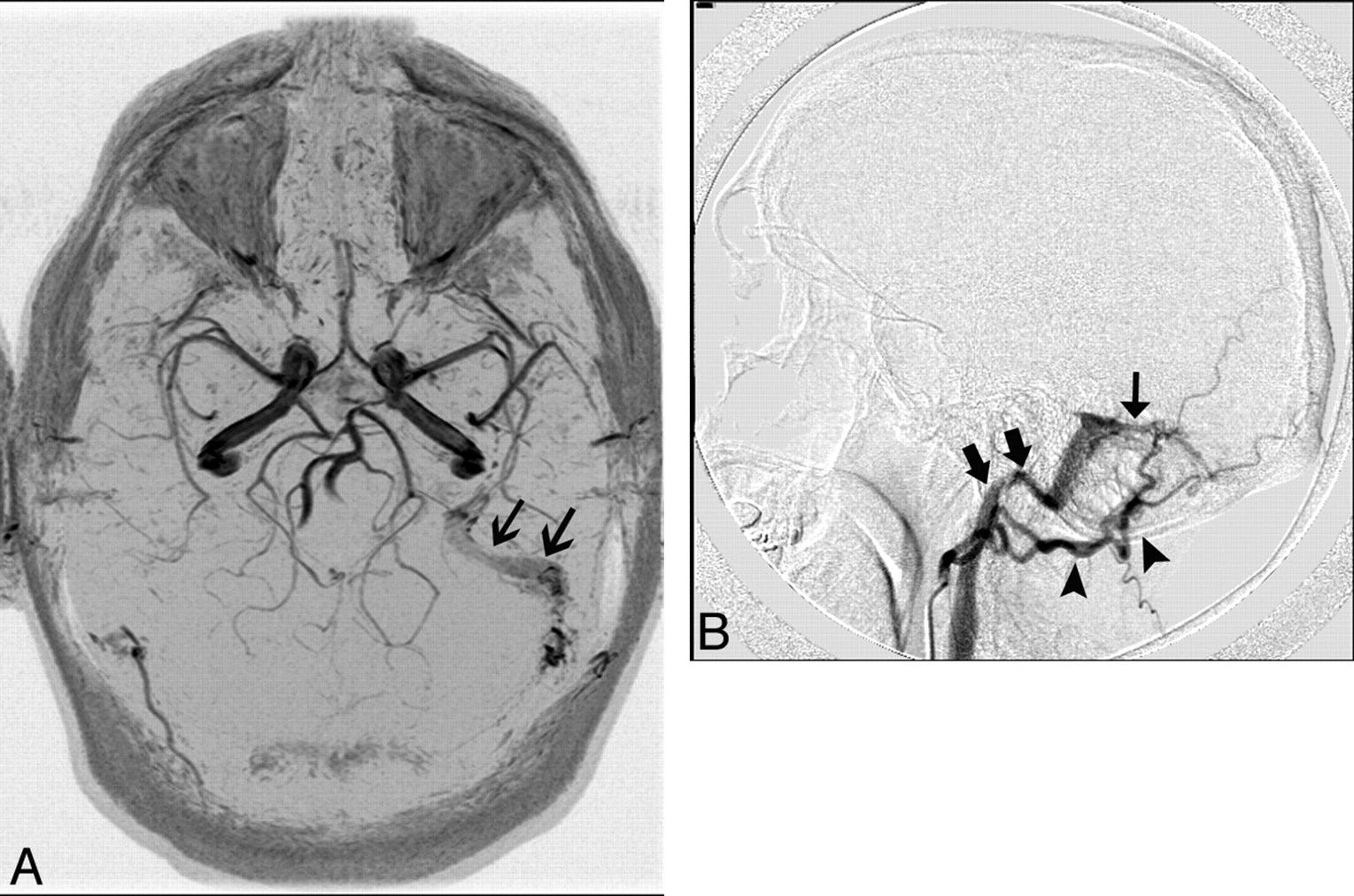
  • Fig 12.
    • Download figure
    • Open in new tab
    • Download powerpoint
    Fig 12.

    A 40-year-old man who presented with left-sided pulsatile tinnitus. A, MR angiogram demonstrates asymmetric increased flow-related enhancement in the left sigmoid sinus (black arrows) and multiple small arterial flow voids within it. B, Lateral projection of DSA shows injection of the left occipital artery (arrowheads) opacifying the AVF (thin black arrow) and rapid opacification of the left sigmoid sinus and internal jugular vein (thick black arrows).

PreviousNext
Back to top

In this issue

American Journal of Neuroradiology: 31 (2)
American Journal of Neuroradiology
Vol. 31, Issue 2
1 Feb 2010
  • Table of Contents
  • Index by author
Advertisement
Print
Download PDF
Email Article
Cite this article
0 Responses
Respond to this article
Share
Bookmark this article
A Compartment-Based Approach for the Imaging Evaluation of Tinnitus
S. Vattoth, R. Shah, J.K. Curé
American Journal of Neuroradiology Feb 2010, 31 (2) 211-218; DOI: 10.3174/ajnr.A1704
del.icio.us logo Twitter logo Facebook logo Mendeley logo
  • Tweet Widget
Purchase

Jump to section

  • Article
    • Abstract
    • Abbreviations
    • Role of Imaging in the Clinical Evaluation of Tinnitus
    • Choice of Imaging Technique and Expected Yield
    • A Compartment-Based Approach to Identify the Etiology of Tinnitus
    • Membranous Labyrinth
    • Skull Base Foramina/Canals
    • Jugular Foramen
    • Brain/Intracranial
    • Neck
    • TMJ
    • Conclusions
    • Acknowledgment
    • Footnotes
    • References
  • Figures & Data
  • Supplemental
  • Info & Metrics
  • Responses
  • References
  • PDF

Related Articles

  • PubMed
  • Google Scholar

Cited By...

  • Temporal Bone CT: Anatomy, Technique, and Associated Pathophysiology
  • CT Angiography as a Screening Tool for Dural Arteriovenous Fistula in Patients with Pulsatile Tinnitus: Feasibility and Test Characteristics
  • Crossref
  • Google Scholar

This article has not yet been cited by articles in journals that are participating in Crossref Cited-by Linking.

More in this TOC Section

  • An Atlas of Neonatal Neurovascular Imaging Anatomy as Depicted with Microvascular Imaging: The Intracranial Arteries
  • An Atlas of Neonatal Neurovascular Imaging Anatomy as Depicted with Microvascular Imaging: The Intracranial Veins
  • Clinical Translation of Hyperpolarized 13C Metabolic Probes for Glioma Imaging
Show more Review Articles

Similar Articles

Advertisement

Indexed Content

  • Current Issue
  • Accepted Manuscripts
  • Article Preview
  • Past Issues
  • Editorials
  • Editor's Choice
  • Fellows' Journal Club
  • Letters to the Editor
  • Video Articles

Cases

  • Case Collection
  • Archive - Case of the Week
  • Archive - Case of the Month
  • Archive - Classic Case

More from AJNR

  • Trainee Corner
  • Imaging Protocols
  • MRI Safety Corner
  • Book Reviews

Multimedia

  • AJNR Podcasts
  • AJNR Scantastics

Resources

  • Turnaround Time
  • Submit a Manuscript
  • Submit a Video Article
  • Submit an eLetter to the Editor/Response
  • Manuscript Submission Guidelines
  • Statistical Tips
  • Fast Publishing of Accepted Manuscripts
  • Graphical Abstract Preparation
  • Imaging Protocol Submission
  • Evidence-Based Medicine Level Guide
  • Publishing Checklists
  • Author Policies
  • Become a Reviewer/Academy of Reviewers
  • News and Updates

About Us

  • About AJNR
  • Editorial Board
  • Editorial Board Alumni
  • Alerts
  • Permissions
  • Not an AJNR Subscriber? Join Now
  • Advertise with Us
  • Librarian Resources
  • Feedback
  • Terms and Conditions
  • AJNR Editorial Board Alumni

American Society of Neuroradiology

  • Not an ASNR Member? Join Now

© 2025 by the American Society of Neuroradiology All rights, including for text and data mining, AI training, and similar technologies, are reserved.
Print ISSN: 0195-6108 Online ISSN: 1936-959X

Powered by HighWire
Email this Article

Thank you for your interest in spreading the word on American Journal of Neuroradiology.

NOTE: We only request your email address so that the person you are recommending the page to knows that you wanted them to see it, and that it is not junk mail. We do not capture any email address.

Enter multiple addresses on separate lines or separate them with commas.
A Compartment-Based Approach for the Imaging Evaluation of Tinnitus
(Your Name) has sent you a message from American Journal of Neuroradiology
(Your Name) thought you would like to see the American Journal of Neuroradiology web site.
CAPTCHA
This question is for testing whether or not you are a human visitor and to prevent automated spam submissions.
Cite this article
S. Vattoth, R. Shah, J.K. Curé
A Compartment-Based Approach for the Imaging Evaluation of Tinnitus
American Journal of Neuroradiology Feb 2010, 31 (2) 211-218; DOI: 10.3174/ajnr.A1704

Citation Manager Formats

  • BibTeX
  • Bookends
  • EasyBib
  • EndNote (tagged)
  • EndNote 8 (xml)
  • Medlars
  • Mendeley
  • Papers
  • RefWorks Tagged
  • Ref Manager
  • RIS
  • Zotero

We use cookies on this site to enhance your user experience. By clicking any link on this page you are giving your consent for us to set cookies.