Advertisement

Main menu

  • Home
  • Content
    • Current Issue
    • Accepted Manuscripts
    • Article Preview
    • Past Issue Archive
    • Video Articles
    • AJNR Case Collection
    • Case of the Week Archive
    • Case of the Month Archive
    • Classic Case Archive
  • Special Collections
    • Advancing NeuroMRI with High-Relaxivity Contrast Agents
    • AJNR Awards
    • Low-Field MRI
    • Alzheimer Disease
    • ASNR Foundation Special Collection
    • Photon-Counting CT
    • View All
  • Multimedia
    • AJNR Podcasts
    • AJNR SCANtastic
    • Trainee Corner
    • MRI Safety Corner
    • Imaging Protocols
  • For Authors
    • Submit a Manuscript
    • Submit a Video Article
    • Submit an eLetter to the Editor/Response
    • Manuscript Submission Guidelines
    • Statistical Tips
    • Fast Publishing of Accepted Manuscripts
    • Graphical Abstract Preparation
    • Imaging Protocol Submission
    • Author Policies
  • About Us
    • About AJNR
    • Editorial Board
    • Editorial Board Alumni
  • More
    • Become a Reviewer/Academy of Reviewers
    • Subscribers
    • Permissions
    • Alerts
    • Feedback
    • Advertisers
    • ASNR Home

User menu

  • Alerts
  • Log in

Search

  • Advanced search
American Journal of Neuroradiology

American Journal of Neuroradiology

ASHNR American Society of Functional Neuroradiology ASHNR American Society of Pediatric Neuroradiology ASSR
  • Alerts
  • Log in

Advanced Search

  • Home
  • Content
    • Current Issue
    • Accepted Manuscripts
    • Article Preview
    • Past Issue Archive
    • Video Articles
    • AJNR Case Collection
    • Case of the Week Archive
    • Case of the Month Archive
    • Classic Case Archive
  • Special Collections
    • Advancing NeuroMRI with High-Relaxivity Contrast Agents
    • AJNR Awards
    • Low-Field MRI
    • Alzheimer Disease
    • ASNR Foundation Special Collection
    • Photon-Counting CT
    • View All
  • Multimedia
    • AJNR Podcasts
    • AJNR SCANtastic
    • Trainee Corner
    • MRI Safety Corner
    • Imaging Protocols
  • For Authors
    • Submit a Manuscript
    • Submit a Video Article
    • Submit an eLetter to the Editor/Response
    • Manuscript Submission Guidelines
    • Statistical Tips
    • Fast Publishing of Accepted Manuscripts
    • Graphical Abstract Preparation
    • Imaging Protocol Submission
    • Author Policies
  • About Us
    • About AJNR
    • Editorial Board
    • Editorial Board Alumni
  • More
    • Become a Reviewer/Academy of Reviewers
    • Subscribers
    • Permissions
    • Alerts
    • Feedback
    • Advertisers
    • ASNR Home
  • Follow AJNR on Twitter
  • Visit AJNR on Facebook
  • Follow AJNR on Instagram
  • Join AJNR on LinkedIn
  • RSS Feeds

AJNR Awards, New Junior Editors, and more. Read the latest AJNR updates


Improved Turnaround Times | Median time to first decision: 12 days

Research ArticlePediatric NeuroimagingF

Cervical and Intracranial Arterial Anomalies in 70 Patients with PHACE Syndrome

C.P. Hess, H.J. Fullerton, D.W. Metry, B.A. Drolet, D.H. Siegel, K.I. Auguste, N. Gupta, A.N. Haggstrom, C.F. Dowd, I.J. Frieden and A.J. Barkovich
American Journal of Neuroradiology November 2010, 31 (10) 1980-1986; DOI: https://doi.org/10.3174/ajnr.A2206
C.P. Hess
  • Find this author on Google Scholar
  • Find this author on PubMed
  • Search for this author on this site
H.J. Fullerton
  • Find this author on Google Scholar
  • Find this author on PubMed
  • Search for this author on this site
D.W. Metry
  • Find this author on Google Scholar
  • Find this author on PubMed
  • Search for this author on this site
B.A. Drolet
  • Find this author on Google Scholar
  • Find this author on PubMed
  • Search for this author on this site
D.H. Siegel
  • Find this author on Google Scholar
  • Find this author on PubMed
  • Search for this author on this site
K.I. Auguste
  • Find this author on Google Scholar
  • Find this author on PubMed
  • Search for this author on this site
N. Gupta
  • Find this author on Google Scholar
  • Find this author on PubMed
  • Search for this author on this site
A.N. Haggstrom
  • Find this author on Google Scholar
  • Find this author on PubMed
  • Search for this author on this site
C.F. Dowd
  • Find this author on Google Scholar
  • Find this author on PubMed
  • Search for this author on this site
I.J. Frieden
  • Find this author on Google Scholar
  • Find this author on PubMed
  • Search for this author on this site
A.J. Barkovich
  • Find this author on Google Scholar
  • Find this author on PubMed
  • Search for this author on this site
  • Article
  • Figures & Data
  • Info & Metrics
  • Responses
  • References
  • PDF
Loading

Article Figures & Data

Figures

  • Tables
  • Fig 1.
    • Download figure
    • Open in new tab
    • Download powerpoint
    Fig 1.

    Representative arterial dysgenesis images are shown as 3D volume renderings from MRA source data, generated by using OsiriX (http://www.osirix-viewer.com/Downloads.html).32 A, Enlargement of the right ICA, MCA, and ACA (white asterisks). There is also an accessory left MCA (red arrow), and the left PCA is duplicated with an anomalous branching pattern (red asterisk). B, Proximal irregularity of a markedly enlarged left ICA (red arrows) and tortuosity of the right PCA (white asterisk). The origin of the right MCA (white arrow) is also anomalous, arising from the cavernous ICA. C, Looping course of the left M1 segment (white arrow) and enlarged left A1/A2 junction (red arrow). D, Marked dysgenesis of the cavernous and supraclinoid right ICA (red arrow) and contralateral dysgenesis of the cavernous and supraclinoid left ICA (white arrows). Note also that the right ICA is not visualized proximal to the cavernous sinus, the right PcomA supplies the right MCA, and the right MCA is markedly narrowed (white asterisks).

  • Fig 2.
    • Download figure
    • Open in new tab
    • Download powerpoint
    Fig 2.

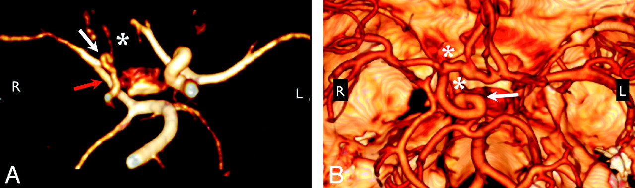
    Anomalous course and/or origin. A, 3D rendering from MRA shows elongation of the right PcomA, an anomalous connection between the right P1 segment and the right MCA (red arrow). The right ICA and left PCA are also diffusely narrow (white arrow), and the right A1 segment is hypoplastic (white asterisk). B, 3D rendering from CTA shows the tortuous looped course of the supraclinoid right ICA (white arrow). Note also the presence of 2 ACAs, a separate artery of the corpus callosum, and aneurysms at the right carotid terminus and distal right A1 segment (white asterisks). Volume renderings were generated by using OsiriX.32

  • Fig 3.
    • Download figure
    • Open in new tab
    • Download powerpoint
    Fig 3.

    Arterial narrowing and nonvisualization. All images are shown as maximum-intensity-projection reconstructions from MRA source data. A, Submentovertex projection showing nonvisualization of the left ICA. B, AP projection shows long-segment narrowing of the entire visualized course of the right ICA (red arrow). C, AP projection shows nonvisualization of the left ICA from the distal cervical to the cavernous segments. The intradural left VA is also absent. D, AP projection shows absence of the entire left ICA and long-segment tapered narrowing and luminal irregularity of the distal cervical right ICA (red arrow).

  • Fig 4.
    • Download figure
    • Open in new tab
    • Download powerpoint
    Fig 4.

    Examples of segmental and pleurisegmental ICA dysgenesis. Lesions of the ICA, especially dysgenesis, narrowing, and nonvisualization, often involve the entire length of the artery (red arrows, A) or relatively long portions of the artery corresponding to developmental segments (red arrow, B). The right proximal MCA shows diminished flow-related enhancement as well (red asterisk, A). There is segmental dysgenesis of the petrous left ICA with irregular enlargement of the artery (B). Note also a markedly enlarged left ECA, which supplies a large hemangioma, and an absent right A1 segment.

Tables

  • Figures
    • View popup
    Table 1:

    Location of arterial abnormalities

    LocationNo. (%)
    ICA53/70 (76%)
        Cervical35
        Petrous33
        Cavernous27
        Supraclinoid32
    MCA13/70 (20%)
    ACA11/70 (16%)
    PCA10/70 (14%)
    BA or VA5/70 (7%)
    • View popup
    Table 2:

    Structural brain abnormalities

    LocationNo.
    Posterior fossa
        Cerebellar hypoplasia18
        Atrophy of cerebral peduncle2
    Malformations of cortical development
        Callosal dysgenesis3
        Hemispheric hypoplasia2
        Subependymal heterotopia2
        Polymicrogyria2
    Extra-axial lesions
        Intracranial hemangioma3
        Lipoma2a
    Other
        Thin corpus callosum2
        Ocular abnormality2b
        Hippocampal malrotation2
        Cranial nerve dysplasia (7/8 complex)1
        Pituitary ectopia1
    • a One interhemispheric lipoma was seen in association with callosal dysgenesis; 1 infracollicular lipoma was seen in isolation.

    • b One case each of coloboma and microphthalmia.

PreviousNext
Back to top

In this issue

American Journal of Neuroradiology: 31 (10)
American Journal of Neuroradiology
Vol. 31, Issue 10
1 Nov 2010
  • Table of Contents
  • Index by author
Advertisement
Print
Download PDF
Email Article

Thank you for your interest in spreading the word on American Journal of Neuroradiology.

NOTE: We only request your email address so that the person you are recommending the page to knows that you wanted them to see it, and that it is not junk mail. We do not capture any email address.

Enter multiple addresses on separate lines or separate them with commas.
Cervical and Intracranial Arterial Anomalies in 70 Patients with PHACE Syndrome
(Your Name) has sent you a message from American Journal of Neuroradiology
(Your Name) thought you would like to see the American Journal of Neuroradiology web site.
CAPTCHA
This question is for testing whether or not you are a human visitor and to prevent automated spam submissions.
Cite this article
C.P. Hess, H.J. Fullerton, D.W. Metry, B.A. Drolet, D.H. Siegel, K.I. Auguste, N. Gupta, A.N. Haggstrom, C.F. Dowd, I.J. Frieden, A.J. Barkovich
Cervical and Intracranial Arterial Anomalies in 70 Patients with PHACE Syndrome
American Journal of Neuroradiology Nov 2010, 31 (10) 1980-1986; DOI: 10.3174/ajnr.A2206

Citation Manager Formats

  • BibTeX
  • Bookends
  • EasyBib
  • EndNote (tagged)
  • EndNote 8 (xml)
  • Medlars
  • Mendeley
  • Papers
  • RefWorks Tagged
  • Ref Manager
  • RIS
  • Zotero
0 Responses
Respond to this article
Share
Bookmark this article
Cervical and Intracranial Arterial Anomalies in 70 Patients with PHACE Syndrome
C.P. Hess, H.J. Fullerton, D.W. Metry, B.A. Drolet, D.H. Siegel, K.I. Auguste, N. Gupta, A.N. Haggstrom, C.F. Dowd, I.J. Frieden, A.J. Barkovich
American Journal of Neuroradiology Nov 2010, 31 (10) 1980-1986; DOI: 10.3174/ajnr.A2206
del.icio.us logo Twitter logo Facebook logo Mendeley logo
  • Tweet Widget
  • Facebook Like
  • Google Plus One
Purchase

Jump to section

  • Article
    • Abstract
    • Abbreviations
    • Materials and Methods
    • Results
    • Discussion
    • Conclusions
    • Footnotes
    • References
  • Figures & Data
  • Info & Metrics
  • Responses
  • References
  • PDF

Related Articles

  • No related articles found.
  • PubMed
  • Google Scholar

Cited By...

  • Arterial Spin-Labeling Perfusion for PHACE Syndrome
  • Utilisation of advanced MRI techniques to understand neurovascular complications of PHACE syndrome: a case of arterial stenosis and dissection
  • Clinical and Imaging Characteristics of Arteriopathy Subtypes in Children with Arterial Ischemic Stroke: Results of the VIPS Study
  • Concomitant carotid aplasia and basilar artery occlusion in a child with PHACES syndrome
  • Enlargement of the Internal Auditory Canal and Associated Posterior Fossa Anomalies in PHACES Association
  • Arteriopathy Diagnosis in Childhood Arterial Ischemic Stroke: Results of the Vascular Effects of Infection in Pediatric Stroke Study
  • Stroke in Children With Posterior Fossa Brain Malformations, Hemangiomas, Arterial Anomalies, Coarctation of the Aorta and Cardiac Defects, and Eye Abnormalities (PHACE) Syndrome: A Systematic Review of the Literature
  • Management of difficult infantile haemangiomas
  • Crossref
  • Google Scholar

This article has not yet been cited by articles in journals that are participating in Crossref Cited-by Linking.

More in this TOC Section

  • Pediatric Glioma:Imaging Clusters,Molecular Traits
  • Clinical SVR of Fetal Brain MRI
  • FRACTURE MR in Congenital Vertebral Anomalies
Show more PEDIATRIC NEUROIMAGING

Similar Articles

Advertisement

Indexed Content

  • Current Issue
  • Accepted Manuscripts
  • Article Preview
  • Past Issues
  • Editorials
  • Editor's Choice
  • Fellows' Journal Club
  • Letters to the Editor
  • Video Articles

Cases

  • Case Collection
  • Archive - Case of the Week
  • Archive - Case of the Month
  • Archive - Classic Case

More from AJNR

  • Trainee Corner
  • Imaging Protocols
  • MRI Safety Corner
  • Book Reviews

Multimedia

  • AJNR Podcasts
  • AJNR Scantastics

Resources

  • Turnaround Time
  • Submit a Manuscript
  • Submit a Video Article
  • Submit an eLetter to the Editor/Response
  • Manuscript Submission Guidelines
  • Statistical Tips
  • Fast Publishing of Accepted Manuscripts
  • Graphical Abstract Preparation
  • Imaging Protocol Submission
  • Evidence-Based Medicine Level Guide
  • Publishing Checklists
  • Author Policies
  • Become a Reviewer/Academy of Reviewers
  • News and Updates

About Us

  • About AJNR
  • Editorial Board
  • Editorial Board Alumni
  • Alerts
  • Permissions
  • Not an AJNR Subscriber? Join Now
  • Advertise with Us
  • Librarian Resources
  • Feedback
  • Terms and Conditions
  • AJNR Editorial Board Alumni

American Society of Neuroradiology

  • Not an ASNR Member? Join Now

© 2025 by the American Society of Neuroradiology All rights, including for text and data mining, AI training, and similar technologies, are reserved.
Print ISSN: 0195-6108 Online ISSN: 1936-959X

Powered by HighWire