
Article Figures & Data
Figures
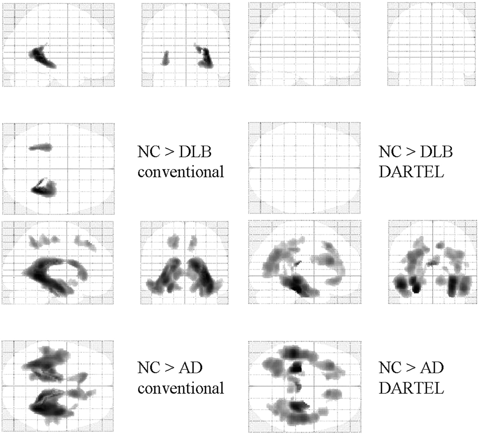
- Fig 1.
Statistical parametric maps comparing the GM volume of patients with that of age-matched healthy controls (NC). Comparisons based on conventional VBM (left) and VBM-DARTEL (right) are both illustrated. Highlighted areas represent regions in which patients have significantly decreased GM compared with controls (P < .001, corrected). The regions in which patients with DLB and those with AD show reductions in GM compared with controls overlapped: They were the medial temporal and frontal lobes and the middle temporal gyri on both sides. The pattern of GM decrease in patients with AD revealed by conventional VBM is more scattered than that identified with VBM-DARTEL.
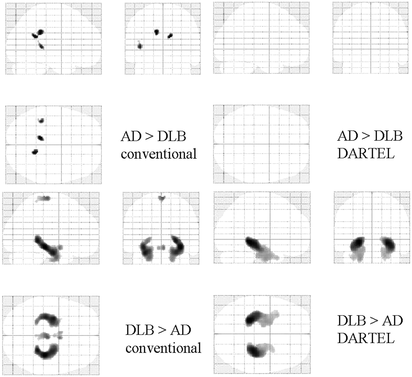
- Fig 2.
Statistical parametric maps comparing the brains of patients with DLB with those with AD. Comparisons based on conventional VBM (left) and VBM-DARTEL (right) are both illustrated. Highlighted areas represent regions in which patients have significantly decreased GM compared with age-matched healthy controls (P < .001, uncorrected). Patients with AD show significant bilateral GM loss in the MTLs. For patients with DLB, the regions in which significant decreases are identified differed between conventional VBM (upper left) and VBM-DARTEL (upper right). While GM decreases are not found in VBM-DARTEL−based comparisons, scattered decreases in the deep brain are identified with conventional VBM.
- Fig 3.
Statistical parametric maps comparing the WM volume of patients with that of age-matched healthy controls (NC). Comparisons based on conventional VBM (left) and VBM-DARTEL (right) are both illustrated. Highlighted areas represent regions in which patients have significantly decreased WM compared with the controls (P < .001, uncorrected). Patients with AD show significant WM loss in the bilateral medial temporal, parieto-occipital, and frontal lobes. For patients with DLB, the regions in which significant decreases were identified differ between conventional VBM (upper left) and VBM-DARTEL (upper right). While WM decreases are not found in VBM-DARTEL−based comparisons, decreases in CSF, like WM, are identified in the deep brain with conventional VBM.
- Fig 4.
Statistical parametric maps comparing the brains of patients with DLB and patients with AD. Highlighted areas represent regions with significantly decreased WM in patients with AD compared with those with DLB (P < .001, uncorrected). The MTLs and parieto-occipital deep brain regions are detected in both conventional VBM (lower left) and VBM-DARTEL (lower right). In comparison with patients with AD, patients with DLB show no regions with significantly decreased WM (P < .001, uncorrected).


Tables
Patients with DLB Patients with AD Controls Median age (yr) 72.7 ± 4.5 72.6 ± 2.9 72.0 ± 3.8 Median MMSE score 19.0 ± 3.5 18.7 ± 4.0 29.6 ± 0.8 Number of women (%) 60% 61% 50% Neuropsychological examinations (%) Visual hallucination: 61% Cognitive fluctuation: 95% Parkinsonism: 84% - Table 2:
Regions in which VBM-DARTEL identified significantly reduced GM: comparisons between patients and healthy controls
Comparison and Brain Region Talairach Coordinates t Value Side X Y Z Patients with DLB < controls Fusiform gyrus Lt −30 −30 −16 8.56 Amygdala Rt 22 2 −15 8.40 Medial frontal lobe 0 37 −9 6.90 Patients with AD < controls Medial temporal lobe Lt −36 −24 −10 12.13 Medial temporal lobe Rt 37 −25 −14 11.82 Anterior cingulate gyrus Rt 10 33 −13 8.08 Patients with DLB > patients with AD Parahippocampal gyrus Lt −23 −36 −2 6.62 Medial temporal lobe Rt 28 −29 −4 5.84 - Table 3:
Regions in which conventional VBM identified significantly reduced GM: comparisons between patients and healthy controls
Comparison and Brain Region Talairach Coordinates t Value Side X Y Z Patients with DLB < controls Amygdala Rt 14 0 −15 9.14 Amygdala Rt −14 0 15 7.84 Inferior temporal gyrus Rt 60 −19 −21 8.07 Patients with AD < controls Medial temporal lobe Rt 38 −26 −9 12.76 Medial temporal lobe Lt −35 −26 −9 11.85 Superior temporal gyrus Lt −61 −50 14 6.90 Patients with DLB > patients with AD Medial temporal lobe Rt 34 −26 −9 5.96 Hippocampus Lt −28 −11 −18 5.14 - Table 4:
Regions in which VBM-DARTEL identified significantly reduced WM: comparisons between patients and normal controls
Comparison and Brain Region Talairach Coordinates t Value Side X Y Z Controls > AD patients Medial temporal lobe Lt −25 −27 −16 6.30 Medial temporal lobe Rt 30 −27 −7 5.88 Fornix Rt 3 −7 13 5.77 Frontal lobe Lt −30 25 19 4.65 Parieto-occipital lobe Lt −21 −54 33 4.33 DLB patients > AD patients Medial temporal lobe Lt −25 −20 −18 7.02 Medial temporal lobe Rt 27 −27 −15 6.09 Fornix Lt −1 −6 11 4.69 Frontal lobe Rt 6 23 53 3.63 Temporal lobe Rt 45 2 −22 3.45 Medial parieto-occipital lobe Rt 9 −50 22 3.28 - Table 5:
Regions in which conventional VBM identified significantly reduced WM: comparisons between patients and normal controls
Comparison and Brain Region Talairach Coordinates t Value Side X Y Z Controls > DLB patients Medial temporal lobe Rt 34 −41 2 4.98 Medial temporal lobe Lt −32 −31 −4 3.97 Controls > AD patients Medial temporal lobe Rt 34 −43 2 7.71 Parietal lobe Lt −16 −46 47 4.74 Frontal lobe Rt 12 35 44 4.50 DLB patients > AD patients Medial temporal lobe Lt −24 −24 −16 −6.76 Medial temporal lobe Rt 24 −17 −21 5.82 Parietal lobe Lt −48 −24 46 3.68


