Advertisement

Main menu

  • Home
  • Content
    • Current Issue
    • Accepted Manuscripts
    • Article Preview
    • Past Issue Archive
    • Video Articles
    • AJNR Case Collection
    • Case of the Week Archive
    • Case of the Month Archive
    • Classic Case Archive
  • Special Collections
    • AJNR Awards
    • Low-Field MRI
    • Alzheimer Disease
    • ASNR Foundation Special Collection
    • Photon-Counting CT
    • View All
  • Multimedia
    • AJNR Podcasts
    • AJNR SCANtastic
    • Trainee Corner
    • MRI Safety Corner
    • Imaging Protocols
  • For Authors
    • Submit a Manuscript
    • Submit a Video Article
    • Submit an eLetter to the Editor/Response
    • Manuscript Submission Guidelines
    • Statistical Tips
    • Fast Publishing of Accepted Manuscripts
    • Graphical Abstract Preparation
    • Imaging Protocol Submission
    • Author Policies
  • About Us
    • About AJNR
    • Editorial Board
    • Editorial Board Alumni
  • More
    • Become a Reviewer/Academy of Reviewers
    • Subscribers
    • Permissions
    • Alerts
    • Feedback
    • Advertisers
    • ASNR Home

User menu

  • Alerts
  • Log in

Search

  • Advanced search
American Journal of Neuroradiology

American Journal of Neuroradiology

ASHNR American Society of Functional Neuroradiology ASHNR American Society of Pediatric Neuroradiology ASSR
  • Alerts
  • Log in

Advanced Search

  • Home
  • Content
    • Current Issue
    • Accepted Manuscripts
    • Article Preview
    • Past Issue Archive
    • Video Articles
    • AJNR Case Collection
    • Case of the Week Archive
    • Case of the Month Archive
    • Classic Case Archive
  • Special Collections
    • AJNR Awards
    • Low-Field MRI
    • Alzheimer Disease
    • ASNR Foundation Special Collection
    • Photon-Counting CT
    • View All
  • Multimedia
    • AJNR Podcasts
    • AJNR SCANtastic
    • Trainee Corner
    • MRI Safety Corner
    • Imaging Protocols
  • For Authors
    • Submit a Manuscript
    • Submit a Video Article
    • Submit an eLetter to the Editor/Response
    • Manuscript Submission Guidelines
    • Statistical Tips
    • Fast Publishing of Accepted Manuscripts
    • Graphical Abstract Preparation
    • Imaging Protocol Submission
    • Author Policies
  • About Us
    • About AJNR
    • Editorial Board
    • Editorial Board Alumni
  • More
    • Become a Reviewer/Academy of Reviewers
    • Subscribers
    • Permissions
    • Alerts
    • Feedback
    • Advertisers
    • ASNR Home
  • Follow AJNR on Twitter
  • Visit AJNR on Facebook
  • Follow AJNR on Instagram
  • Join AJNR on LinkedIn
  • RSS Feeds

AJNR Awards, New Junior Editors, and more. Read the latest AJNR updates

Research ArticleBrain
Open Access

White Matter Microstructure Changes in the Thalamus in Parkinson Disease with Depression: A Diffusion Tensor MR Imaging Study

W. Li, J. Liu, F. Skidmore, Y. Liu, J. Tian and K. Li
American Journal of Neuroradiology November 2010, 31 (10) 1861-1866; DOI: https://doi.org/10.3174/ajnr.A2195
W. Li
  • Find this author on Google Scholar
  • Find this author on PubMed
  • Search for this author on this site
J. Liu
  • Find this author on Google Scholar
  • Find this author on PubMed
  • Search for this author on this site
F. Skidmore
  • Find this author on Google Scholar
  • Find this author on PubMed
  • Search for this author on this site
Y. Liu
  • Find this author on Google Scholar
  • Find this author on PubMed
  • Search for this author on this site
J. Tian
  • Find this author on Google Scholar
  • Find this author on PubMed
  • Search for this author on this site
K. Li
  • Find this author on Google Scholar
  • Find this author on PubMed
  • Search for this author on this site
  • Article
  • Figures & Data
  • Info & Metrics
  • Responses
  • References
  • PDF
Loading

Article Figures & Data

Figures

  • Tables
  • Fig 1.
    • Download figure
    • Open in new tab
    • Download powerpoint
    Fig 1.

    Illustration of regions of difference in patients with DPD and NDPD in the bilateral thalami. A, Standard MNI 152 T1 brain images (in axial and coronal directions) are overlaid with the statistically significant differing regions (in red). FA in the mediodorsal thalamus differs in patients with DPD compared with patients with NDPD. B, Scatterplots show the mean FA values in the detected significant regions for every subject in the DPD and NDPD groups. Blue lines denote the mean values for each group. Asterisks indicate significant differences between these 2 groups with P < .005.
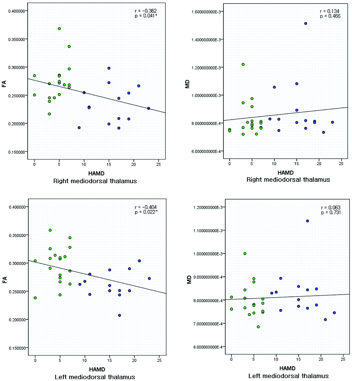
  • Fig 2.
    • Download figure
    • Open in new tab
    • Download powerpoint
    Fig 2.

    Scatterplots with trend lines showing the correlations of diffusion quantities (FA and MD) with HAMD for 32 PD subjects in the right and left mediodorsal thalami. Green represents patients with NDPD; blue represents patients with DPD. The asterisks indicate that a significant negative correlation was found between FA and HAMD.

Tables

  • Figures
    • View popup
    Table 1:

    Clinical and demographic characteristics of patients

    GroupsDPD (n = 14)NDPD (n = 18)P Valuea
    Sex (M/F)4/1010/8NS
    Age (yr)65.28 ± 8.8961.05 ± 10.17NS
    PD duration6.29 ± 5.515.67 ± 2.57NS
    MMSE29.5 ± 0.729.2 ± 1.0NS
    UPDRS39.04 ± 22.2833.83 ± 15.09NS
    H-Y1.96 ± 0.991.83 ± 0.75NS
    HAMD15.64 ± 4.214.44 ± 2.14<.001
    LEDD (mg/day)315 ± 201363 ± 197NS
    • a Unpaired 2-tailed t tests were used for parametric comparison of data between the 2 groups for age, PD duration, MMSE, UPDRS, H-Y, HAMD, and LEDD; The Mann-Whitney U test was used for nonparametric comparison of sex between the 2 groups. Significance was set at P <. 05.

    • View popup
    Table 2:

    Diffusion quantity comparison between patients with DPD and those with NDPD

    Mediodorsal ThalamusmFAt ValueaP ValuemMD (×10−4 mm2/s)t ValueaP Value
    DPDNDPDDPDNDPD
    Right0.23 ± 0.030.27 ± 0.04−3.300.003b8.92 ± 2.068.26 ± 1.221.061.301
    Left0.26 ± 0.020.30 ± 0.03−3.463.002b8.27 ± 1.037.97 ± 0.710.924.365
    • a Indicates unpaired 2-tailed t test.

    • b Indicates statistical significance with P < .005.

    • View popup
    Table 3:

    Pearson correlation between HAMD and diffusion quantities for PDa

    RegionsmFAmMD
    rP ValuerP Value
    Right mediodorsal thalamus−0.362.041b0.134.466
    Left mediodorsal thalamus−0.404.022b0.063.731
    • a SPSS, Version 17.0, was used for correlation analysis. For correlation coefficient calculation, the Pearson test was selected; for the significance test of r, a 2-tailed t test was selected.

    • b Indicates statistical significance with P < .05.

PreviousNext
Back to top

In this issue

American Journal of Neuroradiology: 31 (10)
American Journal of Neuroradiology
Vol. 31, Issue 10
1 Nov 2010
  • Table of Contents
  • Index by author
Advertisement
Print
Download PDF
Email Article

Thank you for your interest in spreading the word on American Journal of Neuroradiology.

NOTE: We only request your email address so that the person you are recommending the page to knows that you wanted them to see it, and that it is not junk mail. We do not capture any email address.

Enter multiple addresses on separate lines or separate them with commas.
White Matter Microstructure Changes in the Thalamus in Parkinson Disease with Depression: A Diffusion Tensor MR Imaging Study
(Your Name) has sent you a message from American Journal of Neuroradiology
(Your Name) thought you would like to see the American Journal of Neuroradiology web site.
CAPTCHA
This question is for testing whether or not you are a human visitor and to prevent automated spam submissions.
Cite this article
W. Li, J. Liu, F. Skidmore, Y. Liu, J. Tian, K. Li
White Matter Microstructure Changes in the Thalamus in Parkinson Disease with Depression: A Diffusion Tensor MR Imaging Study
American Journal of Neuroradiology Nov 2010, 31 (10) 1861-1866; DOI: 10.3174/ajnr.A2195

Citation Manager Formats

  • BibTeX
  • Bookends
  • EasyBib
  • EndNote (tagged)
  • EndNote 8 (xml)
  • Medlars
  • Mendeley
  • Papers
  • RefWorks Tagged
  • Ref Manager
  • RIS
  • Zotero
0 Responses
Respond to this article
Share
Bookmark this article
White Matter Microstructure Changes in the Thalamus in Parkinson Disease with Depression: A Diffusion Tensor MR Imaging Study
W. Li, J. Liu, F. Skidmore, Y. Liu, J. Tian, K. Li
American Journal of Neuroradiology Nov 2010, 31 (10) 1861-1866; DOI: 10.3174/ajnr.A2195
del.icio.us logo Twitter logo Facebook logo Mendeley logo
  • Tweet Widget
  • Facebook Like
  • Google Plus One
Purchase

Jump to section

  • Article
    • Abstract
    • Abbreviations
    • Materials and Methods
    • Results
    • Discussion
    • Conclusions
    • Footnotes
    • References
  • Figures & Data
  • Info & Metrics
  • Responses
  • References
  • PDF

Related Articles

  • No related articles found.
  • PubMed
  • Google Scholar

Cited By...

  • Subcortical imaging-derived phenotypes are associated with the risk of Parkinsons disease: A Mendelian Randomization Study
  • Thalamic white matter macrostructure and subnuclei volumes in Parkinsons disease depression
  • Mediodorsal nucleus and its multiple cognitive functions
  • Thalamic Projection Fiber Integrity in de novo Parkinson Disease
  • Individual Detection of Patients with Parkinson Disease using Support Vector Machine Analysis of Diffusion Tensor Imaging Data: Initial Results
  • White matter abnormalities and illness severity in major depressive disorder
  • Crossref
  • Google Scholar

This article has not yet been cited by articles in journals that are participating in Crossref Cited-by Linking.

More in this TOC Section

  • Evaluating the Effects of White Matter Multiple Sclerosis Lesions on the Volume Estimation of 6 Brain Tissue Segmentation Methods
  • Quiet PROPELLER MRI Techniques Match the Quality of Conventional PROPELLER Brain Imaging Techniques
  • Predictors of Reperfusion in Patients with Acute Ischemic Stroke
Show more Brain

Similar Articles

Advertisement

Indexed Content

  • Current Issue
  • Accepted Manuscripts
  • Article Preview
  • Past Issues
  • Editorials
  • Editor's Choice
  • Fellows' Journal Club
  • Letters to the Editor
  • Video Articles

Cases

  • Case Collection
  • Archive - Case of the Week
  • Archive - Case of the Month
  • Archive - Classic Case

More from AJNR

  • Trainee Corner
  • Imaging Protocols
  • MRI Safety Corner
  • Book Reviews

Multimedia

  • AJNR Podcasts
  • AJNR Scantastics

Resources

  • Turnaround Time
  • Submit a Manuscript
  • Submit a Video Article
  • Submit an eLetter to the Editor/Response
  • Manuscript Submission Guidelines
  • Statistical Tips
  • Fast Publishing of Accepted Manuscripts
  • Graphical Abstract Preparation
  • Imaging Protocol Submission
  • Evidence-Based Medicine Level Guide
  • Publishing Checklists
  • Author Policies
  • Become a Reviewer/Academy of Reviewers
  • News and Updates

About Us

  • About AJNR
  • Editorial Board
  • Editorial Board Alumni
  • Alerts
  • Permissions
  • Not an AJNR Subscriber? Join Now
  • Advertise with Us
  • Librarian Resources
  • Feedback
  • Terms and Conditions
  • AJNR Editorial Board Alumni

American Society of Neuroradiology

  • Not an ASNR Member? Join Now

© 2025 by the American Society of Neuroradiology All rights, including for text and data mining, AI training, and similar technologies, are reserved.
Print ISSN: 0195-6108 Online ISSN: 1936-959X

Powered by HighWire