Advertisement

Main menu

  • Home
  • Content
    • Current Issue
    • Accepted Manuscripts
    • Article Preview
    • Past Issue Archive
    • Video Articles
    • AJNR Case Collection
    • Case of the Week Archive
    • Case of the Month Archive
    • Classic Case Archive
  • Special Collections
    • Advancing NeuroMRI with High-Relaxivity Contrast Agents
    • AJNR Awards
    • Low-Field MRI
    • Alzheimer Disease
    • ASNR Foundation Special Collection
    • Photon-Counting CT
    • View All
  • Multimedia
    • AJNR Podcasts
    • AJNR SCANtastic
    • Trainee Corner
    • MRI Safety Corner
    • Imaging Protocols
  • For Authors
    • Submit a Manuscript
    • Submit a Video Article
    • Submit an eLetter to the Editor/Response
    • Manuscript Submission Guidelines
    • Statistical Tips
    • Fast Publishing of Accepted Manuscripts
    • Graphical Abstract Preparation
    • Imaging Protocol Submission
    • Author Policies
  • About Us
    • About AJNR
    • Editorial Board
    • Editorial Board Alumni
  • More
    • Become a Reviewer/Academy of Reviewers
    • Subscribers
    • Permissions
    • Alerts
    • Feedback
    • Advertisers
    • ASNR Home

User menu

  • Alerts
  • Log in

Search

  • Advanced search
American Journal of Neuroradiology

American Journal of Neuroradiology

ASHNR American Society of Functional Neuroradiology ASHNR American Society of Pediatric Neuroradiology ASSR
  • Alerts
  • Log in

Advanced Search

  • Home
  • Content
    • Current Issue
    • Accepted Manuscripts
    • Article Preview
    • Past Issue Archive
    • Video Articles
    • AJNR Case Collection
    • Case of the Week Archive
    • Case of the Month Archive
    • Classic Case Archive
  • Special Collections
    • Advancing NeuroMRI with High-Relaxivity Contrast Agents
    • AJNR Awards
    • Low-Field MRI
    • Alzheimer Disease
    • ASNR Foundation Special Collection
    • Photon-Counting CT
    • View All
  • Multimedia
    • AJNR Podcasts
    • AJNR SCANtastic
    • Trainee Corner
    • MRI Safety Corner
    • Imaging Protocols
  • For Authors
    • Submit a Manuscript
    • Submit a Video Article
    • Submit an eLetter to the Editor/Response
    • Manuscript Submission Guidelines
    • Statistical Tips
    • Fast Publishing of Accepted Manuscripts
    • Graphical Abstract Preparation
    • Imaging Protocol Submission
    • Author Policies
  • About Us
    • About AJNR
    • Editorial Board
    • Editorial Board Alumni
  • More
    • Become a Reviewer/Academy of Reviewers
    • Subscribers
    • Permissions
    • Alerts
    • Feedback
    • Advertisers
    • ASNR Home
  • Follow AJNR on Twitter
  • Visit AJNR on Facebook
  • Follow AJNR on Instagram
  • Join AJNR on LinkedIn
  • RSS Feeds

AJNR Awards, New Junior Editors, and more. Read the latest AJNR updates


Improved Turnaround Times | Median time to first decision: 12 days

Research ArticleBrainC

Unilateral Calcification of the Caudate and Putamen: Association with Underlying Developmental Venous Anomaly

S. Dehkharghani, W.P. Dillon, S.O. Bryant and N.J. Fischbein
American Journal of Neuroradiology November 2010, 31 (10) 1848-1852; DOI: https://doi.org/10.3174/ajnr.A2199
S. Dehkharghani
  • Find this author on Google Scholar
  • Find this author on PubMed
  • Search for this author on this site
W.P. Dillon
  • Find this author on Google Scholar
  • Find this author on PubMed
  • Search for this author on this site
S.O. Bryant
  • Find this author on Google Scholar
  • Find this author on PubMed
  • Search for this author on this site
N.J. Fischbein
  • Find this author on Google Scholar
  • Find this author on PubMed
  • Search for this author on this site
  • Article
  • Figures & Data
  • Info & Metrics
  • Responses
  • References
  • PDF
Loading

Article Figures & Data

Figures

  • Tables
  • Fig 1.
    • Download figure
    • Open in new tab
    • Download powerpoint
    Fig 1.

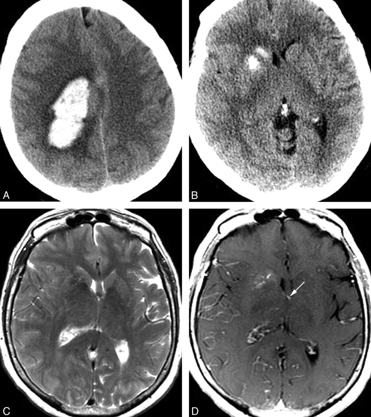
    A 70-year-old woman presenting with acute onset of headache and left-sided weakness. A, Noncontrast head CT scan demonstrates a large right frontoparietal lobar hemorrhage with surrounding vasogenic edema. This hemorrhage was subsequently shown to be due to a right hemispheric arteriovenous malformation with intranidal and flow-related aneurysms. B, CT scan caudal to A demonstrates calcification of the right caudate and anterior putamen, with sparing of the anterior limb of the internal capsule. C, Axial T2 MR image at a similar level demonstrates normal to slightly decreased signal intensity in the right caudate and putamen. Mild sulcal effacement in the right hemisphere is due to mass effect from the more superior parenchymal hematoma. Despite this, the right frontal horn is mildly dilated compared with the left. D, Axial T1 postgadolinium image demonstrates a DVA involving the right caudate and putamen. This DVA drains into an ependymal vein, likely the right thalamostriate vein, which is partly included on this image (arrow).

  • Fig 2.
    • Download figure
    • Open in new tab
    • Download powerpoint
    Fig 2.

    A 50-year-old man presenting with headache. A, Noncontrast CT scan of the brain demonstrates calcification involving the left caudate and putamen without mass effect and with some encroachment on but relative sparing of the anterior limb of the internal capsule. Mild asymmetric prominence of the left frontal ventricular horn is consistent with mild volume loss. B, Coronal gradient recalled-echo image demonstrates signal-intensity loss due to susceptibility effects in the distribution of the basal ganglia mineralization. C, Axial T1 postgadolinium MR image demonstrates enhancing venous radicles in the left basal ganglia, converging on a common venous stem that courses toward the adjacent ventricular surface. Findings of T2- and diffusion-weighted images (not shown) were normal.

  • Fig 3.
    • Download figure
    • Open in new tab
    • Download powerpoint
    Fig 3.

    A 48-year-old woman presenting with headache and seizure. A, An axial source image from CTA demonstrates mineralization of the right caudate and anterior putamen, with sparing of the anterior limb of the internal capsule. The patient's noncontrast head CT (not shown) also demonstrated this finding. An adjacent developmental venous anomaly (white arrows) is demonstrated in the periventricular white matter, coursing toward the midline. B, Lateral-projection venous phase image from a catheter angiogram demonstrates the venous radicles of the DVA converging toward a common venous pouch (P). A focal stenosis (large black arrow) manifested as a caliber transition zone is present where the pouch meets the inferior sagittal sinus; a second possible stenosis is present at the point where the inferior sagittal sinus drains to the Galenic system.

  • Fig 4.
    • Download figure
    • Open in new tab
    • Download powerpoint
    Fig 4.

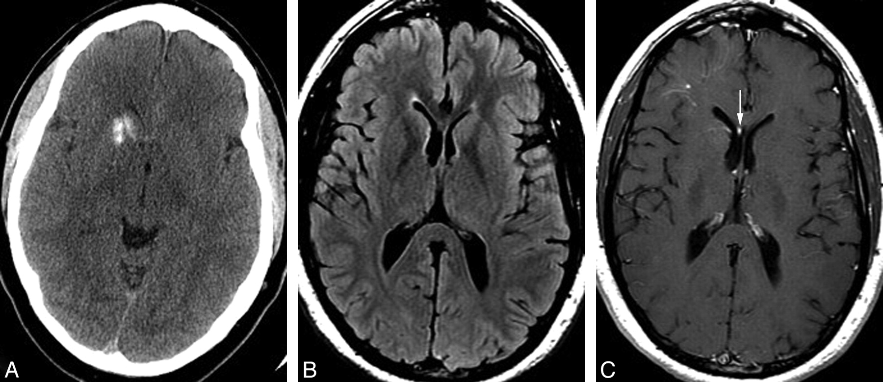
    A 39-year-old man with a history of antithrombin III deficiency, presenting with headache and suspicion for intracranial hemorrhage. Some years earlier, the patient had been told that he had a “right basal ganglia hemorrhage.” A, Noncontrast head CT demonstrates unilateral mineralization of the right caudate and anterior putamen, with relative sparing of the anterior limb of the internal capsule, which can be seen between the caudate and putaminal mineralization. B, Axial fluid-attenuated inversion recovery MR image shows no hyperintensity in the right caudate or putamen but rather a subtle hypointensity due to the calcification. The right frontal horn is mildly dilated, consistent with subtle volume loss in the right basal ganglia. C, Axial T1 postgadolinium image demonstrates a deep DVA involving the right basal ganglia and draining toward a right subependymal vein and then to the right septal vein (arrow). A second more superficial DVA is present in the subcortical right frontal region. Mild asymmetric prominence of the right frontal ventricular horn is again consistent with subtle parenchymal volume loss.

Tables

  • Figures
  • Clinical and imaging characteristics of study population

    CharacteristicsPatient
    123456
    General
    Age (yr)/sex50/M39/M79/F70/F48/F30/M
    PresentationHAHAICHICHHA, SZHA
    CT
        SideLRRRRR
        Ca2+ distributionaC + PC + PbC + PbC + PC + PC + P
    CTANADVA, otherwise negativeNAAVM, DVA; otherwise negativeLarge DVA with venous restrictionNA
    MR imaging
        Mass effect–––––c–
        T2 Change––––NA–
        DVA drainageDeepDeepDeepDeepDeepDeep
    Angiography
        Venous restrictionNA–NA–+NA
    Additional featuresIpsilateral prominence of frontal hornIpsilateral prominence of frontal horn,Ipsilateral prominence of frontal horn,Ipsilateral prominence of frontal horn,
    2nd DVA, periphery right frontal lobeRight frontal lobar ICHRight frontal/parietal lobar ICH
    • Note:— –indicates negative.

    • a Preferential calcification of the caudate and anterior putamen was noted in all cases, with at least partial sparing of the anterior limb of the internal capsule.

    • b Trace high density of the globus pallidus interna was noted, characteristic of senescent mineralization, involving the globus pallidus interna on the left in case 2 and bilaterally in case 3.

    • c MR imaging was not available; cross-sectional assessment was performed by CT and CTA.

PreviousNext
Back to top

In this issue

American Journal of Neuroradiology: 31 (10)
American Journal of Neuroradiology
Vol. 31, Issue 10
1 Nov 2010
  • Table of Contents
  • Index by author
Advertisement
Print
Download PDF
Email Article

Thank you for your interest in spreading the word on American Journal of Neuroradiology.

NOTE: We only request your email address so that the person you are recommending the page to knows that you wanted them to see it, and that it is not junk mail. We do not capture any email address.

Enter multiple addresses on separate lines or separate them with commas.
Unilateral Calcification of the Caudate and Putamen: Association with Underlying Developmental Venous Anomaly
(Your Name) has sent you a message from American Journal of Neuroradiology
(Your Name) thought you would like to see the American Journal of Neuroradiology web site.
CAPTCHA
This question is for testing whether or not you are a human visitor and to prevent automated spam submissions.
Cite this article
S. Dehkharghani, W.P. Dillon, S.O. Bryant, N.J. Fischbein
Unilateral Calcification of the Caudate and Putamen: Association with Underlying Developmental Venous Anomaly
American Journal of Neuroradiology Nov 2010, 31 (10) 1848-1852; DOI: 10.3174/ajnr.A2199

Citation Manager Formats

  • BibTeX
  • Bookends
  • EasyBib
  • EndNote (tagged)
  • EndNote 8 (xml)
  • Medlars
  • Mendeley
  • Papers
  • RefWorks Tagged
  • Ref Manager
  • RIS
  • Zotero
0 Responses
Respond to this article
Share
Bookmark this article
Unilateral Calcification of the Caudate and Putamen: Association with Underlying Developmental Venous Anomaly
S. Dehkharghani, W.P. Dillon, S.O. Bryant, N.J. Fischbein
American Journal of Neuroradiology Nov 2010, 31 (10) 1848-1852; DOI: 10.3174/ajnr.A2199
del.icio.us logo Twitter logo Facebook logo Mendeley logo
  • Tweet Widget
  • Facebook Like
  • Google Plus One
Purchase

Jump to section

  • Article
    • Abstract
    • Abbreviations
    • Materials and Methods
    • Discussion
    • Conclusions
    • References
  • Figures & Data
  • Info & Metrics
  • Responses
  • References
  • PDF

Related Articles

  • No related articles found.
  • PubMed
  • Google Scholar

Cited By...

  • Symptomatic Developmental Venous Anomaly: State-of-the-Art Review on Genetics, Pathophysiology, and Imaging Approach to Diagnosis
  • Crossref
  • Google Scholar

This article has not yet been cited by articles in journals that are participating in Crossref Cited-by Linking.

More in this TOC Section

  • Usefulness of Quantitative Susceptibility Mapping for the Diagnosis of Parkinson Disease
  • White Matter Alterations in the Brains of Patients with Active, Remitted, and Cured Cushing Syndrome: A DTI Study
  • Qualitative and Quantitative Analysis of MR Imaging Findings in Patients with Middle Cerebral Artery Stroke Implanted with Mesenchymal Stem Cells
Show more Brain

Similar Articles

Advertisement

Indexed Content

  • Current Issue
  • Accepted Manuscripts
  • Article Preview
  • Past Issues
  • Editorials
  • Editor's Choice
  • Fellows' Journal Club
  • Letters to the Editor
  • Video Articles

Cases

  • Case Collection
  • Archive - Case of the Week
  • Archive - Case of the Month
  • Archive - Classic Case

More from AJNR

  • Trainee Corner
  • Imaging Protocols
  • MRI Safety Corner
  • Book Reviews

Multimedia

  • AJNR Podcasts
  • AJNR Scantastics

Resources

  • Turnaround Time
  • Submit a Manuscript
  • Submit a Video Article
  • Submit an eLetter to the Editor/Response
  • Manuscript Submission Guidelines
  • Statistical Tips
  • Fast Publishing of Accepted Manuscripts
  • Graphical Abstract Preparation
  • Imaging Protocol Submission
  • Evidence-Based Medicine Level Guide
  • Publishing Checklists
  • Author Policies
  • Become a Reviewer/Academy of Reviewers
  • News and Updates

About Us

  • About AJNR
  • Editorial Board
  • Editorial Board Alumni
  • Alerts
  • Permissions
  • Not an AJNR Subscriber? Join Now
  • Advertise with Us
  • Librarian Resources
  • Feedback
  • Terms and Conditions
  • AJNR Editorial Board Alumni

American Society of Neuroradiology

  • Not an ASNR Member? Join Now

© 2025 by the American Society of Neuroradiology All rights, including for text and data mining, AI training, and similar technologies, are reserved.
Print ISSN: 0195-6108 Online ISSN: 1936-959X

Powered by HighWire