Advertisement

Main menu

  • Home
  • Content
    • Current Issue
    • Accepted Manuscripts
    • Article Preview
    • Past Issue Archive
    • Video Articles
    • AJNR Case Collection
    • Case of the Week Archive
    • Case of the Month Archive
    • Classic Case Archive
  • Special Collections
    • AJNR Awards
    • Low-Field MRI
    • Alzheimer Disease
    • ASNR Foundation Special Collection
    • Photon-Counting CT
    • View All
  • Multimedia
    • AJNR Podcasts
    • AJNR SCANtastic
    • Trainee Corner
    • MRI Safety Corner
    • Imaging Protocols
  • For Authors
    • Submit a Manuscript
    • Submit a Video Article
    • Submit an eLetter to the Editor/Response
    • Manuscript Submission Guidelines
    • Statistical Tips
    • Fast Publishing of Accepted Manuscripts
    • Graphical Abstract Preparation
    • Imaging Protocol Submission
    • Author Policies
  • About Us
    • About AJNR
    • Editorial Board
    • Editorial Board Alumni
  • More
    • Become a Reviewer/Academy of Reviewers
    • Subscribers
    • Permissions
    • Alerts
    • Feedback
    • Advertisers
    • ASNR Home

User menu

  • Alerts
  • Log in

Search

  • Advanced search
American Journal of Neuroradiology

American Journal of Neuroradiology

ASHNR American Society of Functional Neuroradiology ASHNR American Society of Pediatric Neuroradiology ASSR
  • Alerts
  • Log in

Advanced Search

  • Home
  • Content
    • Current Issue
    • Accepted Manuscripts
    • Article Preview
    • Past Issue Archive
    • Video Articles
    • AJNR Case Collection
    • Case of the Week Archive
    • Case of the Month Archive
    • Classic Case Archive
  • Special Collections
    • AJNR Awards
    • Low-Field MRI
    • Alzheimer Disease
    • ASNR Foundation Special Collection
    • Photon-Counting CT
    • View All
  • Multimedia
    • AJNR Podcasts
    • AJNR SCANtastic
    • Trainee Corner
    • MRI Safety Corner
    • Imaging Protocols
  • For Authors
    • Submit a Manuscript
    • Submit a Video Article
    • Submit an eLetter to the Editor/Response
    • Manuscript Submission Guidelines
    • Statistical Tips
    • Fast Publishing of Accepted Manuscripts
    • Graphical Abstract Preparation
    • Imaging Protocol Submission
    • Author Policies
  • About Us
    • About AJNR
    • Editorial Board
    • Editorial Board Alumni
  • More
    • Become a Reviewer/Academy of Reviewers
    • Subscribers
    • Permissions
    • Alerts
    • Feedback
    • Advertisers
    • ASNR Home
  • Follow AJNR on Twitter
  • Visit AJNR on Facebook
  • Follow AJNR on Instagram
  • Join AJNR on LinkedIn
  • RSS Feeds

AJNR Awards, New Junior Editors, and more. Read the latest AJNR updates

Research ArticleSpine Imaging and Spine Image-Guided InterventionsE

Long-Term Outcome in the Repair of Spinal Cord Perimedullary Arteriovenous Fistulas

L. Antonietti, S.A. Sheth, V.V. Halbach, R.T. Higashida, C.F. Dowd, M.T. Lawton, J.D. English and S.W. Hetts
American Journal of Neuroradiology November 2010, 31 (10) 1824-1830; DOI: https://doi.org/10.3174/ajnr.A2236
L. Antonietti
  • Find this author on Google Scholar
  • Find this author on PubMed
  • Search for this author on this site
S.A. Sheth
  • Find this author on Google Scholar
  • Find this author on PubMed
  • Search for this author on this site
V.V. Halbach
  • Find this author on Google Scholar
  • Find this author on PubMed
  • Search for this author on this site
R.T. Higashida
  • Find this author on Google Scholar
  • Find this author on PubMed
  • Search for this author on this site
C.F. Dowd
  • Find this author on Google Scholar
  • Find this author on PubMed
  • Search for this author on this site
M.T. Lawton
  • Find this author on Google Scholar
  • Find this author on PubMed
  • Search for this author on this site
J.D. English
  • Find this author on Google Scholar
  • Find this author on PubMed
  • Search for this author on this site
S.W. Hetts
  • Find this author on Google Scholar
  • Find this author on PubMed
  • Search for this author on this site
  • Article
  • Figures & Data
  • Supplemental
  • Info & Metrics
  • Responses
  • References
  • PDF
Loading

Article Figures & Data

Figures

  • Tables
  • Fig 1.
    • Download figure
    • Open in new tab
    • Download powerpoint
    Fig 1.

    Histogram detailing the age distribution of patients at the time of diagnosis and treatment of PMAVFs in our study cohort.

  • Fig 2.
    • Download figure
    • Open in new tab
    • Download powerpoint
    Fig 2.

    Histogram detailing the location of the PMAVFs of the 29 patients evaluated at our institution.

  • Fig 3.
    • Download figure
    • Open in new tab
    • Download powerpoint
    Fig 3.

    A 27-year-old man with 18 months of progressive bilateral lower extremity weakness, urinary retention, and constipation. At presentation to our hospital, the patient was using crutches, had decreased sensation in both lower extremities, and loss of bowel and bladder function. His ALS scores on admission were 4 for ambulation and 3 for micturition. The patient underwent surgical correction of his type 2 PMAVF. At follow-up clinical evaluation 2 months after surgical correction, the patient had interval improvement in ambulation (ALS ambulation = 3) as well as micturition (ALS micturition = 2). A, From left to right: Myelogram shows serpiginous filling defects in the lower thoracic region. Sagittal T2-weighted MR image demonstrates cord edema extending to the conus medullaris with abnormal surrounding flow-voids. Sagittal T1-weighted postgadolinium MR image again shows abnormal vascular structures along the conus medullaris, worrisome for a spinal vascular malformation. B, Preoperative spinal angiogram. From left to right: Serial angiograms of a selective injection of the right T10 intercostal artery demonstrate supply of the perimedullary fistula via an ASA branch. C, Intraoperative indocyanine green angiography in the arterial (top) and venous (bottom) phases identifies 1 of the dominant feeders (arrow) of the perimedullary fistula. D, Intraoperative photographs again demonstrate 1 of the main feeding arteries (top, arrow) and placement of an arteriovenous malformation clip (bottom). E, First postoperative conventional spinal angiogram. From left to right: Serial angiograms of a selective injection of the right T10 intercostal artery demonstrate opacification of the anterior spinal artery with early shunt at the level of the surgical clips, consistent with a residual perimedullary fistula. The long arrow indicates a residual fistula site; the short arrow, early opacification of anterior spinal vein. F, Intraoperative photographs after rotation of the cord and conus medullaris demonstrate residual fistula along the anterior aspect of the cord fed by an ASA branch (top), which was treated with cauterization and placement of an AVM clip (bottom, arrow).
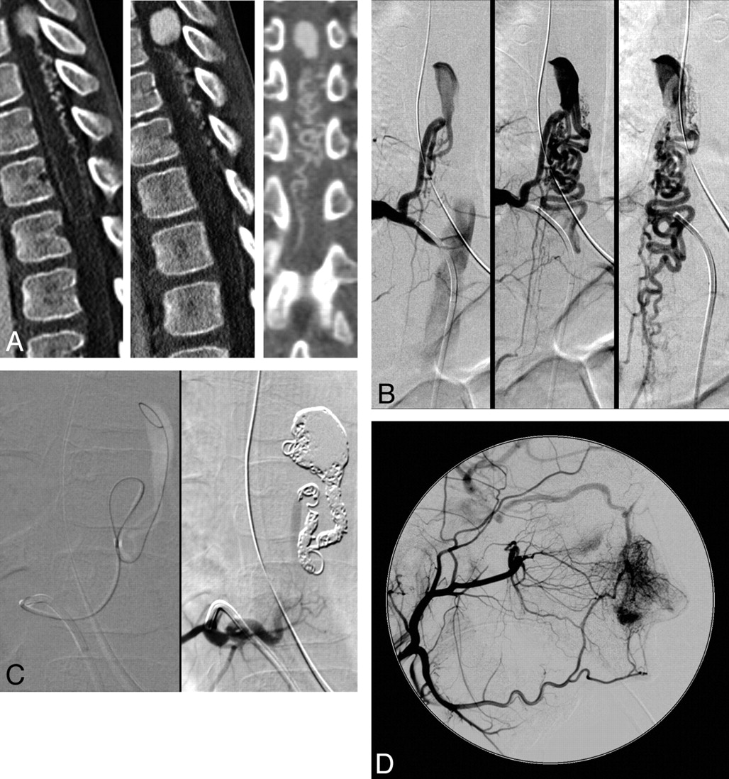
  • Fig 4.
    • Download figure
    • Open in new tab
    • Download powerpoint
    Fig 4.

    A 6-year-old girl with 3 weeks of waxing and waning abdominal pain, followed by headache and neck stiffness and progressive slowing of her gait. Review of the patient's history revealed a strong paternal family history of HHT. On presentation, her neurologic examination findings were unremarkable, except for 4/5 strength dorsiflexion of the bilateral lower extremities (ALS ambulation = 1, ALS micturition = 0). Lumbar puncture findings were consistent with SAH. The patient underwent endovascular correction of her type 3 PMAVF. The patient's neurologic examination findings returned to normal immediately postprocedure and remained normal on clinical follow-up at 4 months. A, Sagittal and coronal maximum-intensity-projection images of a contrast-enhanced CT scan of the spine demonstrate a large varix at the T8–9 level, with a tangle of draining veins extending inferiorly. B, From left to right: Serial angiograms of a selective injection of the right T10 intercostal artery demonstrate opacification of the feeding artery to the single-hole perimedullary fistula with a large partially thrombosed varix, corresponding to the enhancing “mass” identified on CT and the tangle of draining veins extending inferiorly. C, Left: Selective diagnostic catheter placement in the right T10 intercostal artery, with a microcatheter positioned within the varix. Right: Coil mass occupying the varix and the fistula site. Selective injection of the right T10 intercostal artery shows slow filling of the feeding vessel without any residual shunt. D, Lateral view of a selective injection of the right external carotid artery demonstrates exuberant vascularity of the nasal mucosa with microshunts and early opacification of the right ophthalmic vein, suggestive of the diagnosis of HHT.
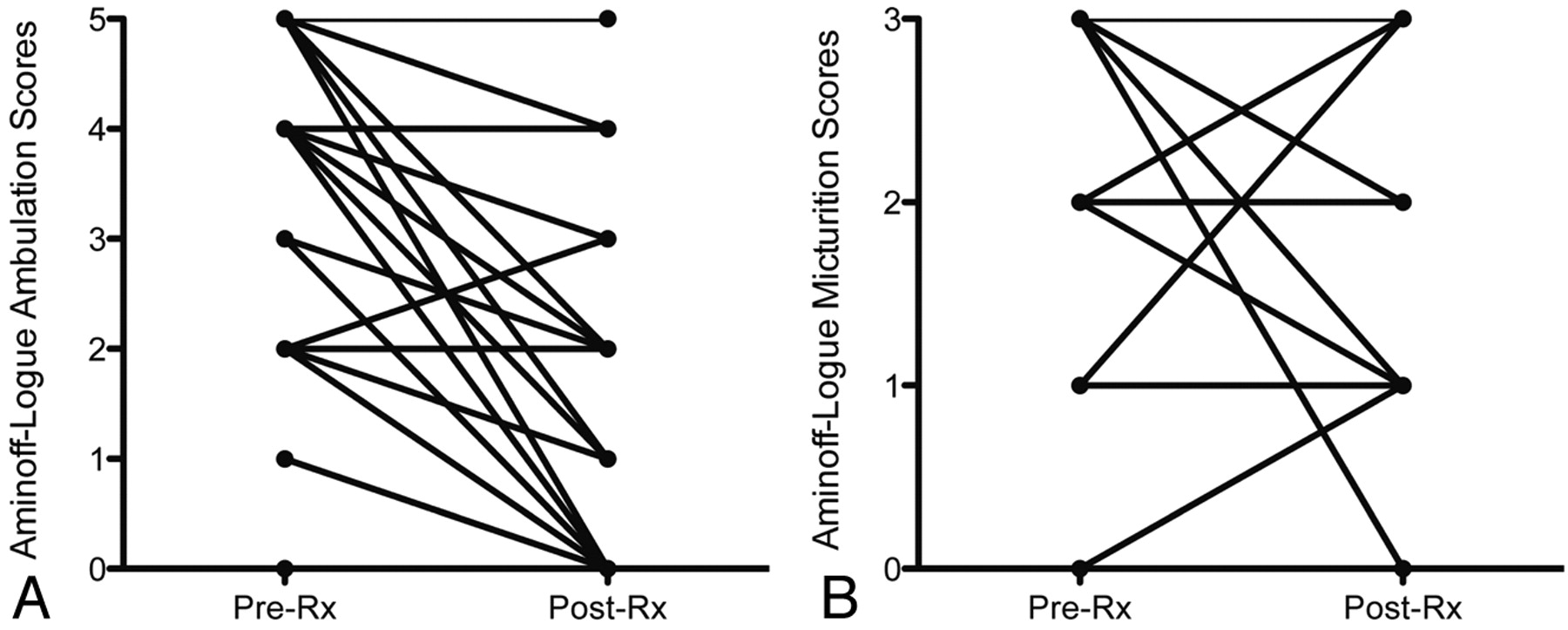
  • Fig 5.
    • Download figure
    • Open in new tab
    • Download powerpoint
    Fig 5.

    Plots of changes in ambulation (A) and micturition (B) ALS scores before and after treatment. Each line may represent >1 individual. Statistical analysis revealed significant improvement in ambulation but not micturition scores after surgical and/or endovascular correction of the PMAVF (P value < .05).

Tables

  • Figures
    • View popup
    Table:

    ALS score of disability

    Classification
    Gait disturbance
        Grade 0Normal gait
        Grade 1Leg weakness or abnormal gait, no restricted activity
        Grade 2Grade 1 with restricted activity
        Grade 3Requires cane or similar support for walking
        Grade 4Requires walker or crutches for walking
        Grade 5Unable to stand, confined to bed or wheelchair
    Micturition
        Grade 0No urinary symptoms
        Grade 1Hesitance, urgency, or frequency
        Grade 2Occasional urinary incontinence or retention
        Grade 3Total urinary incontinence or retention
PreviousNext
Back to top

In this issue

American Journal of Neuroradiology: 31 (10)
American Journal of Neuroradiology
Vol. 31, Issue 10
1 Nov 2010
  • Table of Contents
  • Index by author
Advertisement
Print
Download PDF
Email Article

Thank you for your interest in spreading the word on American Journal of Neuroradiology.

NOTE: We only request your email address so that the person you are recommending the page to knows that you wanted them to see it, and that it is not junk mail. We do not capture any email address.

Enter multiple addresses on separate lines or separate them with commas.
Long-Term Outcome in the Repair of Spinal Cord Perimedullary Arteriovenous Fistulas
(Your Name) has sent you a message from American Journal of Neuroradiology
(Your Name) thought you would like to see the American Journal of Neuroradiology web site.
CAPTCHA
This question is for testing whether or not you are a human visitor and to prevent automated spam submissions.
Cite this article
L. Antonietti, S.A. Sheth, V.V. Halbach, R.T. Higashida, C.F. Dowd, M.T. Lawton, J.D. English, S.W. Hetts
Long-Term Outcome in the Repair of Spinal Cord Perimedullary Arteriovenous Fistulas
American Journal of Neuroradiology Nov 2010, 31 (10) 1824-1830; DOI: 10.3174/ajnr.A2236

Citation Manager Formats

  • BibTeX
  • Bookends
  • EasyBib
  • EndNote (tagged)
  • EndNote 8 (xml)
  • Medlars
  • Mendeley
  • Papers
  • RefWorks Tagged
  • Ref Manager
  • RIS
  • Zotero
0 Responses
Respond to this article
Share
Bookmark this article
Long-Term Outcome in the Repair of Spinal Cord Perimedullary Arteriovenous Fistulas
L. Antonietti, S.A. Sheth, V.V. Halbach, R.T. Higashida, C.F. Dowd, M.T. Lawton, J.D. English, S.W. Hetts
American Journal of Neuroradiology Nov 2010, 31 (10) 1824-1830; DOI: 10.3174/ajnr.A2236
del.icio.us logo Twitter logo Facebook logo Mendeley logo
  • Tweet Widget
  • Facebook Like
  • Google Plus One
Purchase

Jump to section

  • Article
    • Abstract
    • Abbreviations
    • Materials and Methods
    • Results
    • Discussion
    • Conclusions
    • Acknowledgments
    • Footnotes
    • References
  • Figures & Data
  • Supplemental
  • Info & Metrics
  • Responses
  • References
  • PDF

Related Articles

  • PubMed
  • Google Scholar

Cited By...

  • Clarifying the clinical landscape of pediatric spinal arteriovenous shunts: an institutional experience and individual patient-data meta-analysis
  • Spinal Vascular Shunts: A Patterned Approach
  • Pediatric perimedullary arteriovenous fistula: clinical features and endovascular treatments
  • Utility of MRI in spinal arteriovenous fistula
  • Crossref
  • Google Scholar

This article has not yet been cited by articles in journals that are participating in Crossref Cited-by Linking.

More in this TOC Section

  • Dynamic CT Myelography: Patient Positioning
  • Evaluation of SIH MR Scoring Systems in Normals
  • Management Outcomes For VO Spine Biopsy
Show more SPINE IMAGING AND SPINE IMAGE-GUIDED INTERVENTIONS

Similar Articles

Advertisement

Indexed Content

  • Current Issue
  • Accepted Manuscripts
  • Article Preview
  • Past Issues
  • Editorials
  • Editor's Choice
  • Fellows' Journal Club
  • Letters to the Editor
  • Video Articles

Cases

  • Case Collection
  • Archive - Case of the Week
  • Archive - Case of the Month
  • Archive - Classic Case

More from AJNR

  • Trainee Corner
  • Imaging Protocols
  • MRI Safety Corner
  • Book Reviews

Multimedia

  • AJNR Podcasts
  • AJNR Scantastics

Resources

  • Turnaround Time
  • Submit a Manuscript
  • Submit a Video Article
  • Submit an eLetter to the Editor/Response
  • Manuscript Submission Guidelines
  • Statistical Tips
  • Fast Publishing of Accepted Manuscripts
  • Graphical Abstract Preparation
  • Imaging Protocol Submission
  • Evidence-Based Medicine Level Guide
  • Publishing Checklists
  • Author Policies
  • Become a Reviewer/Academy of Reviewers
  • News and Updates

About Us

  • About AJNR
  • Editorial Board
  • Editorial Board Alumni
  • Alerts
  • Permissions
  • Not an AJNR Subscriber? Join Now
  • Advertise with Us
  • Librarian Resources
  • Feedback
  • Terms and Conditions
  • AJNR Editorial Board Alumni

American Society of Neuroradiology

  • Not an ASNR Member? Join Now

© 2025 by the American Society of Neuroradiology All rights, including for text and data mining, AI training, and similar technologies, are reserved.
Print ISSN: 0195-6108 Online ISSN: 1936-959X

Powered by HighWire