
Article Figures & Data
Figures
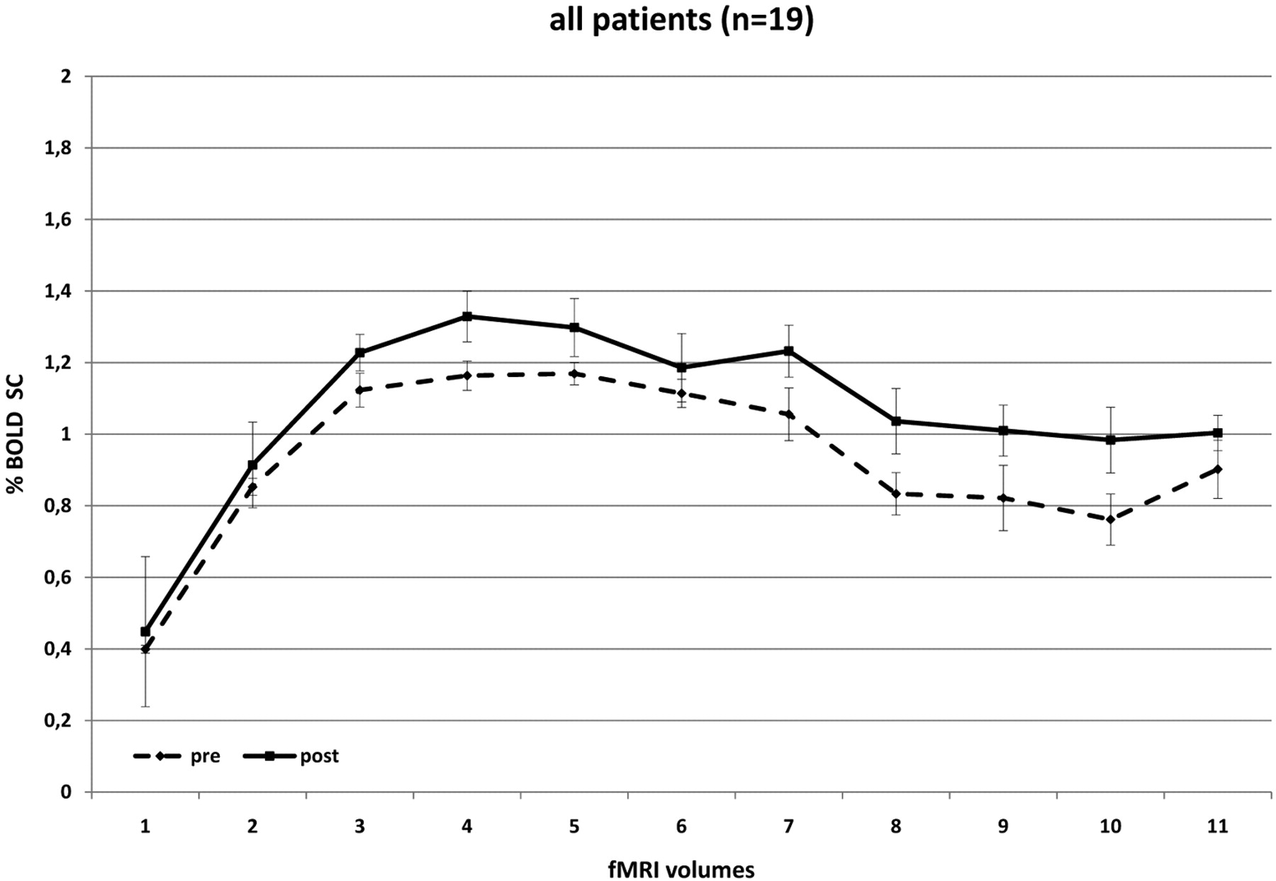
- Fig 1.
BOLD SCs in all patients before and after CEA. There is a significant increase in BOLD SC after revascularization, testifying to an ameliorated cerebrovascular reactivity.
- Fig 2.
Pre- and postoperative BOLD signal-intensity curves of patients with ICA stenosis of ≤70% (A); ≤80% (B), ≤90% (C), and ≤99% (D). Dashed lines demonstrate the preoperative and solid lines, the postoperative data. There are significant differences in pre- and postoperative curves in A and B (low-grade stenoses), and no significant increase in BOLD SC in C and D (high-grade stenoses).
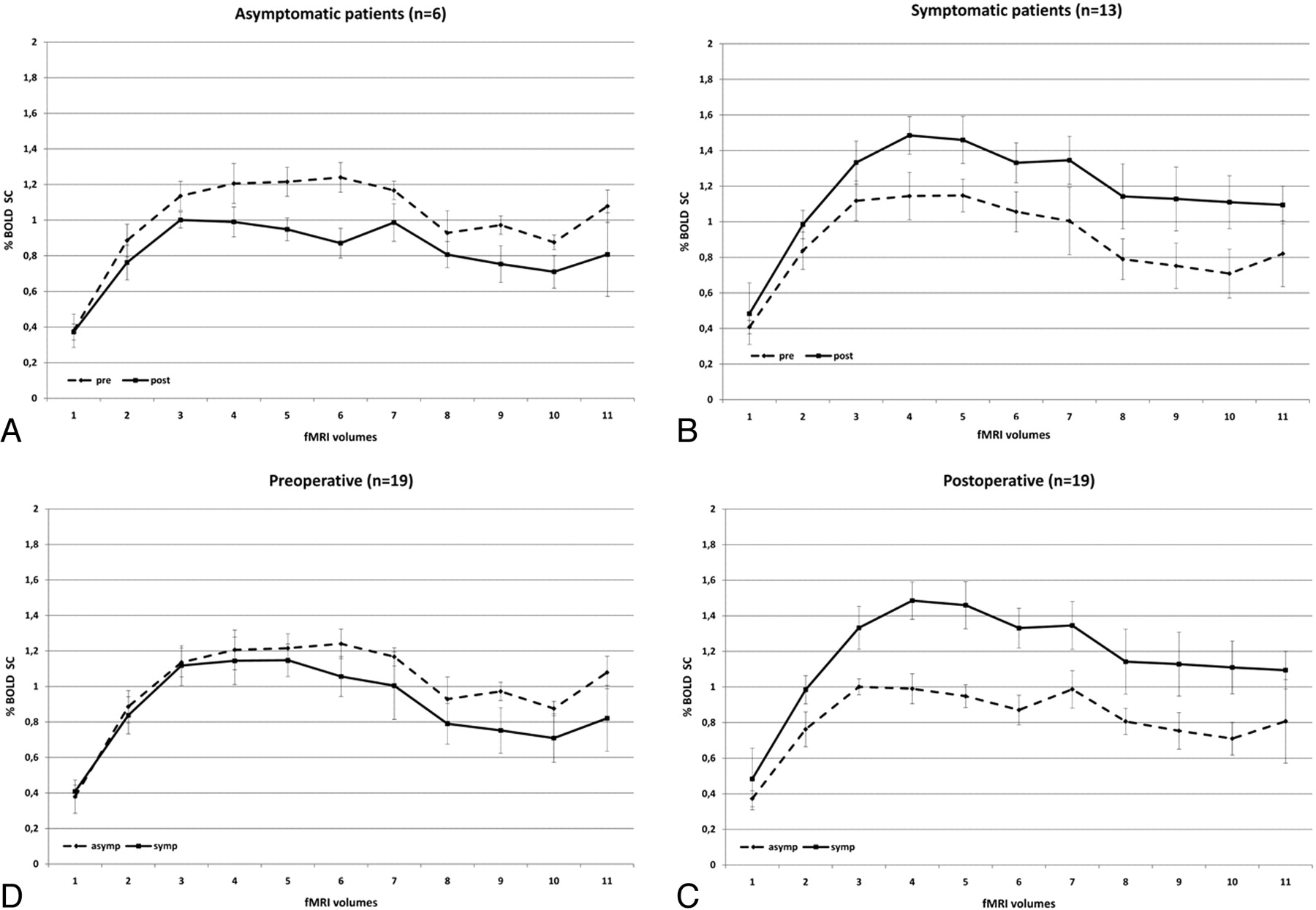
- Fig 3.
BOLD signal-intensity curves of symptomatic (solid line) and asymptomatic (dashed line) patients demonstrates that symptomatic patients have a lower degree of activation than asymptomatic patients preoperatively. Postoperatively, their BOLD signal-intensity curve exceeds that of asymptomatic patients.
- Fig 4.
BOLD signal-intensity curves of patients with minor (A) and severe (B) symptoms of ICA stenosis. Preoperative BOLD SCs are similar in both patient groups. Postoperative changes in BOLD signal intensity are stronger in patients with previous stroke compared with patients with previous TIA.
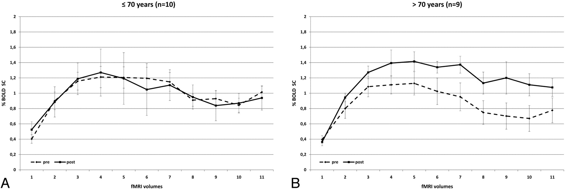
- Fig 5.
Comparison of BOLD signal-intensity curves in patients who are younger (A) and older (B) than 70 years. In contrast to younger patients, older patients present with a significant benefit of CEA and the degree of signal-intensity drop following sustained activity is less pronounced.


Tables
- Table 1:
Patient demographic data and BOLD signal gain and change for percentage ICA stenosis
Subject/ Sex/Age (yr) Presenting Symptoms Days between Presenting Symptoms and First MRI Degree of Stenosis BOLD Signal Gain Pre (BOLD %SC) ± SD Post (BOLD %SC) ± SD T2WI and DWI Findings Symp Side Symp Side 1/F/66 None N/A 90 70 0.02 0.685 ± 0.15 0.704 ± 0.18 Chronic microangiopathy 2/F/57 None N/A 85 70 −0.11 1.618 ± 0.3 1.507 ± 0.35 Chronic microangiopathy 3/M/78 Stroke 10 90 0 0.07 1.4 ± 0.32 1.471 ± 0.53 Acute cortical ischemic stroke and chronic microangiopathy 4/M/77 Stroke 12 80 0 1.1 0.271 ± 0.29 1.361 ± 0.33 Acute deep white matter ischemic stroke 5/M/72 AF 19 80 0 0.16 0.775 ± 0.19 0.932 ± 0.30 Chronic microangiopathy 6/M/62 None N/A 85 0 −0.68 0.543 ± 0.18 -0.139 ± 0.12 Chronic microangiopathy 7/M/62 None N/A 95 85 −0.48 1.021 ± 0.36 0.543 ± 0.18 Chronic microangiopathy 8/M/81 TIA 16 70 0 0.21 1.323 ± 0.32 1.528 ± 0.4 Chronic microangiopathy 9/M/69 AF 14 90 0 −0.12 0.814 ± 0.23 0.693 ± 0.2 Chronic microangiopathy 10/M/71 AF 8 90 0 −0.14 0.832 ± 0.12 0.693 ± 0.2 Acute cortical ischemic stroke and microangiopathy 11/M/72 TIA 19 70 0 0.27 0.924 ± 0.29 1.195 ± 0.41 Chronic microangiopathy 12/M/78 Stroke 25 90 0 0.72 0.569 ± 0.14 1.291 ± 0.23 Chronic microangiopathy 13/M/66 None N/A 80 0 0.1 1.095 ± 0.29 1.193 ± 0.23 Chronic microangiopathy 14/M/80 Stroke 5 60 80 −0.15 0.961 ± 0.71 0.815 ± 0.33 Acute cortical ischemic stroke and microangiopathy 15/F/74 Stroke 6 95 65 0.41 0.632 ± 0.16 1.038 ± 0.27 Acute deep white matter ischemic stroke 16/M/66 None N/A 80 0 0.02 1.085 ± 0.31 1.107 ± .29 Chronic microangiopathy 17/M/67 Stroke 12 70 0 1.32 0.753 ± 0.17 2.076 ± 0.67 Acute deep white matter ischemic stroke 18/M/66 AF 7 95 0 −0.06 1.141 ± 0.27 1.079 ± 0.21 Chronic microangiopathy 19/M/50 Stroke 9 55 0 −0.1 1.172 ± 0.31 1.068 ± 0.23 Acute cortical ischemic stroke Mean BOLD Pre SD Mean BOLD Post SD MSC SD PValue Stenosis ≤70% 1.03 0.44 1.34 0.61 0.31 0.61 .001 ≤80% 0.81 0.43 1.15 0.32 0.34 0.50 .000 ≤90% 0.99 0.44 0.99 0.48 −0.01 0.39 .453 ≤100% 0.77 0.34 0.66 0.61 −0.11 0.50 .100 Stroke/TIA/AF Stroke 0.82 0.49 1.30 0.54 0.55 0.65 .000 TIA/AF 0.97 0.32 1.02 0.41 0.05 0.26 .054 Symp/asymp Asymp 1.01 0.44 0.82 0.58 −0.19 0.35 .000 Symp 0.89 0.42 1.17 0.50 0.28 0.55 .000 Age ≤70 yr 0.99 0.39 0.98 0.64 −0.01 0.56 .429 >70 yr 0.85 0.46 1.15 0.43 0.29 0.48 .000 All 0.93 0.43 1.06 0.55 0.13 0.66 .002


