Advertisement

Main menu

  • Home
  • Content
    • Current Issue
    • Accepted Manuscripts
    • Article Preview
    • Past Issue Archive
    • Video Articles
    • AJNR Case Collection
    • Case of the Week Archive
    • Case of the Month Archive
    • Classic Case Archive
  • Special Collections
    • AJNR Awards
    • Low-Field MRI
    • Alzheimer Disease
    • ASNR Foundation Special Collection
    • Photon-Counting CT
    • View All
  • Multimedia
    • AJNR Podcasts
    • AJNR SCANtastic
    • Trainee Corner
    • MRI Safety Corner
    • Imaging Protocols
  • For Authors
    • Submit a Manuscript
    • Submit a Video Article
    • Submit an eLetter to the Editor/Response
    • Manuscript Submission Guidelines
    • Statistical Tips
    • Fast Publishing of Accepted Manuscripts
    • Graphical Abstract Preparation
    • Imaging Protocol Submission
    • Author Policies
  • About Us
    • About AJNR
    • Editorial Board
    • Editorial Board Alumni
  • More
    • Become a Reviewer/Academy of Reviewers
    • Subscribers
    • Permissions
    • Alerts
    • Feedback
    • Advertisers
    • ASNR Home

User menu

  • Alerts
  • Log in

Search

  • Advanced search
American Journal of Neuroradiology

American Journal of Neuroradiology

ASHNR American Society of Functional Neuroradiology ASHNR American Society of Pediatric Neuroradiology ASSR
  • Alerts
  • Log in

Advanced Search

  • Home
  • Content
    • Current Issue
    • Accepted Manuscripts
    • Article Preview
    • Past Issue Archive
    • Video Articles
    • AJNR Case Collection
    • Case of the Week Archive
    • Case of the Month Archive
    • Classic Case Archive
  • Special Collections
    • AJNR Awards
    • Low-Field MRI
    • Alzheimer Disease
    • ASNR Foundation Special Collection
    • Photon-Counting CT
    • View All
  • Multimedia
    • AJNR Podcasts
    • AJNR SCANtastic
    • Trainee Corner
    • MRI Safety Corner
    • Imaging Protocols
  • For Authors
    • Submit a Manuscript
    • Submit a Video Article
    • Submit an eLetter to the Editor/Response
    • Manuscript Submission Guidelines
    • Statistical Tips
    • Fast Publishing of Accepted Manuscripts
    • Graphical Abstract Preparation
    • Imaging Protocol Submission
    • Author Policies
  • About Us
    • About AJNR
    • Editorial Board
    • Editorial Board Alumni
  • More
    • Become a Reviewer/Academy of Reviewers
    • Subscribers
    • Permissions
    • Alerts
    • Feedback
    • Advertisers
    • ASNR Home
  • Follow AJNR on Twitter
  • Visit AJNR on Facebook
  • Follow AJNR on Instagram
  • Join AJNR on LinkedIn
  • RSS Feeds

AJNR Awards, New Junior Editors, and more. Read the latest AJNR updates

LetterLetter

A Case Of Neuro-Behcet Disease Mimicking Gliomatosis Cerebri

A.O. Varoglu
American Journal of Neuroradiology January 2010, 31 (1) E1; DOI: https://doi.org/10.3174/ajnr.A1868
A.O. Varoglu
  • Find this author on Google Scholar
  • Find this author on PubMed
  • Search for this author on this site
  • Article
  • Figures & Data
  • Info & Metrics
  • Responses
  • References
  • PDF
Loading

Gliomatosis cerebri (GC) is a rare clinical entity characterized by diffuse and infiltrative overgrowth of tumor cells. The most commonly involved areas of GC are in the bilateral hemispheres, at least 2 lobes per hemisphere, and the infratentorial region.1 The neurologic involvement of Behcet disease ranges from 2.2% to 49%.2 We report a case of Neuro-Behcet disease (NBD) mimicking GC. There have been a limited number of case reports describing neurologic pseudotumoral presentation in NBD.3

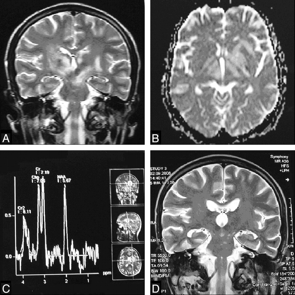
A 38-year-old man was admitted to our clinic with mental status changes, deterioration of speaking and walking, oral and genital mucocutaneous ulcerations, and incontinence. His symptoms had existed for 1 month. On admission, tetraparesis was found on his neurologic examination. Regarding the MR imaging findings, we initially thought that the lesions supported GC (Fig. 1A). MR imaging spectroscopy and diffusion-weighted MR imaging (DWI) investigations did not support tumors (Fig 1B, -C). Eye lesions were not detected. Consultation with a dermatologist resulted in a diagnosis of NBD. Three months after the treatment, the neurologic status had substantially recovered, and the lesions were markedly reduced on MR imaging (Fig 1D).

Fig 1.
  • Download figure
  • Open in new tab
  • Download powerpoint
Fig 1.

A, T2-weighted image reveals high signal intensity in the mesencephalon, bilateral basal ganglions, posterior limb of the internal capsule, and centrum semiovale regions. B, ADC map reveals high signal intensity. C, MR spectroscopy reveals that major peaks are normal, excluding GC. D, T2-weighted images reveal markedly smaller lesions after treatment. Cr indicates creatine.

The differential diagnosis of NBD may be difficult, because NBD can mimic other neurologic disorders, particularly in areas in which the disease is uncommon. The most common lesions of NBD are located at the mesodiencephalic junction, followed by the pontobulbar region. The lesions are bilateral in approximately one-third of the cases, and hemispheric lesions are not commonly seen.2 Therefore, our case was mimicking GC in terms of the unusual location of these lesions. DWI is a useful tool for the differential diagnosis of NBD in distinguishing it from other disease, especially acute ischemic infarcts.3 A stereotactic biopsy is usually used to distinguish NBD from cerebral tumors. There are no published reports concerning the use of MR spectroscopy for this separation. DWI and MR spectroscopy may help in the neoplasms and demyelinating lesions.4 Apparent diffusion coefficient values are elevated for chronic lesions as in our case, though these values may be reduced for acute lesions in a demyelinating disease. The typical DWI features for primary neoplasms are variable. Typical MR spectroscopic features for primary neoplasms are elevated peaks of lipid, lactate, choline, and myo-inositol and reduced N-acetylaspartate signals. In our case, these features were not observed and MR spectroscopy findings were normal.4 After the treatment, the lesions significantly decreased. In conclusion, we suggest that MR spectroscopy and DWI should be used in the differential diagnosis of NBD to avoid unnecessary invasive approaches.

References

  1. 1.↵
    1. Cavaliere RL
    . Low-grade gliomas: an update on pathology and therapy. Lancet Neurol 2005; 4: 460
    PubMed
  2. 2.↵
    1. Akman-Demir G,
    2. Serdaroglu P,
    3. Tasci B
    . Clinical patterns of neurological involvement in Behcet's disease: evaluation of 200 patients—The Neuro-Behcet Study Group. Brain 1999; 122: 2171–82
    Abstract/FREE Full Text
  3. 3.↵
    1. Heo JH,
    2. Lee ST,
    3. Chu K,
    4. et al
    . Neuro-Behcet's disease mimicking multiple brain tumors: diffusion-weighted MR study and literature review. J Neurol Sci 2008; 264: 177–81
    CrossRefPubMed
  4. 4.↵
    1. Al-Okaili RN,
    2. Krejza J,
    3. Wang S,
    4. et al
    . Advanced MR imaging techniques in the diagnosis of intraaxial brain tumors in adults. Radiographics 2006; 26 (suppl 1): 173–89
    CrossRefPubMed
  • Copyright © American Society of Neuroradiology
PreviousNext
Back to top

In this issue

American Journal of Neuroradiology: 31 (1)
American Journal of Neuroradiology
Vol. 31, Issue 1
1 Jan 2010
  • Table of Contents
  • Index by author
Advertisement
Print
Download PDF
Email Article

Thank you for your interest in spreading the word on American Journal of Neuroradiology.

NOTE: We only request your email address so that the person you are recommending the page to knows that you wanted them to see it, and that it is not junk mail. We do not capture any email address.

Enter multiple addresses on separate lines or separate them with commas.
A Case Of Neuro-Behcet Disease Mimicking Gliomatosis Cerebri
(Your Name) has sent you a message from American Journal of Neuroradiology
(Your Name) thought you would like to see the American Journal of Neuroradiology web site.
CAPTCHA
This question is for testing whether or not you are a human visitor and to prevent automated spam submissions.
Cite this article
A.O. Varoglu
A Case Of Neuro-Behcet Disease Mimicking Gliomatosis Cerebri
American Journal of Neuroradiology Jan 2010, 31 (1) E1; DOI: 10.3174/ajnr.A1868

Citation Manager Formats

  • BibTeX
  • Bookends
  • EasyBib
  • EndNote (tagged)
  • EndNote 8 (xml)
  • Medlars
  • Mendeley
  • Papers
  • RefWorks Tagged
  • Ref Manager
  • RIS
  • Zotero
0 Responses
Respond to this article
Share
Bookmark this article
A Case Of Neuro-Behcet Disease Mimicking Gliomatosis Cerebri
A.O. Varoglu
American Journal of Neuroradiology Jan 2010, 31 (1) E1; DOI: 10.3174/ajnr.A1868
del.icio.us logo Twitter logo Facebook logo Mendeley logo
  • Tweet Widget
  • Facebook Like
  • Google Plus One
Purchase

Jump to section

  • Article
    • References
  • Figures & Data
  • Info & Metrics
  • Responses
  • References
  • PDF

Related Articles

  • No related articles found.
  • PubMed
  • Google Scholar

Cited By...

  • No citing articles found.
  • Crossref (7)
  • Google Scholar

This article has been cited by the following articles in journals that are participating in Crossref Cited-by Linking.

  • Pseudotumoural presentation of neuro-Behcet's disease: case series and review of literature
    N. Noel, M. Hutie, B. Wechsler, S. Vignes, D. Le Thi Huong-Boutin, Z. Amoura, D. Dormont, V. Delcey, M. Polivka, P. Cacoub, D. Saadoun
    Rheumatology 2012 51 7
  • Cerebral tumor or pseudotumor?
    D. Leclercq, S. Trunet, A. Bertrand, D. Galanaud, S. Lehéricy, D. Dormont, A. Drier
    Diagnostic and Interventional Imaging 2014 95 10
  • Enfermedad de Behçet pseudotumoral
    Sara Alonso, Anne Riveros-Frutos, Melania Martínez-Morillo, Laia Grau-Ferrer, Cristina Carrato, Alejandro Olivé
    Reumatología Clínica 2016 12 2
  • Pseudotumoral neuro-behcet’s disease: case series and review of literature
    Mohamed Amine Hadj Taieb, Houssem Slimane, Mariem Mhiri, Rihab Ben Dhia, Nizar Daoussi, Mahbouba Frih-Ayed
    Acta Neurologica Belgica 2024 124 2
  • Diffuse Low-Grade Gliomas in Adults
    Catherine Godfraind
    2013
  • Pseudotumoral Behçet's Disease
    Sara Alonso, Anne Riveros-Frutos, Melania Martínez-Morillo, Laia Grau-Ferrer, Cristina Carrato, Alejandro Olivé
    Reumatología Clínica (English Edition) 2016 12 2
  • Tumeur ou pseudotumeur cérébrale ?
    D. Leclercq, S. Trunet, A. Bertrand, D. Galanaud, S. Lehéricy, D. Dormont, A. Drier
    Journal de Radiologie Diagnostique et Interventionnelle 2014 95 10

More in this TOC Section

  • Letter to the Editor regarding “Automated Volumetric Software in Dementia: Help or Hindrance to the Neuroradiologist?”
  • Reply:
  • Brain AVM’s Nidus: What if We Hadn’t Understood Anything?
Show more LETTERS

Similar Articles

Advertisement

Indexed Content

  • Current Issue
  • Accepted Manuscripts
  • Article Preview
  • Past Issues
  • Editorials
  • Editor's Choice
  • Fellows' Journal Club
  • Letters to the Editor
  • Video Articles

Cases

  • Case Collection
  • Archive - Case of the Week
  • Archive - Case of the Month
  • Archive - Classic Case

More from AJNR

  • Trainee Corner
  • Imaging Protocols
  • MRI Safety Corner

Multimedia

  • AJNR Podcasts
  • AJNR Scantastics

Resources

  • Turnaround Time
  • Submit a Manuscript
  • Submit a Video Article
  • Submit an eLetter to the Editor/Response
  • Manuscript Submission Guidelines
  • Statistical Tips
  • Fast Publishing of Accepted Manuscripts
  • Graphical Abstract Preparation
  • Imaging Protocol Submission
  • Evidence-Based Medicine Level Guide
  • Publishing Checklists
  • Author Policies
  • Become a Reviewer/Academy of Reviewers
  • News and Updates

About Us

  • About AJNR
  • Editorial Board
  • Editorial Board Alumni
  • Alerts
  • Permissions
  • Not an AJNR Subscriber? Join Now
  • Advertise with Us
  • Librarian Resources
  • Feedback
  • Terms and Conditions
  • AJNR Editorial Board Alumni

American Society of Neuroradiology

  • Not an ASNR Member? Join Now

© 2025 by the American Society of Neuroradiology All rights, including for text and data mining, AI training, and similar technologies, are reserved.
Print ISSN: 0195-6108 Online ISSN: 1936-959X

Powered by HighWire