
Article Figures & Data
Figures
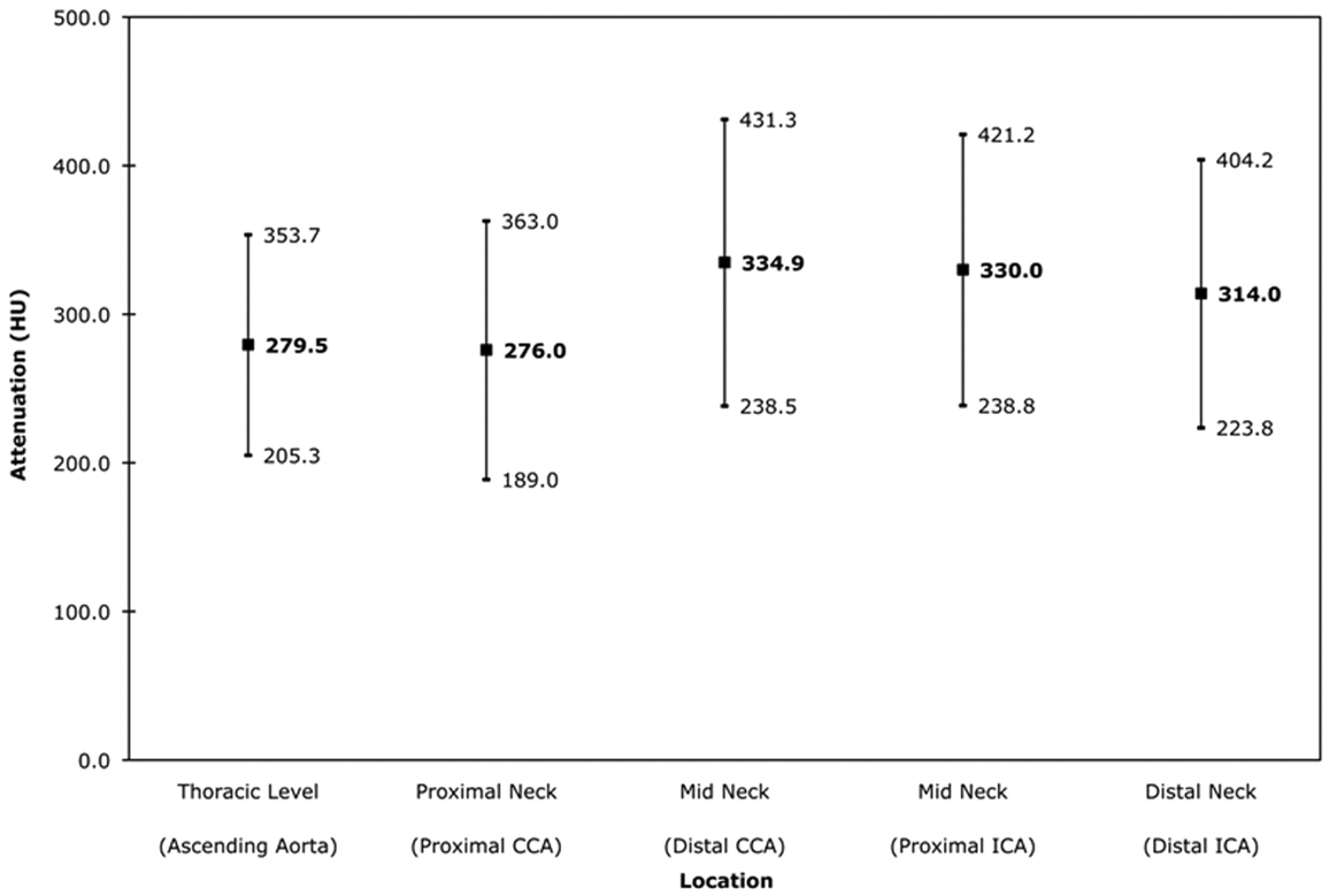
- Fig 1.
Intraluminal attenuation by arterial location. The mean arterial attenuation is shown in bold (center value). Values at 1 SD above and below the mean are also noted. CCA indicates common carotid artery; ICA, internal carotid artery.
- Fig 2.
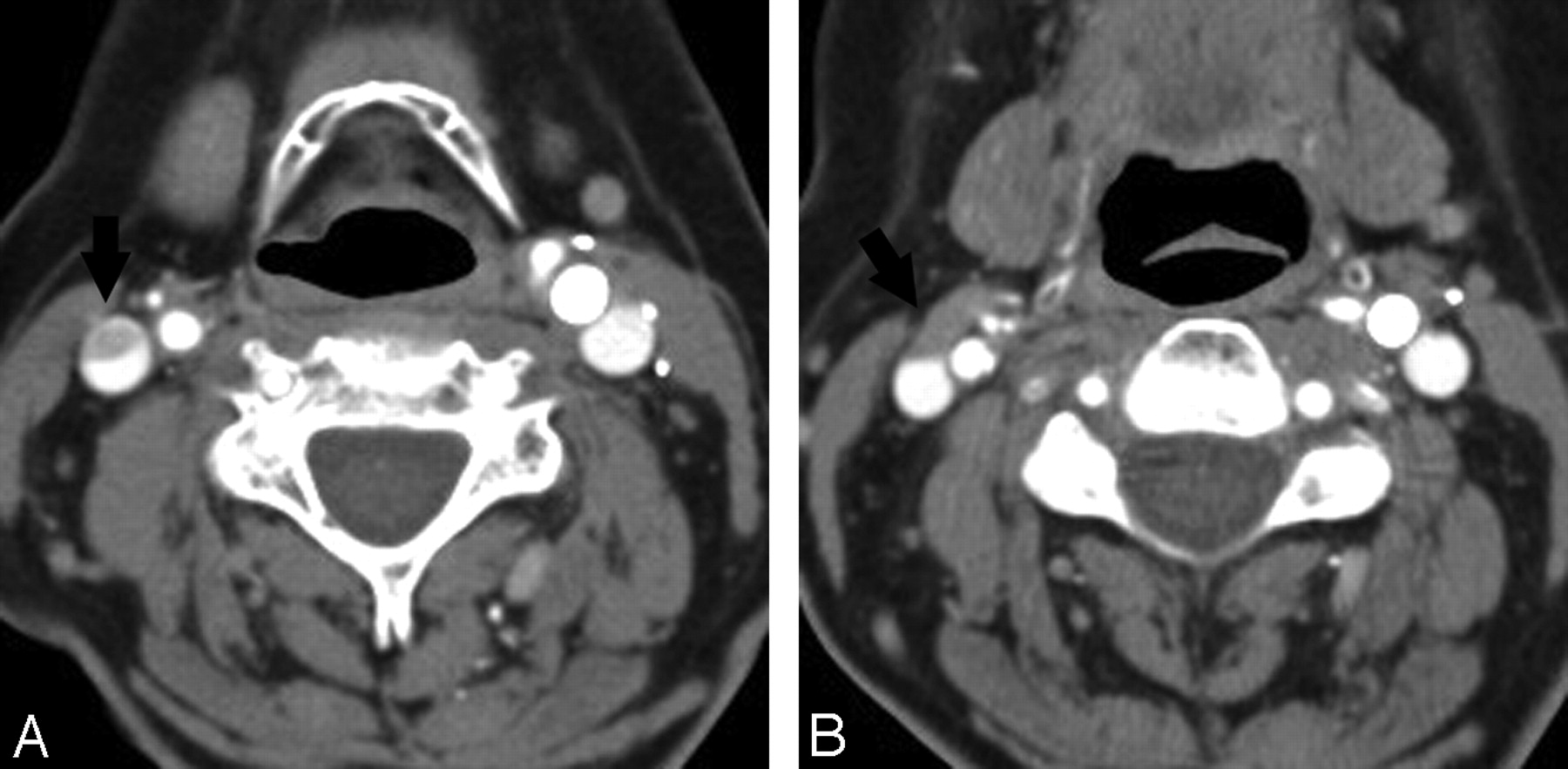
Artifacts interfering with CTA interpretation. A, Contrast reflux into small neck veins obscures evaluation of the vertebral arteries (black arrows). B, Significant streak artifacts from dental hardware limit evaluation of the carotid and vertebral arteries. C, Left-sided injection results in attenuated contrast opacification of the left brachiocephalic vein, which obscures the origin of the great vessels. D, Photon starvation between the shoulders results in streaks of low attenuation that make evaluation of the vertebral arteries difficult.
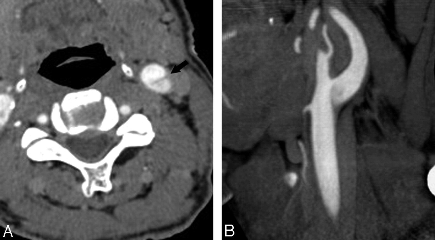
- Fig 3.
A 68-year-old man with pseudodissection of the left carotid bifurcation. A and B, Axial source (A) and sagittal reformatted maximum-intensity-projection (MIP) image from CTA (B) demonstrate faint linear flap extending vertically from the left carotid bifurcation and the appearance of 2 vascular compartments with different degrees of opacification.
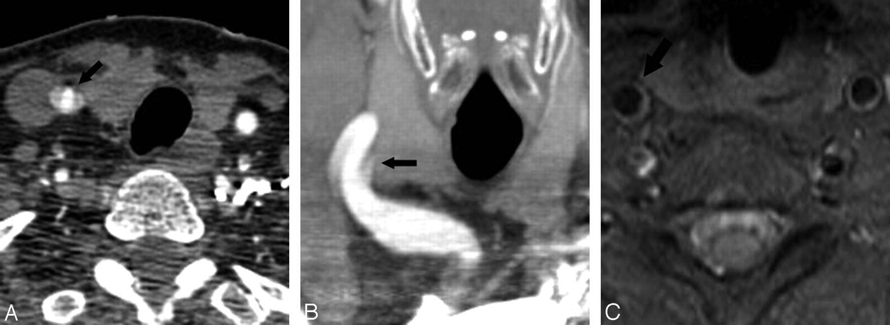
- Fig 4.
A 61-year-old woman with pseudodissection of the proximal right CCA. A and B, Axial source (A) and coronal reformatted MIP image from CTA (B) shows a trilaminar appearance of the proximal right CCA (arrow), disconcerting for dissection. C, Axial T1-weighted image using a fat saturation technique through the cervical carotid artery (arrow), obtained immediately after the CT study, shows no evidence of dissection. The finding on CTA is considered to represent pulsation artifacts.
- Fig 5.
A 79-year-old woman with left hemiparesis. A−C, CTA shows nonopacification and apparent occlusion of the entire right ICA, from its origin to the carotid terminus. Compare the normally opacified left ICA (black arrows) with the nonopacified right side (white arrows in C denote the expected course of the right ICA). D and E, Axial contrast-enhanced images, however, show opacification and confirm patency of the right ICA at comparable levels. F, Conventional angiogram obtained 4 hours later (anteroposterior [AP] view right carotid injection) confirms patency of the entire intracranial ICA but reveals an abrupt cutoff of the proximal M1 segment (arrow), consistent with occlusion. Nonopacification on CTA is attributed to the sluggish flow resulting from the M1 occlusion on the right side.
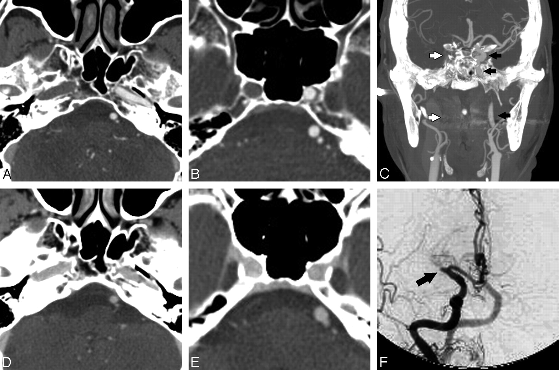
- Fig 6.
A 71-year-old woman with left-sided hemiparesis. A, Axial source CTA image shows opacification but decreased attenuation of the right supraclinoid ICA (large arrow) compared with the normal left side (small arrow). B, Axial reformatted MIP image again demonstrates overall decreased attenuation of the right supraclinoid ICA (large black arrow) compared with the normal left ICA and middle cerebral artery (MCA, small black arrows). A large filling defect is noted in the right MCA (white arrow). C, Angiogram, AP-view right carotid injection, shows an abrupt cutoff (black arrow) in the right MCA consistent with occlusion. No hemodynamically significant stenoses were observed at the origin or along the course of the right carotid artery to account for impaired blood flow and decreased arterial attenuation, which was instead attributed to the presence of a distal thrombus.
- Fig 7.
A 41-year-old man with apparent thrombosis of the entire right internal jugular vein (IJV). A, Coronal reformatted MIP image from CTA shows nonopacification of the entire visualized right internal jugular vein (IJV, arrows) compared with the opacified left IJV (arrowheads). B, Axial source CTA image again shows no contrast in the right IJV (arrow). C, Contrast-enhanced CT at the same level as B shows delayed contrast filling of the IJV (black arrow), suggesting sluggish flow but patency of the superior IJV at the skull base. More inferior portions of the IJV in the neck could not be assessed on contrast-enhanced CT, which images only the head, so it is unclear whether the remainder of the IJV was thrombosed or patent but with slow flow. No follow-up imaging was available in this patient, who had a right central venous catheter (not shown), which may have provided a nidus for thrombosis or impeded venous drainage causing slow flow.
- Fig 8.
A 69-year-old woman with venous pseudothrombosis. A, Axial source CTA image shows an apparent eccentric filling defect in the right IJV (arrow). B, Nonopacified blood in a small vein (black arrow) draining into the IJV is responsible for causing these flow artifacts.


Tables
Artifacts and problems limiting artery evaluation
Type of Artifacts Description No. of Cases (incidence) on 64-Section CTA No. of Cases (incidence) on 16-Section CTA P Value (Fisher exact test) Patient motion artifacts Gross motion during scan acquisition 0 (0%) 3 (2.7%) .247 Streak artifacts of venous origin Dense undiluted contrast in SCV, BCV; SVC obscures great vessel origins 32 (32%) 29 (25.7%) .363 Shoulder streak artifacts Photon starvation between shoulders obscures CCA, VA, IJV in lower neck 28 (28%) 33 (29.2%) .880 Metallic streak artifacts Dental hardware, cardiac defibrillator leads, spinal fusion hardware obscure adjacent arteries 26 (26%) 32 (28.3%) .759 Contrast material reflux Contrast reflux into veins of neck obscures adjacent VA 16 (16%) 17 (15%) .852 Flow artifacts: Altered flow dynamics (eg, slow flow) and rapid scanning time create appearance of dissection flap or nonopacification of patent vessel 14a (14%) 0 (0%) <.001 Arterial Pseudo-dissection 5 Pseudo-occlusion 6 Differential attenuation 1 Venous Pseudo-thrombosis 3 -
Note:—CCA indicates common carotid artery; BCV, brachiocephalic vein; IJV, internal jugular vein; SCV, subclavian vein; SVC, superior vena cava; VA, vertebral artery; CTA, CT angiography.
-
a One patient had both pseudodissection and pseudo-occlusion.
-


