
Article Figures & Data
Figures
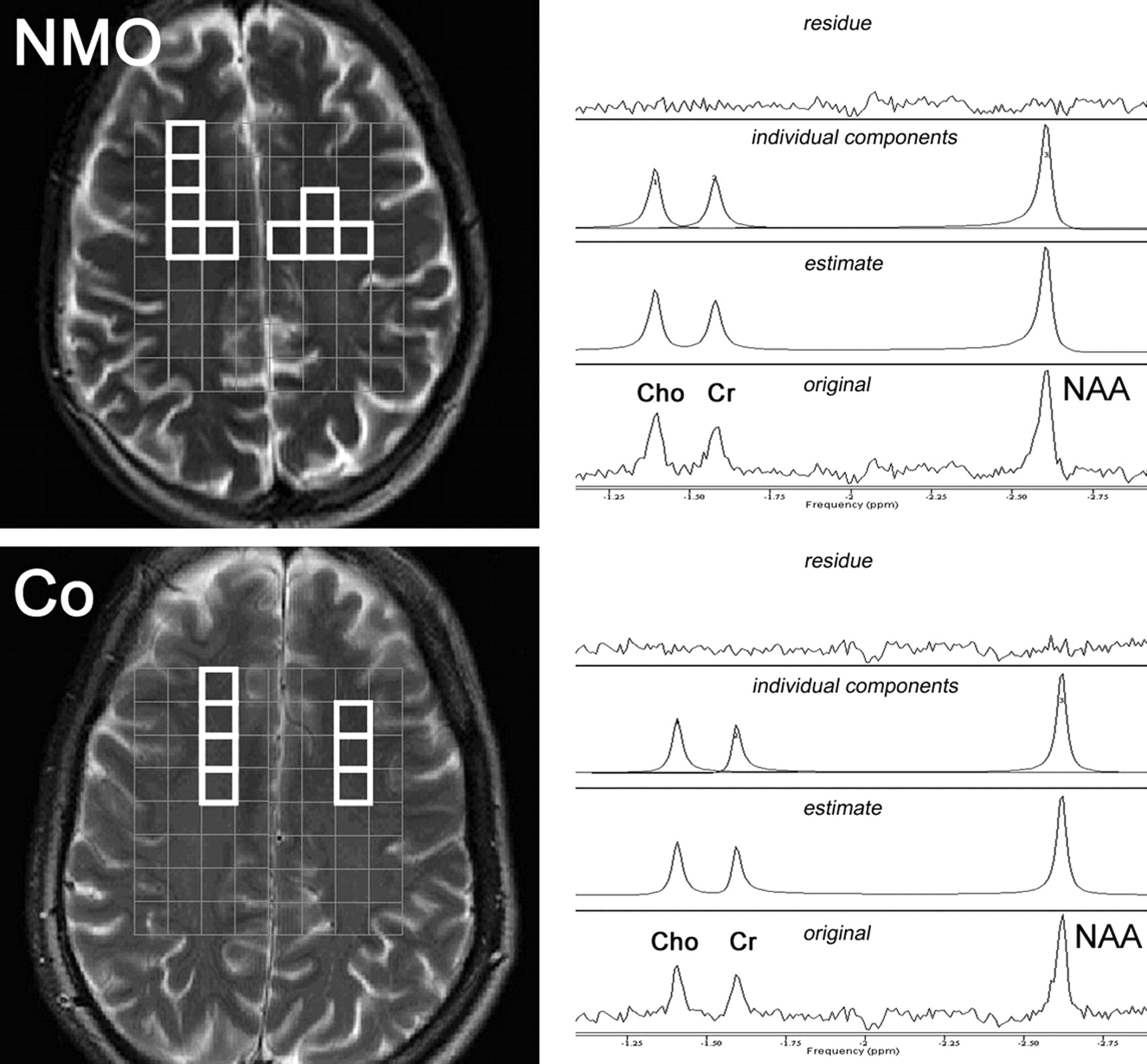
- Fig 1.
Spectral region including resonance lines of choline (Cho), creatine (Cr), and N-acetylaspartate (NAA). Left: Anatomic T2-weighted MR images with the depiction of 1H-MR 2D spectroscopic imaging point-resolved spectroscopy sequence box volume of interest (gray, 8 × 8 cm) and voxels in the normal-appearing white matter (NAWM) chosen for the spectroscopic data evaluation. Right: Representative MR spectra from a single voxel with original spectra (lower trace); advanced method for accurate, robust, and efficient spectral fitting estimate (second trace); individual components (third trace); and fit residue (upper trace). NMO indicates neuromyelitis optica; Co, controls.
- Fig 2.
MR spectroscopy ratios of brain metabolites in the NAWM of patients with NMO and controls. White indicates patients with NMO; gray, controls; squares, NAA:Cr ratio; circles, NAA:Cho ratio; triangles, Cho:Cr ratio; bars, means.
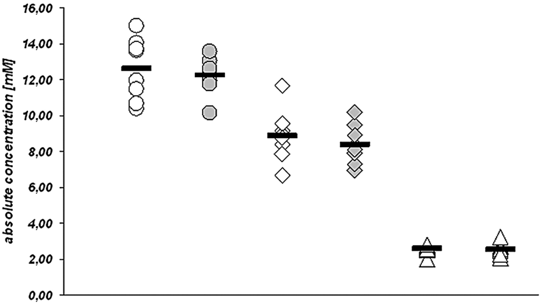
- Fig 3.
MR spectroscopy absolute concentrations of brain metabolites in the NAWM of patients with NMO and controls. White indicates patients with NMO; gray, controls; circles, NAA absolute concentrations; squares, Cr absolute concentrations; triangles, Cho absolute concentrations; bars, means.


Tables
Clinical data of patients with NMO and controls
Neuromyelitis Optica Controls No. Sex Age at MRS (yr) CSF Disease Duration (mo) Relapses No. Sex Age at MRS (yr) Cells/μL Eosinophils, Neutrophils Oligoclonal Bands Total LETM ON Brain Lesion 1 F 17.0 181 Yes Negative 32 4 3 1 1 1 F 17.0 2 F 28.5 52 Yes Positive 95 9 4 5 0 2 F 28.5 3 F 38.5 147 Yes Negative 17 2 2 0 0 3 F 40.0 4 F 42.0 17 – Negative 60 8 4 1 3 4 F 42.0 5 F 51.0 3 – Negative 67 4 4 0 0 5 F 51.0 6 F 63.5 18 – Positive 123 13 11 0 2 6 F 65.0 7 F 65.5 44 Yes Negative 54 3 3 0 0 7 F 63.0 8 M 64.5 59 – Negative 54 5 5 0 0 8 M 64.0 -
Note:—MRS indicates MR spectroscopy; LETM, longitudinal extensive transverse myelitis; ON, optic neuritis; CSF, cerebrospinal fluid.
-
In this issue


Jump to section
Related Articles
Cited By...
- MR T2-relaxation time as an indirect measure of brain water content and disease activity in NMOSD
- An optimal 1H-MRS technique at 7T: Proof-of-principle in Chronic Multiple Sclerosis and Neuromyelitis Optica Brain Lesions and Normal Appearing Brain Tissue
- Quantitative 7T MRI does not detect occult brain damage in neuromyelitis optica
- Neuromyelitis Optica: A Diffusional Kurtosis Imaging Study
- Conventional and Advanced Imaging in Neuromyelitis Optica
- Serum and CSF N-acetyl aspartate levels differ in multiple sclerosis and neuromyelitis optica
- Comment on "Global N-Acetylaspartate Declines Even in Benign Multiple Sclerosis"