
Article Figures & Data
Figures
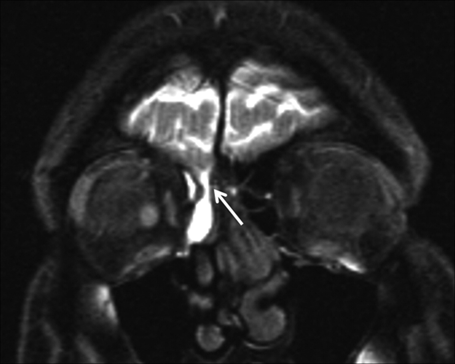
- Fig 1.
CSF rhinorrhea following head trauma in a 42-year-old woman. Coronal T1-weighted MR cisternogram obtained after intrathecal administration of gadopentetate dimeglumine (Gd-DTPA) shows contrast leakage (arrow) extending from the cranial subarachnoid space into the ethmoid air cell region from a defect in the right side of the cribriform plate.
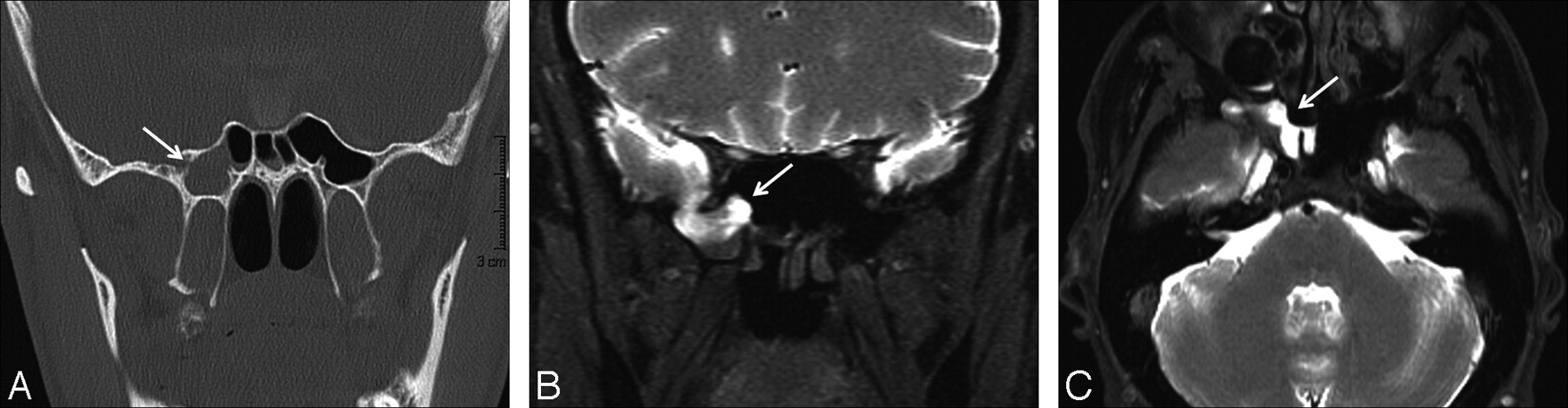
- Fig 2.
CSF rhinorrhea in a 39-year-old man after sellar region surgery. A, Coronal thin-section CT scan reveals a defect in the right side of the sphenoid sinus (arrow) and opacification of the right sphenoid sinus. B and C, Coronal and axial T1-weighted fat-saturated MR cisternograms obtained after the intrathecal administration of Gd-DTPA show contrast leakage (arrows) extending from the cranial subarachnoid space into the right sphenoid sinus.
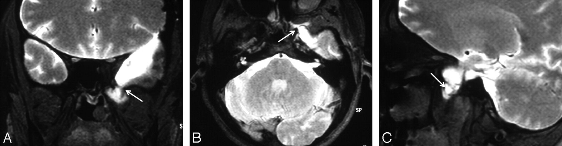
- Fig 3.
CSF rhinorrhea and otorrhea following head trauma in a 15-year-old adolescent boy. A, Axial thin-section CT scan shows a defect in the left petrous temporal bone (arrow) and opacification of the left middle ear cavity. B and C, Coronal and axial T1-weighted fat-saturated MR cisternograms show contrast leakage in the middle ear cavity and eustachian tube (arrows).
- Fig 4.
CSF rhinorrhea following head trauma in a 32-year-old woman. A–C, Axial, coronal, and left parasagittal T1-weighted fat-saturated MR cisternograms obtained after the intrathecal administration of Gd-DTPA show contrast leakage (arrows) extending from the cranial subarachnoid space into the left infratemporal fossa.
- Fig 5.
CSF rhinorrhea following head trauma in a 35-year-old man. A, Coronal thin-section CT scan reveals a defect in the roof of the sphenoid sinus (arrow) and opacification of the right sphenoid sinus. B, A coronal T1-weighted fat-saturated MR cisternogram obtained after the intrathecal administration of Gd-DTPA shows contrast leakage (arrow) extending from the cranial subarachnoid space into the right sphenoid sinus. C, After repair of the dural rupture, suspected CSF rhinorrhea recurred 1 week later and the patient underwent control MR cisternography. Images obtained in the first hour show probable leakage (arrow). D and E, Leakage becomes obvious in late images taken in the third and fifth hours (arrows).


In this issue


Jump to section
Related Articles
Cited By...
- Prospective Safety Study of Intrathecal Gadobutrol in Different Doses
- Prospective Safety Study of Intrathecal Gadobutrol in Different Doses
- High-Resolution Gadolinium-Enhanced MR Cisternography Using Compressed-Sensing T1 SPACE Technique for Detection of Intracranial CSF Leaks
- Intrathecal Use of Gadobutrol for Glymphatic MR Imaging: Prospective Safety Study of 100 Patients
- Imaging of maxillofacial and skull base trauma
- Intrathecal Gadolinium-Enhanced MR Cisternography: A Comprehensive Review
- The Role of MR Myelography with Intrathecal Gadolinium in Localization of Spinal CSF Leaks in Patients with Spontaneous Intracranial Hypotension