
Article Figures & Data
Figures
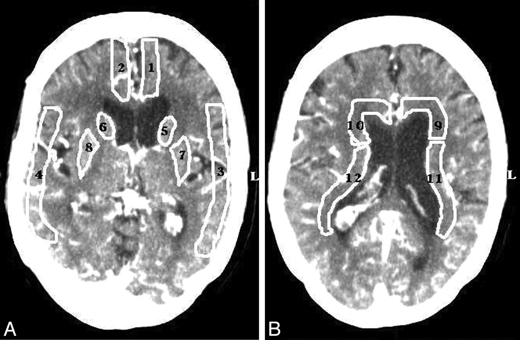
- Fig 1.
A, ROIs that were drawn in the flow territories of the anterior cerebral artery (cortex: ROIs 1 and 2, basal ganglia: ROIs 5 and 6) and the middle cerebral artery (cortex: ROIs 3 and 4, basal ganglia: ROIs 7 and 8). B, In the periventricular white matter, ROIs were drawn anterior (ROIs 9 and 10) and on either side (ROIs 11 and 12) of the lateral ventricles.

Tables
Characteristic Hydrocephalus No Hydrocephalus No. of patients (%) 49 (36) 89 (65) Mean age (SD) 59.8 (13) 54.1 (14) Women (%) 31 (63) 65 (73) Admission WFNS score (%) I 20 (41) 47 (53) II 7 (14) 12 (14) III 7 (14) 9 (10) IV 11 (22) 12 (13) V 4 (8) 9 (10) Day (after SAH) of admission scan (%) 0 33 (67) 56 (63) 1 10 (20) 24 (27) 2 4 (8) 5 (6) 3 2 (4) 4 (5) Amount of blood (Hijdra score) Median cisternal sum score 24 22 Median ventricular sum score 2 0 Mean bicaudate index (SD) 0.22 (0.3) 0.14 (0.3) Mean relative bicaudate index (SD) 1.2 (0.1) 0.75 (0.2) Aneurysm location (%) Anterior communicatinga 22 (45) 41 (46) Internal carotid artery 6 (12) 14 (16) Middle cerebral artery 6 (12) 17 (19) Posteriorb 15 (31) 17 (19) -
Note:—WFNS indicates World Federation of Neurologic Surgeons; SAH, subarachnoid hemorrhage.
-
a Includes the anterior cerebral artery.
-
b Basilar or vertebral arteries.
-
Hydro-cephalus Cortex Difference of Means (95% CI; P Value) Basal Ganglia Difference of Means (95% CI; P Value) Periventricular White Matter Difference of Means (95% CI; P Value) Mean CBV (mL/100 g) (SD) No 4.0 (1.2) −0.1 (−0.4 to 0.1; P = .41) 4.1 (1.2) −0.2 (−0.5 to 0.2; P = .35) 3.1 (0.9) 0.1 (−0.2 to 0.3; P = .61) Yes 4.1 (1.0) 4.3 (1.3) 3.0 (0.9) Mean CBF (mL/100 g/min) (SD) No 53.4 (18.2) 1.8 (−2.8 to 6.4; P = .43) 62.7 (21.1) 6.2 (1.6 to 11.0; P = .014) 32.4 (11.0) 3.8 (0.9 to 6.8; P = .005) Yes 51.6 (18.6) 56.5 (17.2) 28.6 (10.3) Mean MTT (seconds) (SD) No 4.9 (1.8) −0.5 (−1.1 to −0.1; P = .048) 4.3 (1.7) −0.7 (−1.1 to −0.1; P = .008) 6.0 (1.9) −0.9 (−1.6 to −0.2; P = .004) Yes 5.4 (2.7) 5.0 (1.9) 6.9 (2.8) Mean TTP (seconds) (SD) No 23.4 (5.5) −1.6 (−3.0 to −0.1; P = .28) 23.1 (5.6) −1.4 (−2.9 to 0.1; P = .056) 25.7 (4.6) −1.7 (−3.1 to −0.3; P = .034) Yes 25.0 (6.0) 24.5 (5.9) 27.4 (5.4) -
Note:—CI indicates confidence interval; CBV, cerebral blood volume; CBF, cerebral blood flow; MTT, mean transit time; TTP, time-to- peak.
-
In this issue


Jump to section
Related Articles
Cited By...
- Prospective Multicenter Study of Changes in MTT after Aneurysmal SAH and Relationship to Delayed Cerebral Ischemia in Patients with Good- and Poor-Grade Admission Status
- Evaluating CT Perfusion Deficits in Global Cerebral Edema after Aneurysmal Subarachnoid Hemorrhage
- Subarachnoid Hemorrhage-Induced Hydrocephalus in Rats
- Diagnostic Threshold Values of Cerebral Perfusion Measured With Computed Tomography for Delayed Cerebral Ischemia After Aneurysmal Subarachnoid Hemorrhage