
Article Figures & Data
Figures
- Fig 1.
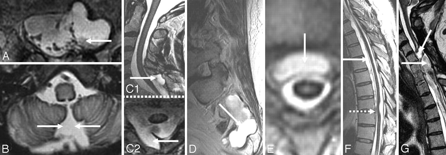
A-I, Axial T2-weighted brain MR images from patients with SS show hemosiderin deposition along the cerebellar folia (A), vermis (B) and around the midbrain (C), pons (D), medulla (E), Sylvian fissure (F), interhemispheric fissure (G), cerebral convexity (H), and the course of the eighth cranial nerve (I). J, Sagittal T1-weighted brain MR image from a patient with SS shows severe cerebellar atrophy.
- Fig 2.
T2-weighted (A) and gradient-echo (T2*) (B) MR images from a patient with SS demonstrate superiority of the gradient-echo technique in detecting the characteristic T2 hypointensity, shown here along the Sylvian and interhemispheric fissures. Both images are from the same patient and were obtained at the same point in time. Reprinted with permission from Kumar N. Superficial siderosis: associations and therapeutic implications. Arch Neurol 2007;64:491–96 (Copyright 2007, American Medical Association)
- Fig 3.
A and B, Sagittal (A) and axial (B) T2-weighted spinal cord MR images show hemosiderin deposition along (A) and around (B) the cord surface. Note associated severe cord atrophy (A) (dotted arrow). C and D, Axial T2-weighted MR images at the level of the cauda equina from patients with SS show peripheralization (C) and clumping (D) of the nerve roots due to arachnoiditis. E, An axial cut at the lumbar levels on a CT myelogram from a patient with SS shows clumping of nerve roots of the cauda equina due to arachnoiditis. F, T2-weighted sagittal MR image of the lumbosacral area from a patient with SS shows a lesion that was suspected of being a possible source of the chronic bleeding. A biopsy was performed, and blood products and fibrous tissue were detected. C and E reprinted with permission from Kumar N. Superficial siderosis: associations and therapeutic implications. Arch Neurol 2007;64:491–96 (Copyright 2007, American Medical Association).
- Fig 4.
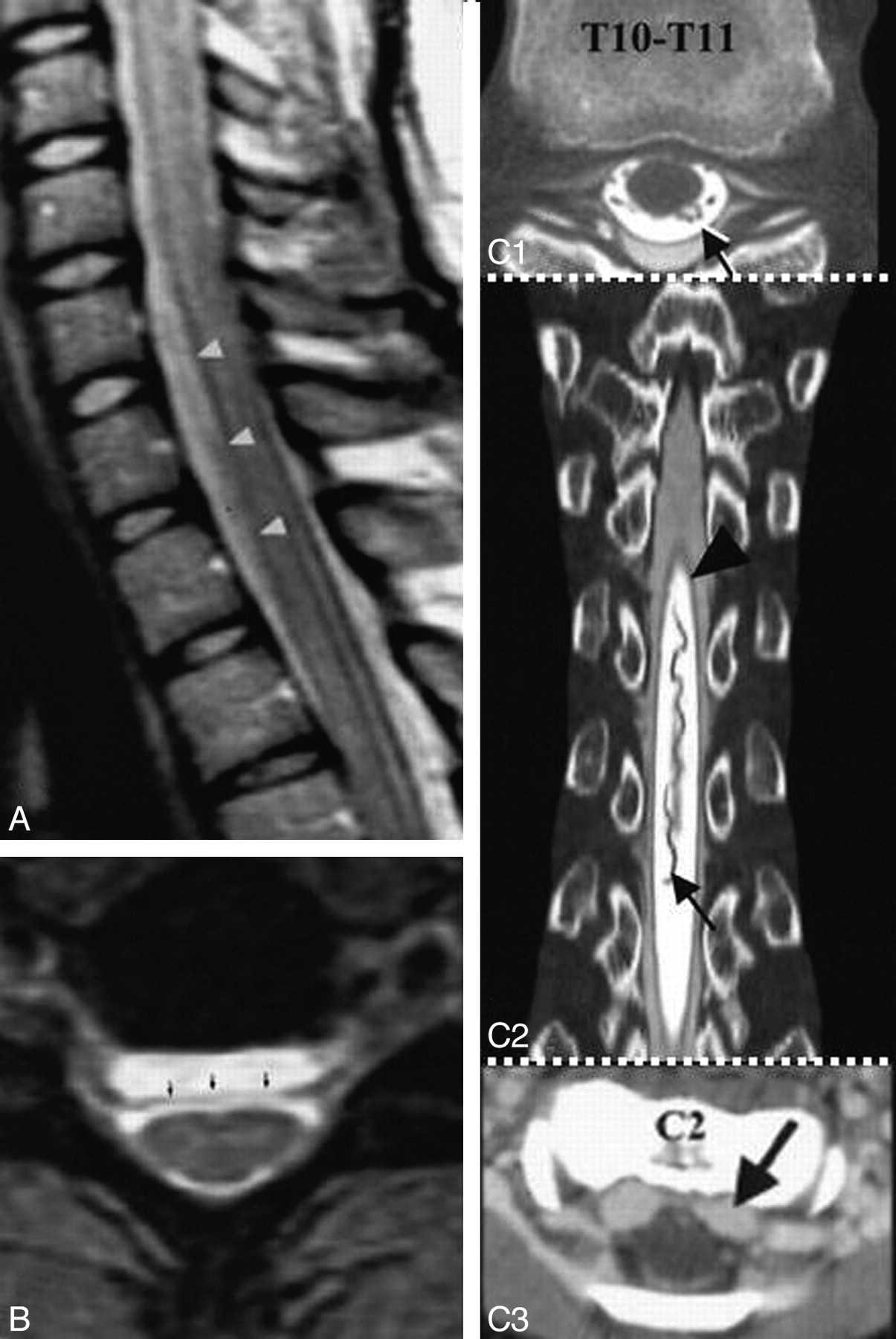
A, Axial T2-weighted spine MR image from a patient with SS shows a left T12 pseudomeningocele. B, Axial T2-weighted brain MR image from a patient with SS shows a posterior fossa fluid-filled collection. C1 and C2, Sagittal (C1) and axial (C2) T2-weighted cervical MR images from a patient with SS show a right C7 pseudomeningocele (same patient as the one shown in Fig 10B). D, Sagittal T2-weighted spine MR image from a patient with SS shows an intrasacral meningocele. E, Axial T2-weighted thoracic cord MR image shows a fluid-filled collection anterior to the spinal cord. F, Sagittal T2-weighted MR image shows a longitudinally extensive fluid collection ventral to the cord. The dotted arrow shows tethering of the cord at T9. G, Sagittal T2-weighted spine MR image from a patient with SS shows multiple intraspinal fluid-filled loculations. C1 and C2 reprinted with permission from Kumar N, Cohen-Gadol AA, Wright RA, et al. Superficial siderosis. Neurology 2006;66:1144–52 (Copyright 2006, Wolters Kluwer Health). F reprinted with permission from Wilden JA, Kumar N, Murali HR, et al. Unusual neuroimaging in superficial siderosis. Neurology 2005;65:489 (Copyright 2005, Wolters Kluwer Health).
- Fig 5.
A and B, Axial (A) and coronal postcontrast (B) T1-weighted MR images from a patient with SS show diffuse pachymeningeal enhancement similar to that reported in patients with craniospinal hypotension. (same patient as shown in 11A1 and A2). C, Axial CT scan from a patient with SS shows increased attenuation around the pons due to presumed calcification of the hemosiderin deposition. A and B reprinted with permission from Kumar N, McKeon A, Rabinstein AA, et al. Superficial siderosis and CSF hypovolemia: the defect (dural) in the link. Neurology 2007;69:925–26 (Copyright 2007, Wolters Kluwer Health).
- Fig 6.
A and B, Pre- (A1 and A2) and postoperative (B1 and B2) sagittal (A1 and B1) and axial (A2 and B2) T2-weighted MR images from a patient with SS show a significant decrease in a longitudinally extensive fluid-filled collection anterior to the spinal cord after repair of a dural defect at T11. The site of the dural defect was localized by a dynamic CT myelogram. A1 and B1 reprinted with permission from Kumar N. Superficial siderosis: associations and therapeutic implications. Arch Neurol 2007;64:491–96 (Copyright 2007, American Medical Association).
- Fig 7.
A1 and A2, Sagittal (A1) and axial (A2) T2-weighted MR images from a patient with SS show a cervicothoracic epidural fluid-filled collection (white arrows) and a T5–6 disk extrusion (black arrowhead) that displaces the dura (white arrowhead) posteriorly. B1 and B2, Corresponding sagittal (reformatted) (B1) and axial (B2) postmyelography CT images demonstrate opacification of the ventral epidural fluid by intrathecal contrast to the same degree as the CSF, thus confirming an active leak. Note partial calcification of the disk (black arrowhead) and the presence of a subarachnoid clot (black arrow). C, Lateral view of the thoracic spine acquired during digital substraction myelography with the patient in the prone position. Note cephalad extension of the contrast in the thecal sac (black arrowhead), focal extravasation of the contrast through a ventral dural tear at T5–6 (white arrow) into the epidural collection (black arrow), and onward cephalad extension in the epidural fluid collection. The asterisk indicates increased attenuation of the contrast because of the smaller volume of the epidural space compared with the subarachnoid space. In this patient, a ventral dural tear at T5–6 was surgically repaired, and a thoracic spine MR imaging performed 3 months later showed resolution of the ventral epidural fluid collection.36 Adapted with permission from J.M. Hoxworth.36
- Fig 8.
A, Axial T2-weighted MR image shows interruption of the rim of hypointensity around the spinal cord at the site of root avulsion. The interruption is likely due to the absence of the spinal cord pia mater at the site of root avulsion. B, C7 to T1 pseudomeningocele due to root avulsion seen on a cervical myelogram (same patient as the one shown in Fig 8A). C, T2-weighted sagittal cervical spine MR image from a patient with SS shows evidence of a prior odontoid fracture (same patient as the one shown in Fig 10C). D, Sagittal T2-weighted cervical spine MR image from a patient with SS shows an intramedullary T2 hypointensity due to myelomalacia secondary to prior trauma. E, T2* MR image from a patient with cerebral amyloid angiopathy shows a right frontal intracerebral hemorrhage (thick arrow) and hemosiderin deposition similar to that seen in superficial siderosis (thin arrows). C reprinted with permission from Kumar N, Cohen-Gadol AA, Wright RA, et al. Superficial siderosis. Neurology 2006;66:1144–52 (Copyright 2006, Wolters Kluwer Health). B and D reprinted with permission from Kumar N. Superficial siderosis: associations and therapeutic implications. Arch Neurol 2007;64:491–96 (Copyright 2007, American Medical Association). E adapted with permission from J. Linn.45
- Fig 9.
A, Sagittal T2-weighted cervicothoracic spine MR image from a patient with intracranial hypotension shows a ventral extradural collection from C6 to T2 (arrowhead), which is isointense with CSF on all imaging sequences. B, Axial T2-weighted gradient-echo MR image at C6–7 in a patient with intracranial hypotension shows a ventral extradural fluid collection separated from the thecal sac by a hypointense dura (arrows). A and B adapted with permission from B.M. Rabin.38 C1−C3, A patient with craniospinal hypotension who had an epidural pseudomeningocele caused by a CSF leak at T8. C1, Axial CT myelogram of the thoracic spine shows the dura marginating an epidural pseudomeningocele (black arrow). C2, Coronal reformatted CT myelogram of the thoracic spine shows a tortuous dilated posterior thoracic spinal vein (black arrow), reminiscent of a dural arteriovenous fistula, and dura (arrowheads) separating intradural and epidural CSF. C3, Contrast-enhanced axial CT scan shows a dilated cervical epidural venous plexus. C1−C3 adapted with permission from J.L. Ulmer.61
- Fig 10.
A, Fluid-filled intraspinal collection anterior to the cord on an axial cut of a thoracic spine CT myelogram. A transdural leak between C7 and T6 was present. A dynamic CT myelogram can localize the exact site of the defect and help direct the laminectomy site. B, Axial CT scan with bone windows from a patient with SS shows a bilaminar C7 fracture (same as patient shown in 4C1 and C2). C, Axial cervical spine CT myelogram shows avulsed C2 nerve roots, which are directed in an anteroposterior direction and are seen as linear streaks with surrounding contrast (same patient as the one shown in Fig 8C). D, Dynamic CT myelogram from a patient with SS and a cervicothoracic epidural fluid-filled collection shows leakage of contrast through a dural tear at T1–2.29 E1, Dynamic CT myelogram shows leakage of contrast (arrow); the dotted arrow points to the intrathecal contrast. E2, Dynamic CT myelogram shows calcified disk protrusion immediately caudal to the dural defect shown in E; the dotted arrow points to intrathecal contrast. This patient (E1 and E2) had diffuse pachymeningeal enhancement, a cervicothoracic epidural fluid collection, and CSF RBCs and xanthochromia, all of which resolved after repair of a dural defect identified at T7–8. F1, Reformatted sagittal cuts from a dynamic CT myelogram obtained in a patient with low-pressure headache without SS show a high-flow CSF leak (arrow) through a ventral midline defect located on the right side of a bilobed spiculated midline osteophyte at T2–3. F2, The osteophyte is shown on an axial thoracic spine CT. This patient also had a ventral epidural fluid-filled collection into which the contrast leaked through the dural defect (dotted arrow). C reprinted with permission from Kumar N. Superficial siderosis: associations and therapeutic implications. Arch Neurol 2007;64:491–96 (Copyright 2007, American Medical Association) and Kumar N, Lindell EP, Wilden JA, et al. Role of dynamic CT myelography in identifying the etiology of superficial siderosis. Neurology 2005;65:486–88 (Copyright 2005, Wolters Kluwer Health). E1 and E2 reprinted with permission from Kumar N, Lane JI, Piepgras DG. Superficial siderosis: sealing the defect. Neurology 2009;72:671–73 (Copyright 2009, Wolters Kluwer Health).
- Fig 11.
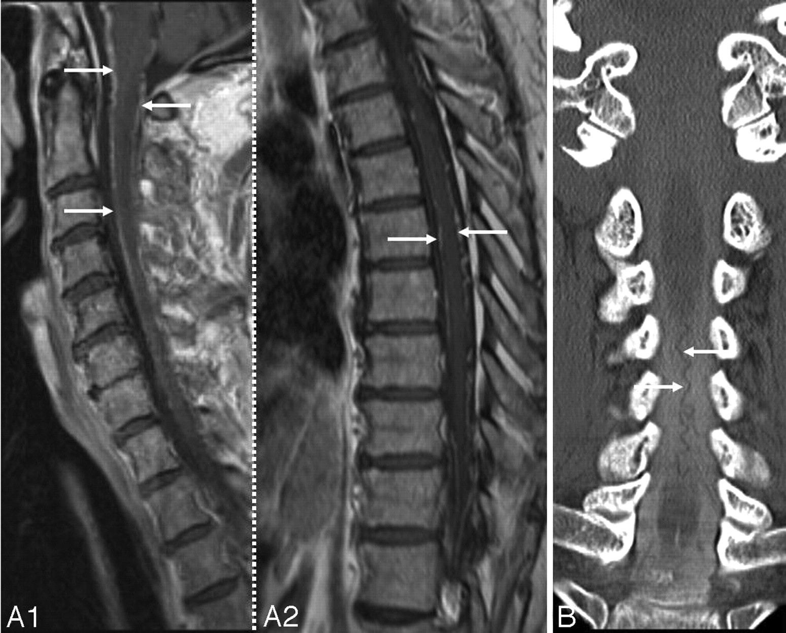
A1 and A2, Postcontrast sagittal T1-weighted cervical (A1) and thoracic (A2) MR images in a patient with SS show abnormal linear enhancement along the ventral and dorsal cord surface (same as the patient shown in 5A and B). B, CT myelogram in a patient with SS shows a posterior dilated pial venous plexus extending from the foramen magnum to C7. This prompted a cerebral and spinal angiography which showed no abnormality. A reprinted with permission from Kumar N, McKeon A, Rabinstein AA, et al. Superficial siderosis and CSF hypovolemia: the defect (dural) in the link. Neurology 2007;69:925–26 (Copyright 2007, Wolters Kluwer Health)


In this issue


Jump to section
Related Articles
Cited By...
- Diskogenic Dural Defect Is the Reason for the Ventral Location of the Epidural Spinal Fluid Collection Seen in Superficial Siderosis
- Dysarthria and ptosis
- Spontaneous Intracranial Hypotension: Atypical Radiologic Appearances, Imaging Mimickers, and Clinical Look-Alikes
- Frequency, Extent, and Correlates of Superficial Siderosis and Ependymal Siderosis in Premature Infants with Germinal Matrix Hemorrhage: An SWI Study
- Presumed superficial haemosiderosis presenting with subarachnoid haemorrhage
- Superficial Siderosis after Germinal Matrix Hemorrhage
- Cortical superficial siderosis: Prevalence and biomarker profile in a memory clinic population
- Cerebral Amyloid Angiopathy as an Etiology for Cortical Superficial Siderosis: An Unproven Hypothesis
- Postoperative MR Imaging of Spontaneous Transdural Spinal Cord Herniation: Expected Findings and Complications
- Diagnostic Significance of Cortical Superficial Siderosis for Alzheimer Disease in Patients with Cognitive Impairment
- Superficial siderosis of the central nervous system
- Prevalence of cortical superficial siderosis in a memory clinic population
- Beyond superficial siderosis: Introducing "duropathies"
- Pilot Safety Trial of Deferiprone in 10 Subjects With Superficial Siderosis
- Ventral intraspinal fluid-filled collection secondary to CSF leak presenting as bibrachial amyotrophy
- Deferiprone Reduces Hemosiderin Deposits in the Brain of a Patient with Superficial Siderosis
- Further In-Depth Look at Superficial Siderosis (and Intracranial Hypotension)
- Superficial Siderosis in Cerebral Amyloid Angiopathy